. 2021 Jul 19:12:681671.
doi: 10.3389/fimmu.2021.681671. eCollection 2021.

AeMOPE-1, a Novel Salivary Peptide From Aedes aegypti, Selectively Modulates Activation of Murine Macrophages and Ameliorates Experimental Colitis

Affiliations

AeMOPE-1, a Novel Salivary Peptide From Aedes aegypti, Selectively Modulates Activation of Murine Macrophages and Ameliorates Experimental Colitis

Priscila G Lara et al. Front Immunol. .

Abstract

The sialotranscriptomes of Aedes aegypti revealed a transcript overexpressed in female salivary glands that codes a mature 7.8 kDa peptide. The peptide, specific to the Aedes genus, has a unique sequence, presents a putative secretory nature and its function is unknown. Here, we confirmed that the peptide is highly expressed in the salivary glands of female mosquitoes when compared to the salivary glands of males, and its secretion in mosquito saliva is able to sensitize the vertebrate host by inducing the production of specific antibodies. The synthetic version of the peptide downmodulated nitric oxide production by activated peritoneal murine macrophages. The fractionation of a Ae. aegypti salivary preparation revealed that the fractions containing the naturally secreted peptide reproduced the nitric oxide downmodulation. The synthetic peptide also selectively interfered with cytokine production by murine macrophages, inhibiting the production of IL-6, IL-12p40 and CCL2 without affecting TNF-α or IL-10 production. Likewise, intracellular proteins associated with macrophage activation were also distinctively modulated: while iNOS and NF-κB p65 expression were diminished, IκBα and p38 MAPK expression did not change in the presence of the peptide. The anti-inflammatory properties of the synthetic peptide were tested in vivo on a dextran sulfate sodium-induced colitis model. The therapeutic administration of the Ae. aegypti peptide reduced the leukocytosis, macrophage activity and nitric oxide levels in the gut, as well as the expression of cytokines associated with the disease, resulting in amelioration of its clinical signs. Given its biological properties in vitro and in vivo, the molecule was termed Aedes-specific MOdulatory PEptide (AeMOPE-1). Thus, AeMOPE-1 is a novel mosquito-derived immunobiologic with potential to treat immune-mediated disorders.

Keywords: AeMOPE-1; Aedes aegypti; experimental colitis; immunomodulation; macrophages.


Conflict of interest statement

The authors declare that the research was conducted in the absence of any commercial or financial relationships that could be construed as a potential conflict of interest.

Figures

Figure 1
Figure 1
Ae. aegypti 7.8 kDa peptide is secreted in the mosquito saliva and triggers the production of specific antibodies following exposure to mosquitoes. (A) Amino acid sequence and alignment of Ae. aegypti (Accession number AAL76011.1) and Ae. albopictus (Accession number AAV90679.1) 7.8 kDa salivary peptides. The alignment was performed using MUSCLE method and graphically edited using BioEdit software. Gray highlights the identical amino acid residues (68% identity) and bold underlined amino acid residues indicate the signal peptide. (B) Ae. aegypti salivary gland extract (SGE - 10 and 40 µg) and 7.8 kDa salivary peptide (0.1, 0.5 and 1 µg) were submitted to electrophoresis on a 15% polyacrylamide gel under reducing conditions. Western blot was performed according to Material and Methods to test the reactivity of pre-immune and immune serum. The immunoreactive bands were revealed with luminescence substrate and acquired by an imaging system. (C) Sera of mice repeatedly exposed to female Ae. aegypti bites recognize the salivary peptide. Plates were coated with the salivary peptide (0.5 µg/well), incubated with serum from animals exposed 0 to 10 times to female mosquito bites (1:100 dilution) and then with secondary antibody anti-mouse IgG conjugated with HRP. The plates were revealed after substrate addition and read in a spectrophotometer at a 450 nm wavelength. White circles in (C) indicate individual sera for each exposure (n = 8). The mean ± SEM are indicated. ***≤ 0.001 versus group “0” (not exposed to mosquito bites) – One-way ANOVA.
Figure 2
Figure 2
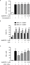
Syn-AeMOPE-1 decreases macrophage production of nitric oxide but does not affect proliferation of T lymphocytes or dendritic cell maturation. (A) Total spleen cells were pre-incubated with syn-AeMOPE-1 (0.25 - 2 µM), stimulated with concanavalin A (0.5 µg/ml) for 72 hours, and the proliferation of T lymphocytes was measured by a colorimetric assay. Results are expressed as the mean ± SEM from one representative experiment of three independent experiments assayed in sextuplicate. (B) Bone marrow-derived dendritic cells were pre-incubated with syn-AeMOPE-1 (0.25 - 2 µM), stimulated with LPS (100 ng/ml) for 24 hours, and the percentage of MHC class II+ CD80+ or MHC class II+ CD86+ cells were evaluated by flow cytometry. Gating strategy and representative plots are presented in Supplementary Figure 2 . Results are expressed as the mean ± SEM from one representative experiment of four independent experiments assayed in quadruplicate. (C) Thioglycolate-elicited peritoneal macrophages were pre-incubated with syn-AeMOPE-1 (0.25 - 2 µM), stimulated with LPS+IFN-γ (10 ng/mL of each) for 48 hours, and nitric oxide production was measured by Griess reaction. Results are expressed as the mean ± SEM from one representative experiment of five independent experiments assayed in duplicate. **≤ 0.01 and ***≤ 0.001 versus unstimulated/untreated cells; # ≤ 0.05 versus LPS+IFN-γ-stimulated cells – One-way ANOVA.
Figure 3
Figure 3
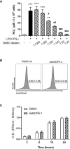
Neither DMSO nor syn-AeMOPE-1 are cytotoxic to macrophages at the dilution used in the experiments. (A) Thioglycolate-elicited peritoneal macrophages were pre-incubated with increasing concentrations of DMSO (indicated in the figure), stimulated with LPS+IFN-γ (10 ng/mL of each) for 48 hours, and nitric oxide production was measured by Griess reaction. (B) Thioglycolate-elicited peritoneal macrophages were incubated with DMSO (1:1000) or AeMOPE-1 (2 µm) for 4 hours, followed by staining with Live/Dead Viability/Cytotoxicity Assay Kit and evaluation by flow cytometry. Gating strategy and representative plots are presented in Supplementary Figure 3A . (C) Thioglycolate-elicited peritoneal macrophages were incubated with DMSO (1:1000) or AeMOPE-1 (2 µm) for 24 hours and the basal metabolic activity was monitored at different time points by a colorimetric assay. Results are expressed as the mean ± SEM from three (A, B) or five (C) independent experiments assayed in duplicates (A, B) or triplicates (C). **≤ 0.01 and ***≤ 0.001 versus unstimulated/untreated cells; # ≤ 0.05, ## ≤ 0.01 and ### ≤ 0.001 versus LPS+IFN-γ-stimulated cells – One-way ANOVA.
Figure 4
Figure 4
Native salivary AeMOPE-1 inhibits nitric oxide production by macrophages. (A) SGE was fractionated by HPLC using a column that resolves peptides, fractions were eluted at 0.5 ml/minute and their absorbance monitored at 280 nm wavelength. The arrow indicates the range of low molecular fractions (F33 to F68). (B) Western Blot of F33 to 68 revealed with serum from animals immunized with syn-AeMOPE-1. (C) Macrophages were pre-incubated with each fraction from F33 to F38 (representing the low molecular weight fractions), stimulated with LPS + IFN-γ (10 ng/mL of each) for 48 hours, and nitric oxide production was measured by Griess reaction. Results are expressed as the mean ± SEM from three independent experiments assayed in duplicate. **≤ 0.01 and ***≤ 0.001 versus unstimulated/untreated cells; ## ≤ 0.01 and ### ≤ 0.001 versus LPS+IFN-γ-stimulated cells – One-way ANOVA.
Figure 5
Figure 5
Syn-AeMOPE-1 selectively modulates macrophage activation. Macrophages were pre-incubated with syn-AeMOPE-1 (2 µM) and stimulated with LPS + IFN-γ (10 ng/mL of each). After 48 hours the production of cytokines (A) IL-6, (B) IL-12p40, (C) CCL2, and (D) TNF-α were detected in supernatants by ELISA. Values are expressed as the mean ± SEM (n = 4). *≤ 0.0001 versus unstimulated/untreated cells; # ≤ 0.0001 versus LPS+IFN-γ-stimulated cells – One-way ANOVA. Representative Western blots and densitometry for (E, I) iNOS, (F, J) IκBα, (G, K) NF-κBp65, and (H, L) p38-MAPK performed with cell lysates at different time points. Results are expressed as the mean ± SEM from three independent experiments. # ≤ 0.05 versus LPS+IFN-γ-stimulated cells –Student’s t test.
Figure 6
Figure 6
Treatment of mice with syn-AeMOPE-1 ameliorates the clinical signs of experimental colitis. Mice were divided into two groups and received 3% DSS in their drinking water for 6 days. One group was treated daily with syn-AeMOPE-1 (1 µg/animal) while the other received an equal dilution of DMSO, both in 0.9% saline, at the beginning of the clinical signs, as indicated by the arrows. (A) Clinical score was assessed daily until the time of euthanasia of animals while (B) overall clinical score and (C) post-mortem score were evaluated on the day of euthanasia. (D) Total circulating leukocytes, (E) lymphocytes, (F) monocytes and (G) neutrophils were counted on the day of euthanasia. Results are expressed as the mean ± SEM (n = 10 animals/group). Data from one representative experiment of two independent experiments are shown. *≤ 0.05 and **≤ 0.01 versus “Vehicle” group –Student’s t test. Expression of (H) IL-6, (I) IFN-γ and (J) CCL2 by RT-qPCR, as well as the (K) activity of macrophages by NAG quantification and (L) nitric oxide levels by Griess reaction, were evaluated in gut homogenates after euthanasia. Results are expressed as the mean ± SEM (n = 4-5 animals/group) from one representative experiment of two independent experiments. *≤ 0.05 and **≤ 0.01 versus “Vehicle” group –Student’s t test.
Figure 7
Figure 7
Immunomodulatory activities of syn-AeMOPE-1 in vitro and in vivo. The AeMOPE-1 transcript was identified in Aedes aegypti sialotranscriptomes. The mature secreted form of the peptide was synthetized and evaluated in murine macrophage cultures and in DSS-induced colitis. The immunomodulatory activities of the peptide are depicted in the figure. (Created with BioRender.com).

Similar articles

Cited by

References

    1. Roy S, Saha TT, Zou Z, Raikhel AS. Regulatory Pathways Controlling Female Insect Reproduction. Annu Rev Entomol (2018) 63:489–511. 10.1146/annurev-ento-020117-043258 - DOI - PubMed
    1. Gulia-Nuss M, Elliot A, Brown MR, Strand MR. Multiple Factors Contribute to Anautogenous Reproduction by the Mosquito Aedes aegypti . J Insect Physiol (2015) 82:8–16. 10.1016/j.jinsphys.2015.08.001 - DOI - PMC - PubMed
    1. Vogel KJ, Brown MR, Strand MR. Ovary Ecdysteroidogenic Hormone Requires a Receptor Tyrosine Kinase to Activate Egg Formation in the Mosquito Aedes aegypti . Proc Natl Acad Sci U S A (2015) 112(16):5057–62. 10.1073/pnas.1501814112 - DOI - PMC - PubMed
    1. Champagne DE, Smartt CT, Ribeiro JM, James AA. The Salivary Gland-Specific Apyrase of the Mosquito Aedes aegypti Is a Member of the 5’-Nucleotidase Family. Proc Natl Acad Sci U S A (1995) 92(3):694–8. 10.1073/pnas.92.3.694 - DOI - PMC - PubMed
    1. Calvo E, Tokumasu F, Marinotti O, Villeval JL, Ribeiro JM, Francischetti IM. Aegyptin, A Novel Mosquito Salivary Gland Protein, Specifically Binds to Collagen and Prevents Its Interaction With Platelet Glycoprotein VI, Integrin alpha2beta1, and Von Willebrand Factor. J Biol Chem (2007) 282(37):26928–38. 10.1074/jbc.M705669200 - DOI - PMC - PubMed

Publication types

MeSH terms

LinkOut - more resources