Optical coherence tomography-measured retinal nerve fiber layer thickness values compensated with a multivariate model and discrimination between stable and progressing glaucoma suspects
- PMID: 34350469
- PMCID: PMC8763932
- DOI: 10.1007/s00417-021-05329-3
Optical coherence tomography-measured retinal nerve fiber layer thickness values compensated with a multivariate model and discrimination between stable and progressing glaucoma suspects
Abstract
Purpose: Our previously introduced multivariate model, compensating for intersubject variability, was applied to circumpapillary retinal nerve fiber layer (RNFL) values measured with optical coherence tomography in glaucoma suspects with or without prior progressive optic disc (OD) change in a series of confocal scanning laser tomography (CSLT, HRT III) measurements.
Methods: In this prospective study, OD change during CSLT follow-up was determined with strict, moderate, and liberal criteria of the topographic change analysis (TCA). Model compensation (MC) as well as age compensation (AC) was applied to RNFL sectors (RNFLMC vs. RNFLAC). Diagnostic performance of RNFLMC vs. RNFLAC was tested with an area under the receiver operating characteristic (AUROC) and was compared between methods.
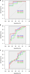
Results: Forty-two glaucoma suspects were included. Patients without prior progressive OD change during the CSLT follow-up (= stable) had thicker RNFL thickness values in most areas and for all progression criteria. RNFLMC AUROC for the global RNFL (0.719) and the inferior quadrant (0.711) performed significantly better compared with RNFLAC AUROC (0.594 and 0.631) to discriminate between stable and progressive glaucoma suspects as defined by the moderate criteria of CSLT progression analysis (p = 0.028; p = 0.024).
Conclusion: MC showed a slight but significant improvement in detection of subjects with prior progressive OD change among a group of glaucoma suspects, when compared to AC, which is the compensation method commonly used during OCT data evaluation in daily routine. Further studies are warranted to validate the present results.
Keywords: Glaucoma; HRT/CSLT; Imaging; OCT; Optic nerve.
© 2021. The Author(s).
Conflict of interest statement
The authors declare no competing interests.
Figures

References
-
- Kerrigan-Baumrind LA, Quigley HA, Pease ME, et al. Number of ganglion cells in glaucoma eyes compared with threshold visual field tests in the same persons. Invest Ophthalmol Vis Sci. 2000;41:741–748. - PubMed
MeSH terms
LinkOut - more resources
Full Text Sources
Medical

