. 2021 Aug 5;17(8):e1009686.
doi: 10.1371/journal.pgen.1009686. eCollection 2021 Aug.

A ubiquitin-like protein encoded by the "noncoding" RNA TINCR promotes keratinocyte proliferation and wound healing

Affiliations

A ubiquitin-like protein encoded by the "noncoding" RNA TINCR promotes keratinocyte proliferation and wound healing

Akihiro Nita et al. PLoS Genet. .

Abstract

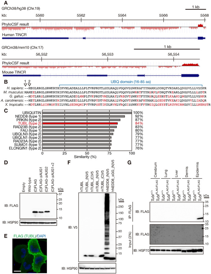
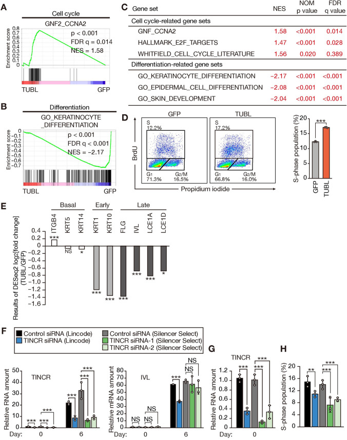
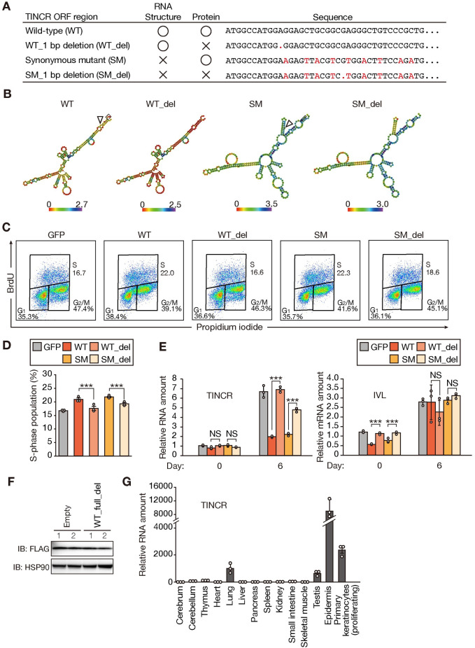
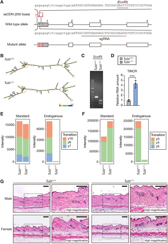
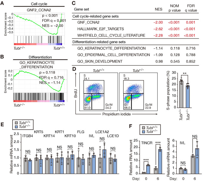
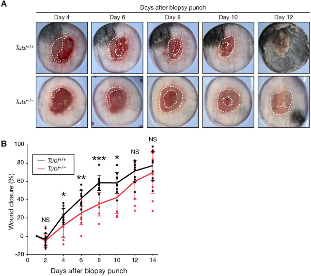
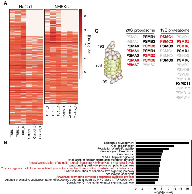
Although long noncoding RNAs (lncRNAs) are transcripts that do not encode proteins by definition, some lncRNAs actually contain small open reading frames that are translated. TINCR (terminal differentiation-induced ncRNA) has been recognized as a lncRNA that contributes to keratinocyte differentiation. However, we here show that TINCR encodes a ubiquitin-like protein that is well conserved among species and whose expression was confirmed by the generation of mice harboring a FLAG epitope tag sequence in the endogenous open reading frame as well as by targeted proteomics. Forced expression of this protein promoted cell cycle progression in normal human epidermal keratinocytes, and mice lacking this protein manifested a delay in skin wound healing associated with attenuated cell cycle progression in keratinocytes. We termed this protein TINCR-encoded ubiquitin-like protein (TUBL), and our results reveal a role for TINCR in the regulation of keratinocyte proliferation and skin regeneration that is dependent on TUBL.


Conflict of interest statement

The authors have declared that no competing interests exist.

Figures

Fig 1
Fig 1. TINCR encodes the ubiquitin-like protein TUBL.
(A) Smoothed PhyloCSF peaks for human and mouse TINCR. (B) Comparison of the predicted amino acid sequence of human TUBL with that of the mouse, avian, reptile, and amphibian proteins. Arrowheads indicate the first and second methionines of mouse TUBL. Amino acids that differ between species are shown in red. (C) Amino acid sequence similarity for human TUBL and other human ubiquitin-like proteins compared with human ubiquitin. (D) Immunoblot (IB) analysis of HEK293T cells transiently transfected with expression vectors for mouse TINCR with a FLAG epitope tag sequence inserted at the COOH-terminus (C) of the TUBL ORF or for its ΔAUG mutants. The analysis was performed with M2 antibodies to FLAG and antibodies to HSP90 (loading control). (E) Immunofluorescence analysis of FLAG in HeLa cells expressing FLAG epitope–tagged mouse TUBL. Nuclei were stained with 4′,6-diamidino-2-phenylindole (DAPI). Scale bar, 20 μm. (F) Immunoblot analysis of HEK293T cells transiently transfected with expression vectors for human (h) or mouse (m) TUBL with or without an NH2 (N)–or COOH (C)–terminal V5 epitope tag or for human NEDD8 with an NH2-terminal V5 tag or its mutant (ΔGG) lacking the diglycine motif. (G) Lysates of tissues from wild-type (Tubl+/+) mice or mice harboring a FLAG epitope tag sequence at the COOH-terminus of the TUBL ORF (TublFLAG/FLAG mice) were subjected to immunoprecipitation (IP) with antibodies to FLAG, and the resulting precipitates as well as the original lysates (3% of input for immunoprecipitation) were subjected to immunoblot analysis with antibodies to FLAG and to HSP70 (loading control).
Fig 2
Fig 2. TUBL expression promotes cell cycle progression and inhibits differentiation in human keratinocytes.
(A, B) GSEA plots for gene sets related to the cell cycle (A) or keratinocyte differentiation (B) constructed from RNA-seq data obtained for NHEKs expressing GFP or human TUBL. FDR, false discovery rate; NES, normalized enrichment score. (C) Summary of GSEA results for gene sets related to the cell cycle or keratinocyte differentiation. NOM, nominal. (D) Flow cytometric traces and quantification of BrdU incorporation for NHEKs stably expressing GFP or TUBL. Data in the right panel are means ± SD (n = 3 independent experiments). ***p < 0.005 (Student’s t test). (E) RNA-seq results for differentially expressed genes related to basal, early, or late keratinocyte differentiation. The results for NHEKs expressing TUBL were normalized by those for NHEKs expressing GFP. *p < 0.05, ***p < 0.005; NS, not significant (adjusted p values). (F, G) RT-qPCR analysis of TINCR and involucrin (IVL) mRNA abundance in NHEKs transfected with the indicated control or TINCR siRNAs for 1 day and then subjected to calcium-induced differentiation for 0 or 6 days (F). The results for TINCR abundance at day 0 of differentiation are expanded in (G). Data are means ± SD (n = 3 independent experiments). ***p < 0.005, NS (Student’s t test). (H) Quantification of BrdU incorporation for NHEKs transfected with the indicated control or TINCR siRNAs for 2 days. Data are means ± SD (n = 3 independent experiments). **p < 0.01, ***p < 0.005 (Student’s t test).
Fig 3
Fig 3. TUBL expression promotes proliferation of mouse primary keratinocytes in a manner independent of the secondary structure of TINCR RNA.
(A) Summary of mutations introduced into the mouse TUBL ORF. (B) Predicted secondary structure and minimal free energy for WT, WT_del, SM, and SM_del. (C, D) Flow cytometric traces (C) and quantification (D) of BrdU incorporation for mouse primary keratinocytes stably expressing either GFP or WT, WT_del, SM, or SM_del forms of TINCR. Data in (D) are means ± SD (n = 3 independent experiments). ***p < 0.005 (Student’s t test). (E) RT-qPCR analysis of TINCR and involucrin (IVL) mRNA abundance in mouse primary keratinocytes stably expressing either GFP or WT, WT_del, SM, or SM_del forms of TINCR and subjected to calcium-induced differentiation in vitro for 0 or 6 days. Data are means ± SD (n = 3 independent experiments). ***p < 0.005, NS (Student’s t test). (F) Immunoblot analysis of HEK293T cells transiently transfected both with an expression vector for the mouse TUBL ORF with a COOH-terminal FLAG epitope tag and with either an expression vector for an almost full-length form of mouse TINCR with a 1-bp deletion in the TUBL ORF (WT_full_del) or the corresponding empty vector. Two replicates (lanes 1 and 2) are shown. (G) RT-qPCR analysis of TINCR in adult mouse tissues and actively proliferating primary mouse keratinocytes. Data are means ± SD (n = 3 independent experiments).
Fig 4
Fig 4. Generation of TUBL-deficient mice.
(A) Schematic representation of the WT TINCR allele, the single-stranded oligodeoxynucleotide (ssODN), and the mutant allele after homologous recombination. Exons are denoted by numbered boxes. The single guide RNA (sgRNA) for the CRISPR-Cas9 system and its protospacer adjacent motif (PAM) are indicated by contiguous black and red underlines, respectively. The TUBL ORF is represented by the gray shading in the box corresponding to exon 1 of TINCR. (B) Predicted secondary structure and minimal free energy for WT TINCR and the mutant form generated by the CRISPR-Cas9 system for establishment of Tubl−/− mice. The triangle indicates the 5’ end of the transcript. (C) PCR analysis of genomic DNA from the tail of mice of the indicated genotypes. The PCR products were digested with EcoRI before electrophoresis. (D) RT-qPCR analysis of TINCR in the epidermis of Tubl+/+ and Tubl−/− mice. Data are means ± SD (n = 3 independent experiments). ***p < 0.005 (Student’s t test). (E, F) Signal intensity of extracted ion chromatograms for mouse TUBL peptides in MRM analysis. The analysis detected two different peptides derived from mouse TUBL with the amino acid sequences AQLVGQGVSSWR (E) and DTLSDLR (F). “Standard” indicates an internal standard corresponding to stable isotope–labeled recombinant mouse TUBL. “Endogenous” indicates the endogenous TUBL peptides in epidermal lysates prepared from Tubl+/+ or Tubl−/− mice. Tryptic peptides derived from endogenous TUBL were mixed with tryptic peptides derived from the isotopically labeled recombinant protein and were then applied to MS analysis. Extracted ion chromatograms for each transition are shown in S1 Fig. (G) Hematoxylin-eosin staining of the skin of Tubl+/+ or Tubl−/− mice at 8 weeks of age. The boxed regions in the left panels of each set are shown at higher magnification in the right panels. Scale bars, 300 μm.
Fig 5
Fig 5. TUBL deficiency delays cell cycle progression in mouse primary keratinocytes.
(A, B) GSEA plots for gene sets related to the cell cycle (A) or keratinocyte differentiation (B) constructed from RNA-seq data for the epidermis of Tubl+/+ or Tubl−/− mice at 8 weeks of age. FDR, false discovery rate; NES, normalized enrichment score. (C) Results of GSEA for gene sets related to the cell cycle or keratinocyte differentiation. NOM, nominal. (D) Flow cytometric traces and quantification of BrdU incorporation in primary keratinocytes from Tubl+/+ or Tubl−/− mice. Data in the right panel are means ± SD (n = 7 or 8 independent experiments). **p < 0.01 (Student’s t test). (E) RT-qPCR analysis of keratinocyte differentiation–related gene expression in the epidermis of Tubl+/+ and Tubl−/− mice. Data are means ± SD (n = 4 independent experiments). NS, Student’s t test. (F) RT-qPCR analysis of TINCR and involucrin mRNA abundance in mouse primary keratinocytes established from Tubl+/+ or Tubl−/− mice and subjected to calcium-induced differentiation in vitro for 0 or 6 days. Data are means ± SD (n = 3 or 4 independent experiments). ***p < 0.005, NS (Student’s t test).
Fig 6
Fig 6. Loss of TUBL results in delayed recovery after skin injury.
(A) Representative macroscopic views of cutaneous ulcers of Tubl+/+ and Tubl−/− mice at 4, 6, 8, 10, and 12 days after wounding with a biopsy punch. The area demarcated by white dashed lines indicates the area of re-epithelialization. (B) Time course of wound healing in Tubl+/+ (n = 11) and Tubl−/− (n = 10) mice. Data are means ± SD. *p < 0.05, **p < 0.01, ***p < 0.005, NS versus corresponding value for Tubl–/–mice (Student’s t test).
Fig 7
Fig 7. Identification of TUBL binding proteins.
(A) Heat map of iBAQ (intensity-based absolute quantification) values for HaCaT cells or NHEKs transiently expressing human TUBL with a COOH-terminal FLAG tag or transfected with the corresponding empty vector (Control). Immunoprecipitates prepared from the cells with antibodies to FLAG were subjected to semiquantitative LC-MS/MS analysis. Data are shown for three biological replicates. (B) GO analysis of identified TUBL binding proteins. Representative findings are presented. (C) Schematic representation of the proteasome complex (left) and lists of proteasome subunits identified as TUBL binding proteins (right). 19S and 20S indicate the 19S regulatory particle and 20S catalytic particle of the proteasome, respectively. Red and black characters indicate subunits identified in both HaCaT cells and NHEKs or in one of the two cell types, respectively.

References

    1. Pasparakis M, Haase I, Nestle FO. Mechanisms regulating skin immunity and inflammation. Nat Rev Immunol. 2014;14(5):289–301. doi: 10.1038/nri3646 - DOI - PubMed
    1. Zomer HD, Trentin AG. Skin wound healing in humans and mice: Challenges in translational research. J Dermatol Sci. 2018;90(1):3–12. doi: 10.1016/j.jdermsci.2017.12.009 - DOI - PubMed
    1. Blanpain C, Fuchs E. Epidermal homeostasis: a balancing act of stem cells in the skin. Nat Rev Mol Cell Biol. 2009;10(3):207–17. doi: 10.1038/nrm2636 - DOI - PMC - PubMed
    1. Eming SA, Martin P, Tomic-Canic M. Wound repair and regeneration: Mechanisms, signaling, and translation. Sci Transl Med. 2014;6(265):265sr6. doi: 10.1126/scitranslmed.3009337 - DOI - PMC - PubMed
    1. Gurtner GC, Werner S, Barrandon Y, Longaker MT. Wound repair and regeneration. Nature. 2008;453(7193):314–21. doi: 10.1038/nature07039 - DOI - PubMed

Publication types