. 2021 Oct 11;33(10):3367-3385.
doi: 10.1093/plcell/koab198.

The 5-formyl-tetrahydrofolate proteome links folates with C/N metabolism and reveals feedback regulation of folate biosynthesis

Affiliations

The 5-formyl-tetrahydrofolate proteome links folates with C/N metabolism and reveals feedback regulation of folate biosynthesis

Weichao Li et al. Plant Cell. .

Abstract

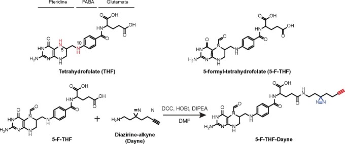
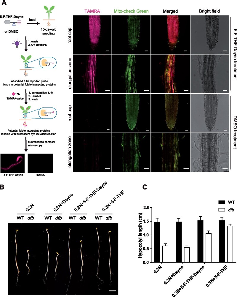
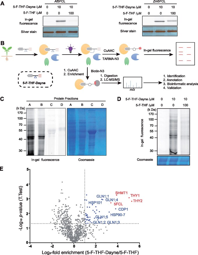
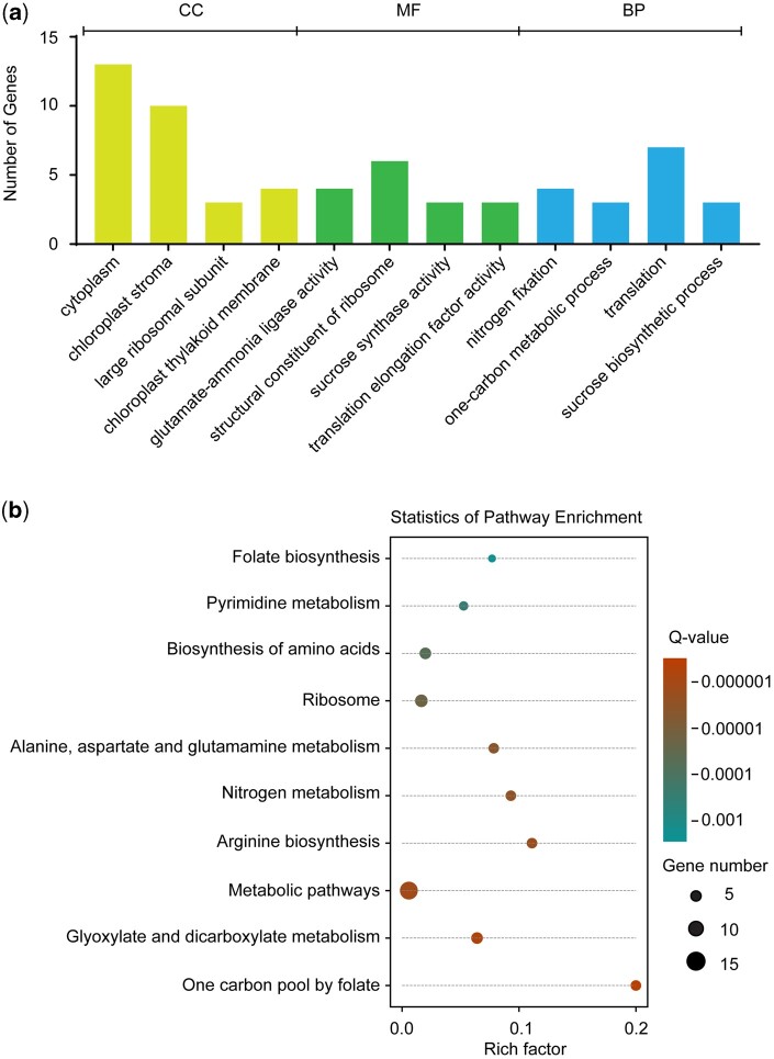
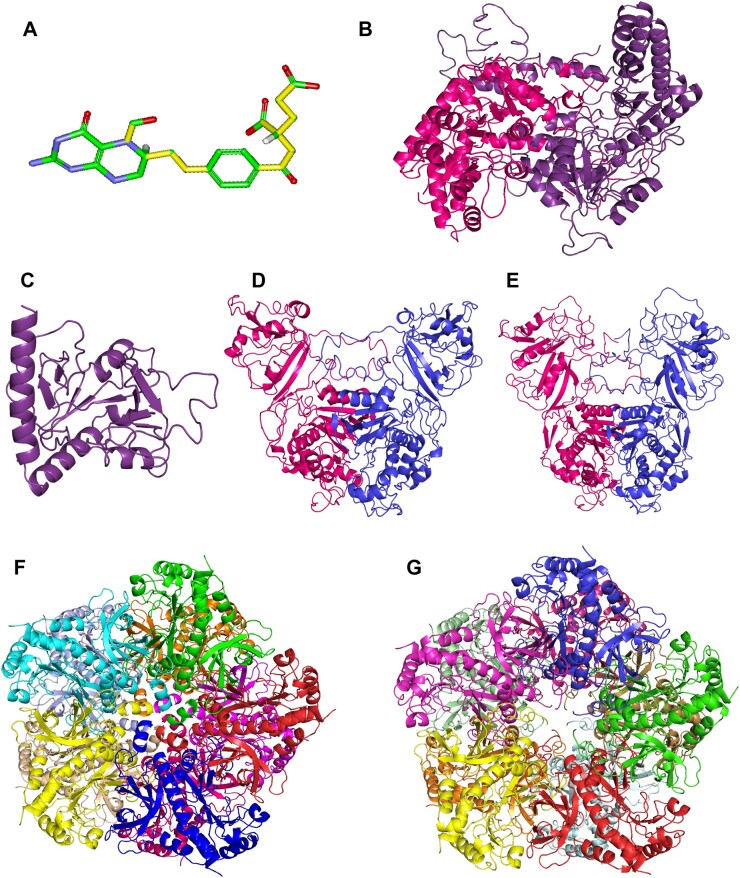
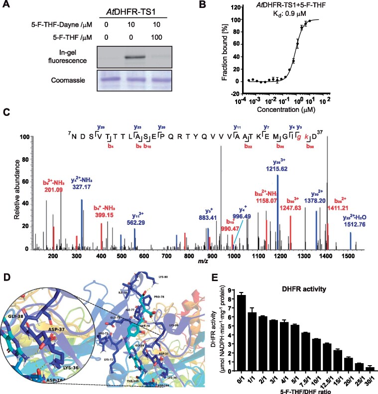
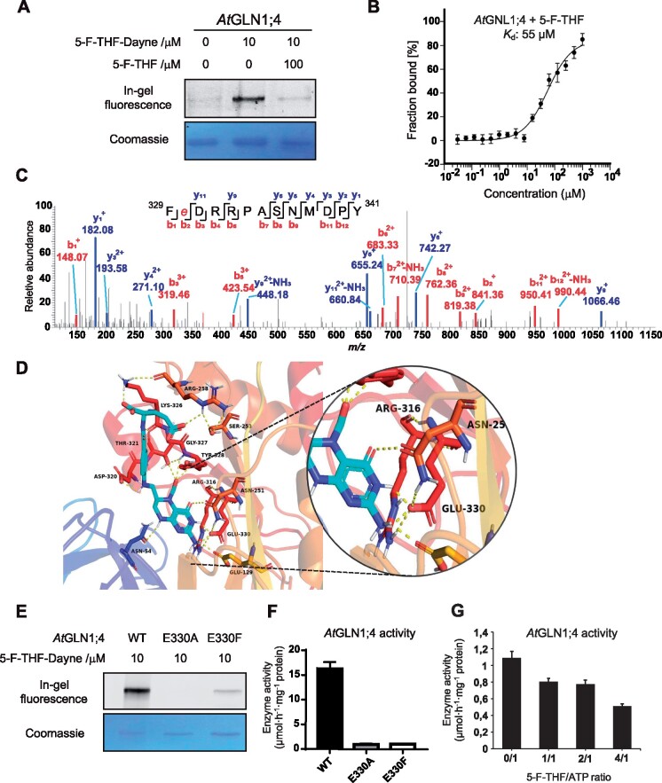
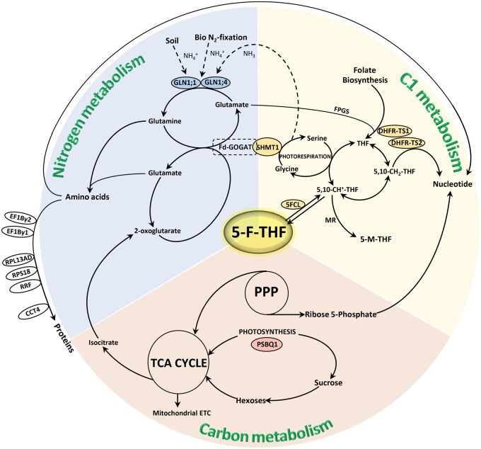
Folates are indispensable for plant development, but their molecular mode of action remains elusive. We synthesized a probe, "5-F-THF-Dayne," comprising 5-formyl-tetrahydrofolate (THF) coupled to a photoaffinity tag. Exploiting this probe in an affinity proteomics study in Arabidopsis thaliana, we retrieved 51 hits. Thirty interactions were independently validated with in vitro expressed proteins to bind 5-F-THF with high or low affinity. Interestingly, the interactors reveal associations beyond one-carbon metabolism, covering also connections to nitrogen (N) metabolism, carbohydrate metabolism/photosynthesis, and proteostasis. Two of the interactions, one with the folate biosynthetic enzyme DIHYDROFOLATE REDUCTASE-THYMIDYLATE SYNTHASE 1 (AtDHFR-TS1) and another with N metabolism-associated glutamine synthetase 1;4 (AtGLN1;4), were further characterized. In silico and experimental analyses revealed G35/K36 and E330 as key residues for the binding of 5-F-THF in AtDHFR-TS1 and AtGLN1;4, respectively. Site-directed mutagenesis of AtGLN1;4 E330, which co-localizes with the ATP-binding pocket, abolished 5-F-THF binding as well as AtGLN1;4 activity. Furthermore, 5-F-THF was noted to competitively inhibit the activities of AtDHFR-TS1 and AtGLN1;4. In summary, we demonstrated a regulatory role for 5-F-THF in N metabolism, revealed 5-F-THF-mediated feedback regulation of folate biosynthesis, and identified a total of 14 previously unknown high-affinity binding cellular targets of 5-F-THF. Together, this sets a landmark toward understanding the role of folates in plant development.


Figures

Figure 1
Figure 1
Schematic structure of THF, 5-F-THF, and 5-F-THF-Dayne probe. THF consists of three moieties: pteridine, PABA, and glutamate. The N5 and N10 positions in THF are marked in red. In the 5-F-THF-Dayne probe, a “diazirine-alkyne” (“Dayne”) photoaffinity tag was coupled to the γ-carboxyl group. The diazo part “N=N” is indicated in blue and the alkyne moiety in red. For detailed synthesis procedure see “Methods” section. HOBt, hydroxyl-benzotriazole; DIPEA, N,N-diisopropyl-ethylamine.
Figure 2
Figure 2
Confirmation of the in vivo biological activity of the 5-F-THF-Dayne probe. A, In vivo fluorescence imaging of 5-F-THF-Dayne in root tips of 10-day-old Arabidopsis WT seedlings. The stepwise workflow is shown at the left. Briefly, root tips were fed with 5-F-THF-Dayne probe for 3 h, followed by a click reaction with the TAMRA azide fluorophore. The tissue was then analyzed under a confocal microscope. Confocal images at the right show active uptake of the probe by Arabidopsis roots. The red signal of TAMRA (shown in magenta) shows the distribution of the probe in the root cap and elongation zone. Root tips incubated with DMSO as a control showed no fluorescence postclick reaction with TAMRA. Mito-check green, mitochondrion marker. Bar = 25 μm. B and C, Complementation of the dfb mutant showing active utilization of 5-F-THF-Dayne by the mutant seedlings. B, Hypocotyl growth (bar = 0.2 cm) and (C) length of 6-day-old Arabidopsis WT and dfb mutant grown in darkness on low N (0.3 N) medium, supplemented with Dayne, 5-F-THF (50 μM) and 5-F-THF-Dayne (50 μM), respectively.
Figure 3
Figure 3
Confirmation of in vitro activity of the 5-F-THF-Dayne and chemoproteomics profiling of 5-F-THF-binding proteins in Arabidopsis. A, In vitro interaction of 5-F-THF-Dayne with 5FCL from Arabidopsis (At5FCL) and maize (Zm5FCL) as indicated by in-gel fluorescence imaging. 5-F-THF-Dayne, 5-F-THF probe. B, Workflow for profiling of 5-F-THF-Dyane interacting proteins. The irregular grapes in yellow, blue, and green colors represent extracted proteins. In brief, 5-F-THF-Dayne probe was incubated with the Arabidopsis protein extract; subsequently, the photoreactive diazirine moiety was covalently linked with the interacting proteins after irradiation by UV light. Eventually, the bound proteins were visualized by in-gel fluorescence detection or enriched for LC–MS/MS identification through a copper-catalyzed azide–alkyne cycloaddition reaction with TAMRA-N3 or biotin-N3, respectively. c, In-gel fluorescence labeling of four protein fractions (A, B, C, and D) of the Arabidopsis proteome by 5-F-THF-Dayne (left) and Coomassie staining of the gel (right). D, Competitive labeling of the Arabidopsis proteome by 5-F-THF-Dayne. E, Quantitative mass spectrometry-based profiling of the 5-F-THF-Dayne binding proteins. Blue and red dots depict selected targets that were outcompeted by 5-F-THF (criteria: t-test difference log2-fold enrichment ≥1 and − log10 (P-value) ≥1.33). Red dots represent the identified enzymes involved in C1 metabolism.
Figure 4
Figure 4
Bioinformatics analysis of the identified 5-F-THF-binding proteins utilizing GO platform. A, Gene function enrichment of 5-F-THF-binding proteins. CC, cellular component; B, KEGG pathway analysis of the identified 5-F-THF-binding proteins.
Figure 5
Figure 5
Structure of the ligand 5-F-THF and homology models of the selected FFBPs. 3D structure of 5-F-THF (A) and homology models of the two known FFBPs—SHMT1 (B) and 5FCL (C), and four high-affinity FFBPs –AtDHFR-TS1 (D), AtDHFR-TS2 (E), AtGLN1;1 (F) and AtGLN1;4 (G). The 5-F-THF molecule shown in stick model and its 11 rotatable bonds are presented in yellow (color of atoms: green for C, red for oxygen, blue for N, and white for hydrogen).
Figure 6
Figure 6
In vitro characterization of the binding of 5-F-THF with the recombinant Arabidopsis AtDHFR-TS1. A, Gel-based labeling results showing the binding of 5-F-THF-Dayne (10 μM) with AtDHFR-TS1. B, Dose-response curve reflecting binding of the recombinant AtDHFR-TS1 with 5-F-THF as assessed by MST analysis. The curve was generated by NanoTemper Analysis version 1.2.231. Normalized fluorescence (hot fluorescence/initial fluorescence) was plotted as a function of 5-F-THF concentration. Three independent thermophoresis measurements were performed; results are shown as mean � SE of the three repetitions. C and D, Identification of the binding site of 5-F-THF in AtDHFR-TS1. C, LC–MS/MS revealed that G35 or K36 is the potential binding position of 5-F-THF-Dayne in AtDHFR-TS1. G, glycine; K, lysine. D, Molecular docking prediction of the binding of 5-F-THF with AtDHFR-TS1. 5-F-THF is represented in light blue and hydrogen bonds are represented with dotted yellow lines. E, Enzymatic activity of the native AtDHFR-TS1 under conditions of varying 5-F-THF/ DHF ratio (50 �M DHF was used). DHFR activity values are means � se of three independent reactions. For the corresponding enzyme kinetics curve showing NADPH absorbance at 340 nm, see Supplemental Figure S8.
Figure 7
Figure 7
In vitro characterization of the binding of 5-F-THF with the recombinant Arabidopsis AtGLN1;4. A, Gel-based labeling results showing the binding of 5-F-THF-Dayne (10 μM) with AtGLN1;4. B, Dose-response curve of the recombinant AtGLN1;4 with 5-F-THF obtained by MST analysis. The binding curve was generated by NanoTemper Analysis version 1.2.231. Normalized fluorescence (hot fluorescence/initial fluorescence) was plotted as a function of 5-F-THF concentration. Three independent thermophoresis measurements were performed; results are shown as mean � SE of the three repetitions. C and D, Identification of the binding site of 5-F-THF in AtGLN1;4. C, LC–MS/MS identification of E330 as the potential binding position of 5-F-THF-Dayne in AtGLN1;4. E, glutamate. D, Molecular docking prediction of the binding of 5-F-THF with AtGLN1;4. 5-F-THF is represented in light blue and hydrogen bonds are represented with dotted yellow lines. E, The gel-based labeling of native and mutant AtGLN1;4s (E330A/F) by 5-F-THF-Dayne. F, Enzymatic activities of native and mutated (E330A/F) AtGLN1;4s. G, Enzymatic activity of native AtGLN1;4 under conditions of varying 5-F-THF/ATP ratio (400 �M ATP was used, Km value as indicated by Ishiyama et al., 2004).
Figure 8
Figure 8
Broad schematic representation of the C1, N, and C metabolism, reflecting 5-F-THF as a nexus between the three metabolisms. Known- and high-affinity FFBPs/enzymes are depicted. The two N metabolism-related high-affinity FFBPs, the GSs—GLN1;1 and GLN1;4 are encircled with blue fill, the C metabolism-related high-affinity FFBP, PSBQ1, with pink fill, and the four C1 metabolism-related FFBPs, DHFR-TS1 and 2( high-affinity), SHMT1 (known) and 5FCL (known), with yellow fill. The encircled high-affinity FFBPs indicated on the left hand of the scheme have broad association with N metabolism, more specifically in amino acid and protein metabolism—RPS18, RPL12-A, RPL12-C, S11 family protein (RPS14C), RPS7A, RPL13AD, translation elongation factor gamma chain (EF1Bγ1 and EF1Bγ2), RRF, and CCT4. The scheme is proposed as a reference road map for future studies involving characterization of these bindings as well as functional relevance in folate-mediated plant development.

Similar articles

Cited by

References

    1. Blancquaert D, De Steur H, Gellynck X, Van Der Straeten D (2014) Present and future of folate biofortification of crop plants. J Exp Bot 65: 895–906 - PubMed
    1. Blancquaert D, Van Daele J, Strobbe S, Kiekens F, Storozhenko S, De Steur H, Gellynck X, Lambert W, Stove C, Van Der Straeten D (2015) Improving folate (vitamin B-9) stability in biofortified rice through metabolic engineering. Nat Biotechnol 33: 1076. - PubMed
    1. Bouvier F, Linka N, Isner JC, Mutterer J, Weber APM, Camara B (2006) Arabidopsis SAMT1 defines a plastid transporter regulating plastid biogenesis and plant development. Plant Cell 18: 3088–3105 - PMC - PubMed
    1. Cao L, Henty-Ridilla JL, Blanchoin L, Staiger CJ (2016) Profilin-dependent nucleation and assembly of actin filaments controls cell elongation in Arabidopsis. Plant Physiol 170: 220–233. - PMC - PubMed
    1. Conway LP, Li WC, Parker CG, , (2021) Chemoproteomic-enabled phenotypic screening. Cell Chem Biol 28: 371–393 - PubMed

Publication types