. 2021 Sep:136:104704.
doi: 10.1016/j.compbiomed.2021.104704. Epub 2021 Jul 29.

Learning-to-augment strategy using noisy and denoised data: Improving generalizability of deep CNN for the detection of COVID-19 in X-ray images

Affiliations

Learning-to-augment strategy using noisy and denoised data: Improving generalizability of deep CNN for the detection of COVID-19 in X-ray images

Mohammad Momeny et al. Comput Biol Med. 2021 Sep.

Abstract

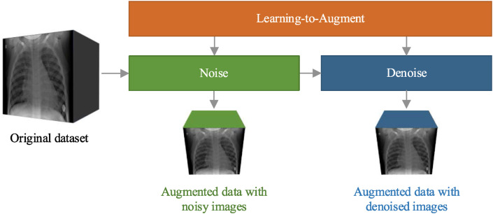
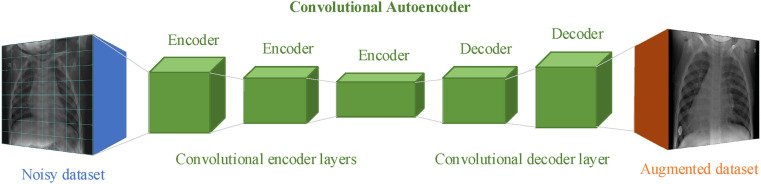
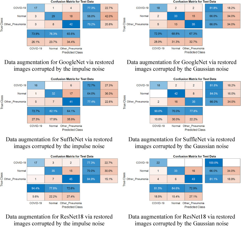
Chest X-ray images are used in deep convolutional neural networks for the detection of COVID-19, the greatest human challenge of the 21st century. Robustness to noise and improvement of generalization are the major challenges in designing these networks. In this paper, we introduce a strategy for data augmentation using the determination of the type and value of noise density to improve the robustness and generalization of deep CNNs for COVID-19 detection. Firstly, we present a learning-to-augment approach that generates new noisy variants of the original image data with optimized noise density. We apply a Bayesian optimization technique to control and choose the optimal noise type and its parameters. Secondly, we propose a novel data augmentation strategy, based on denoised X-ray images, that uses the distance between denoised and original pixels to generate new data. We develop an autoencoder model to create new data using denoised images corrupted by the Gaussian and impulse noise. A database of chest X-ray images, containing COVID-19 positive, healthy, and non-COVID pneumonia cases, is used to fine-tune the pre-trained networks (AlexNet, ShuffleNet, ResNet18, and GoogleNet). The proposed method performs better results compared to the state-of-the-art learning to augment strategies in terms of sensitivity (0.808), specificity (0.915), and F-Measure (0.737). The source code of the proposed method is available at https://github.com/mohamadmomeny/Learning-to-augment-strategy.

Keywords: COVID-19; Classification; Data augmentation; Deep learning; Learning-to-augment; Noise; X-ray images.


Figures

Fig. 1
Fig. 1
Schematic pipeline of the proposed learning-to-augment strategy using noisy and denoised data.
Fig. 2
Fig. 2
Flowchart of our noise-based image data augmentation approach.
Fig. 3
Fig. 3
Flowchart of the autoencoder-restored image-based data augmentation approach.
Fig. 4
Fig. 4
The proposed convolutional autoencoder learns to denoise images.
Fig. 5
Fig. 5
Sample chest X-ray images for (a) COVID-19, (b) healthy, and (c) non-COVID pneumonia cases [56].
Fig. 6
Fig. 6
Noise added to the chest X-ray images.
Fig. 7
Fig. 7
Noise-added and restored COVID-19 positive chest X-ray images.
Fig. 8
Fig. 8
Noise-added and restored healthy chest X-ray images.
Fig. 9
Fig. 9
Noise-added and restored chest X-ray images of non-COVID pneumonia types.
Fig. 10
Fig. 10
The Bayesian optimizer evaluated the data augmentation policies for restored images, initially corrupted by impulse noise (impulse noisy density d=17%, MSE = 0.288).
Fig. 11
Fig. 11
Training and validation accuracy curves of ResNet18 in the X-ray image classification task.
Fig. 12
Fig. 12
Training and validation loss curves of ResNet18 in the X-ray image classification task.
Fig. 13
Fig. 13
Confusion matrices for X-ray image classification by the proposed learning-to-augment approach using restored images corrupted by noise. Here, ‘Normal’ represents the ‘Healthy’ subjects and ‘Other_Pneumonia’ represents the ‘non-COVID pneumonia’ patients.
Fig. 14
Fig. 14
Bar plot showing the accuracies of COVID-19 classification for different augmentation approaches.
Fig. 15
Fig. 15
Evaluation of the classification performance by identifying the positive and negative classes in three steps.
Fig. 16
Fig. 16
Comparison of the sensitivity, specificity, and F-Measure of COVID-19 classification for different augmentation strategies.

References

    1. Varela-Santos S., Melin P. "A new approach for classifying coronavirus COVID-19 based on its manifestation on chest X-rays using texture features and neural networks. Information sciences. 2021;545:403–414. - PMC - PubMed
    1. Bhattacharya S., Maddikunta P.K.R., Pham Q.V., Gadekallu T.R., Chowdhary C.L., Alazab M., Piran M.J. "Deep learning and medical image processing for coronavirus (COVID-19) pandemic: a survey. Sustainable cities and society. 2021;65:102589. - PMC - PubMed
    1. Ahmed I., Ahmad M., Rodrigues J.J., Jeon G., Din S. "A deep learning-based social distance monitoring framework for COVID-19. Sustainable Cities and Society. 2021;65:102571. - PMC - PubMed
    1. Hussain E., Hasan M., Rahman M.A., Lee I., Tamanna T., Parvez M.Z. "CoroDet: a deep learning based classification for COVID-19 detection using chest X-ray images. Chaos, Solitons & Fractals. 2021 doi: 10.1016/j.chaos.2020.110495. 142, p. 110495, 2021/01/01. - DOI - PMC - PubMed
    1. Lee E.Y., Ng M.-Y., Khong P.-L. "COVID-19 pneumonia: what has CT taught us?,". Lancet Infect. Dis. 2020;20(4):384–385. - PMC - PubMed