Review
. 2021 Apr;100(S 01):S1-S36.
doi: 10.1055/a-1337-5703. Epub 2021 Apr 30.

Rare Diseases of Larynx, Trachea and Thyroid

[Article in English, German]
Affiliations
Review

Rare Diseases of Larynx, Trachea and Thyroid

[Article in English, German]
Stephan Hackenberg et al. Laryngorhinootologie. 2021 Apr.

Abstract

This review article covers data on rare diseases of the larynx, the trachea and the thyroid. In particular, congenital malformations, rare manifestations of inflammatory laryngeal disorders, benign and malignant epithelial as well as non-epithelial tumors, laryngeal and tracheal manifestations of general diseases and, finally, thyroid disorders are discussed. The individual chapters contain an overview of the data situation in the literature, the clinical appearance of each disorder, important key points for diagnosis and therapy and a statement on the prognosis of the disease. Finally, the authors indicate on study registers and self-help groups.

Der Übersichtsartikel beinhaltet eine Zusammenstellung seltener Erkrankungen von Larynx, Trachea und Schilddrüse. Im Speziellen werden angeborene Fehlbildungen, seltene Formen der entzündlichen Larynxerkrankungen, gutartige und bösartige epitheliale sowie nicht-epitheliale Tumoren, laryngeale und tracheale Manifestationen von Allgemeinerkrankungen und schließlich seltene Erkrankungen der Schilddrüse besprochen. Die einzelnen Kapitel beinhalten eine Übersicht über die Datenlage in der Literatur, das jeweilige klinische Erscheinungsbild, wichtige Stichpunkte zur Diagnostik und zur Therapie und eine abschließende Stellungnahme zur Prognose der Erkrankung. Des Weiteren finden sich Hinweise zu Studienregistern und Selbsthilfegruppen.


Conflict of interest statement

Die Autorinnen/Autoren geben an, dass kein Interessenkonflikt besteht.

Figures

Fig. 1
Fig. 1
Laryngeal clefts. Type 1 The laryngeal cleft only concerns the postcricoid region and ends without cartilage involvement above the level of the vocal folds. Type 2 The laryngeal cleft draws into the cricoid cartilage and ends below the level of the vocal folds. Type 3 There is also a cleft of the trachea, which ends above the thoracic aperture. Type 4 The tracheal part of the cleft draws beyond the thoracic aperture just above the bifurcation.
Fig. 2
Fig. 2
Tracheoesophageal fistulas. Gross A/Vogt II An atresia of the esophagus is found without development of tracheoesophageal fistula. In this case, no air is accumulated in the stomach. Gross B/Vogt IIIa Esophageal atresia is found with tracheoesophageal fistula at the upper segment. The lower segment ends blindly. Gross C/Vogt IIIb Esophageal atresia is found with tracheoesophageal fistula. The upper segment ends blindly. This is the most frequently observed type of esophageal atresia. Gross D/Vogt IIIc Esophageal atresia is found with fistula development and upper segment. Gross E/Vogt IV Tracheoesophageal fistula is found without presence of esophageal atresia (so-called H fistula).
Fig. 3
Fig. 3
Tracheal agenesis. Type 1 A tracheal stump is found above the bifurcation into which a fistula opens from the cervical esophagus; incidence of 20%. Type 2 A tracheal stump does not exist, the bifurcation is directly connected with the esophagus; incidence of 60%. Type 3 In this case, also the bifurcation is missing, both main bronchi open separately into the esophagus; incidence of 20%.
Fig. 4
Fig. 4
a Tracheal agenesis in a newborn. The epiglottis is developed, the laryngeal lumen is closed. Ventilation of the patient via the esophagus and the esophago-bronchial fistula is performed. b Tracheal agenesis in a newborn. Now with uploaded epiglottis and visibly free esophageal lumen and completely closed laryngeal lumen.
Fig. 5
Fig. 5
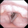
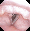
Papillomatosis. (*) Laryngeal papillomatosis (here: HPV type 6) with pathognomonic vascular loops; flexible laryngoscopy (chip/tip).
Fig. 6
Fig. 6
Rhabdomyoma. a Histology reveals a tumor with large polygonal long cells with abundant granular eosinophilic cytoplasm which is partly striated and small round nuclei with distinct cell borders. Mitoses only rarely occur or are completely missing. b In higher amplification, a striation of the tumor cells is seen. c Immunohistochemistry reveals a high expression of desmin. d Myogenin reacts positively in single nuclei.
Fig. 7
Fig. 7
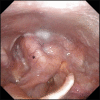
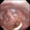
Schwannoma. (*) Plexiform laryngeal schwannoma of the left supraglottis; flexible laryngoscopy (chip/tip).
Fig. 8
Fig. 8
Schwannoma. The figures show a plexiform schwannoma. a Schwannomas are nerve sheath tumors that consist entirely or nearly entirely of differentiated neoplastic Schwann cells. In the overview, a characteristic nodular growth is observed that is typical for plexiform schwannomas. b/c In higher amplification, a monotone, cell-rich spindle-cell tumor without atypia, necrosis or increased mitotic activity as indication of a benign tumor is seen in contrast to classic schwannomas, that have a growth pattern changing between Antoni a and a .
Fig. 9
Fig. 9
Granular cell tumor. (*) Granular cell tumor of the posterior third of the right vocal fold; flexible laryngoscopy (chip/tip).
Fig. 10
Fig. 10
Granular cell tumor. a Microscopic examination shows a tumor that has grown in large accumulations with large tumor cells that are not sharply defined and have a granular cytoplasm. The nuclei are mostly centrally located with homogenous, slightly hyperchromatic and enlarged vesicular nuclei. In immunohistochemistry, the tumor cells react positively on b S100 and c S0X10 as indication for neuroectodermal origin. d Furthermore, tumor cells react positively on CD68.
Fig. 11
Fig. 11
Hemangioma. (*) Congenital subglottic hemangioma on the left with multiple midline hemangiomas; flexible laryngoscopy (chip/tip).
Fig. 12
Fig. 12
Adenoid cystic carcinoma. (*) Adenoid cystic carcinoma of the left subglottis; flexible laryngoscopy (chip/tip).
Fig. 13
Fig. 13
Adenoid cystic carcinoma. a The adenoid cystic carcinoma is a salivary gland carcinoma consisting of epithelial and myoepithelial cells and showing a characteristic tubular, cribriform and solid growth. In the tumor glands, often an amorphous basophilic matrix is found. Due to the typical morphology, the diagnosis is mostly possible with conventional morphological preparation. Extended tumor infiltrates are seen under the laryngeal mucosa. The carcinoma develops perineural sheath infiltration (see b ) and lymphangiosis carcinomatosa (see c ). Furthermore, an infiltration of the parathyroid gland ( d ) and the thyroid gland is revealed.
Fig. 14
Fig. 14
Chondrosarcoma. (*) low-grade (G1) chondrosarcoma at the right arytenoid and cricoid; flexible laryngoscopy (chip/tip).
Fig. 15
Fig. 15
Chondrosarcoma. a Chondrosarcomas of the larynx are malignant mesenchymal tumors of the hyaline cartilage. In the overview, a tumor with lobular structure and chrondroid matrix and increased cellularity is seen. The tumor is located under the laryngeal superficial epithelium. b In higher amplification, the nuclear pleomorphism and hyperchromatism of the neoplastic chondrocytes is displayed in contrast to non-neoplastic cartilaginous tissue (right side). There are also cartilaginous lacunae with two nuclei.
Fig. 16
Fig. 16
Embryonic rhabdomyosarcoma. a The embryonic rhabdomyosarcoma is a malignant soft tissue tumor with morphological and immunophenotypical properties of embryonic skeleton muscles. Histologically predominantly differentiating rhabdomyoblasts develop with strongly eosinophilic and elongated cytoplasm that may be increasingly observed after previous therapy (such as in this case). The nuclei are clearly enlarged and hyperchromatic. b In higher amplification single tumor cells with multiple nuclei (arrow) and sporadic striation. c Immunohistochemically, the tumor cells react positively on desmin and myogenin.
Fig. 17
Fig. 17
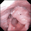
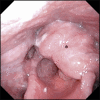
Laryngeal amyloidosis. (*) Laryngeal AL amyloidosis of the kappa light chain type left hemilarynx , flexible laryngoscopy (chip/tip).
Fig. 18
Fig. 18
Amyloidosis. a Laryngeal mucosa with subepithelial, amorphous, extracellular, eosinophilic deposits that in Congo red staining (see b ) have a characteristic red reaction, corresponding to amyloid.
Fig. 19
Fig. 19
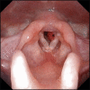
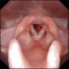
Laryngeal sarcoidosis. Manifestation of sarcoidosis at the larynx with typical deformity of the epiglottis.
Abb. 1
Abb. 1
Larynxspalten. Typ 1 Die Larynxspalte betrifft nur die postkrikoidale Region und endet ohne Knorpelbeteiligung oberhalb der Stimmlippenebene. Typ 2 Die Larynxspalte zieht in den Ringknorpel und endet unterhalb der Stimmlippenebene. Typ 3 Es liegt auch eine Spalte der Trachea vor, welche allerdings oberhalb der Thoraxapertur endet. Typ 4 Der tracheale Anteil der Spalte zieht über die Thoraxapertur hinaus bis knapp über die Bifurkation.
Abb. 2
Abb. 2
Tracheoösophageale Fisteln. Gross A/Vogt II Es zeigt sich eine Atresie des Ösophagus ohne Ausbildung einer tracheoösophagealen Fistel. In diesem Fall zeigen sich keine Luftansammlungen im Magen. Gross B/Vogt IIIa Es besteht eine Ösophagusatresie mit tracheoösophagealer Fistel am oberen Segment. Das untere Segment endet als Blindsack. Gross C/Vogt IIIb Es besteht eine Ösophagusatresie mit tracheoösophagealer Fistel am unteren Segment. Das obere Segment endet als Blindsack. Dies ist die häufigste Form der Ösophagusatresie. Gross D/Vogt IIIc Es besteht eine Ösophagusatresie mit Fistelbildung am unteren und oberen Segment. Gross E/Vogt IV Hier besteht eine tracheoösophageale Fistel ohne Vorliegen einer Ösophagusatresie (sogenannte „H-Fistel“)
Abb. 3
Abb. 3
Trachealagenesie. Typ 1 Hier findet sich ein Trachealstumpf oberhalb der Bifurkation, in den eine Fistel aus dem zervikalen Ösophagus mündet. Häufigkeit 20%. Typ 2 Ein Trachealstumpf existiert nicht, die Bifurkation ist direkt mit dem Ösophagus verbunden. Häufigkeit 60%. Typ 3 In diesem Fall fehlt auch die Bifurkation, die beiden Hauptbronchi münden separat in den Ösophagus. Häufigkeit 20%.
Abb. 5
Abb. 5
Papillomatose. (*) Larynxpapillomatose (hier HPV Typ 6) mit pathognomonischen Gefäßschlingen; flexible Laryngoskopie (chip/tip).
Abb. 6
Abb. 6
Rhabdomyom. a Histologisch sieht man einen Tumor mit großen polygonalen, länglichen Zellen mit reichlich granulärem eosinophilem Zytoplasma, welches teilweise eine Querstreifung erkennen lässt, und kleinen, rundlichen Zellkernen mit distinkten Zellgrenzen. Mitosen kommen sehr selten vor oder fehlen vollständig. b Bei stärkerer Vergrößerung sieht man eine Querstreifung der Tumorzellen. c Immunhistochemisch sieht man eine kräftige Expression von Desmin. d Myogenin reagiert in einzelnen Zellen nukleär positiv.
Abb. 7
Abb. 7
Schwannom. (*) plexiformes Larynxschwannom supraglottisch links; flexible Laryngoskopie (chip/tip)Schwannom.
Abb. 8
Abb. 8
Schwannom. Die Abbildungen zeigen ein plexiformes Schwannom. a Schwannome sind Nervenscheidentumore, die vollständig oder fast vollständig aus differenzierten neoplastischen Schwann‘schen Zellen bestehen. In der Übersicht sieht man ein für plexiforme Schwannome charakteristisches mehrknotiges Wachstum. b / c ) Bei stärkerer Vergrößerung erkennt man, im Gegensatz zu klassischen Schwannomen, die einen Wechsel zwischen Antoni a und a Wachstumsmuster zeigen, einen etwas monotoneren, recht zellreichen, spindelzelligen Tumor ohne Atypien, Nekrosen oder eine gesteigerte mitotische Aktivität als Zeichen eines benignen Tumors.
Abb. 9
Abb. 9
Granularzelltumor. (*) Granularzelltumor hinteres Drittel Stimmlippe rechts; flexible Laryngoskopie (chip/tip).
Abb. 10
Abb. 10
Granularzelltumor. a Bei der mikroskopischen Untersuchung sieht man einen in großen Verbänden gewachsenen Tumor mit großleibigen, unscharf begrenzten eosinophilen Tumorzellen, die ein granuläres Zytoplasma zeigen. Die Zellkerne sind meist zentral gelegen mit gleichförmigen, leicht hyperchromatischen und vergrößerten vesikulären Zellkernen. In der Immunhistochemie reagieren die Tumorzellen positiv für b S100 und c SOX10 als Hinweis für einen neuroektodermalen Ursprung. d Darüber hinaus reagieren die Tumorzellen positiv für CD68.
Abb. 11
Abb. 11
Hämangiom. (*) Kongenitales Hämangiom subglottisch links bei multiplen Mittellinienhämangiomen; flexible Laryngoskopie (chip/tip).
Abb. 12
Abb. 12
Adenoidzystisches Karzinom. (*) Adenoidzystisches Karzinom subglottisch links; flexible Laryngoskopie (chip/tip).
Abb. 13
Abb. 13
adenoidzystisches Karzinom. a Das adenoidzystische Karzinom ist ein Speicheldrüsenkarzinom, das aus epithelialen sowie myoepithelialen Zellen besteht und ein charakteristisches tubuläres, kribriformes und solides Wachstum zeigt. In den Tumordrüsen findet sich häufig eine amorphe basophile Matrix. Aufgrund der typischen Morphologie ist meist die Diagnose am konventionell-morphologischen Präparat möglich. Hier sieht man ausgedehnte Tumorinfiltrate unter der laryngealen Schleimhaut. Das Karzinom bildet eine Perineuralscheideninfiltration (siehe b ) und eine Lymphangiosis carcinomatosa (siehe c ) aus. Zudem zeigt sich hier eine Infiltration der Nebenschilddrüse (in d ) und Schilddrüse (in e ).
Abb. 14
Abb. 14
Chondrosarkom. (*) low-grade (G1) Chondrosarkom am Aryhöcker und Krikoid rechts; flexible Laryngoskopie (chip/tip).
Abb. 15
Abb. 15
Chondrosarkom. a Chondrosarkome des Larynx sind maligne mesenchymale Tumoren des hyalinen Knorpels. In der Übersicht erkennt man einen lobulär aufgebauten Tumor mit chondroider Matrix und erhöhter Zellularität. Der Tumor liegt unter dem laryngealen Oberflächenepithel. b Bei stärkerer Vergrößerung zeichnet sich die nukleäre Pleomorphie und Hyperchromasie der neoplastischen Chondrozyten im Gegensatz zum nicht neoplastischen Knorpelgewebe ab (rechte Bildhälfte). Es finden sich auch Knorpellakunen mit zwei Zellkernen.
Abb. 16
Abb. 16
Embryonales Rhabdomyosarkom. a Das embryonale Rhabomyosarkom ist ein maligner Weichgewebstumor mit morphologischen und immunphänotypischen Eigenschaften der embryonalen Skelettmuskulatur. Histologisch bilden sich hier überwiegend differenzierende Rhabdomyoblasten mit kräftig eosinophilem und elongiertem Zytoplasma aus, die verstärkt nach Vortherapie beobachtet werden können (wie in diesem Fall). Die Zellkerne sind deutlich vergrößert und hyperchromatisch. b Bei stärkerer Vergrößerung sieht man einzelne mehrkernige Tumorzellen (Pfeil) und ganz vereinzelt eine Querstreifung. c Immunhistochemisch reagieren die Tumorzellen positiv für Desmin und d Myogenin.
Abb. 17
Abb. 17
Larynxamyloidose. (*) Laryngeale AL-Amyloidose vom kappa-Leichtkettentyp Hemilarynx links; flexible Laryngoskopie (chip/tip).
Abb. 18
Abb. 18
Amyloidose. a Laryngeale Schleimhaut mit subepithelial, amorphen, extrazellulären eosinophilen Ablagerungen, die in der Kongorotfärbung (siehe b ) eine charakteristische rötliche Reaktion, entsprechend Amyloid erkennen lassen
Abb. 19
Abb. 19
Larynxsarkoidose. Manifestation einer Sarkoidose am Larynx mit typischer Verplumpung der Epiglottis
Abb. 4
Abb. 4
a Trachealagenesie bei einem Neugeborenen. Die Epiglottis ist angelegt, das Larynxlumen verschlossen. Beatmung des Patienten über den Ösophagus und die ösophagobronchiale Fistel. b Trachealagenesie bei einem Neugeborenen. Nun bei aufgeladener Epiglottis mit sichtbarem freien Ösophaguslumen und komplett verschlossenem Kehlkopflumen.

Similar articles

Cited by

References

    1. Holinger L D. Etiology of Stridor in the Neonate, Infant and Child. Ann Oto Rhinol Laryn. 1980;89:397–400. - PubMed
    1. Friedman E M, Vastola A P, Mcgill TJ I et al.Chronic Pediatric Stridor – Etiology and Outcome. Laryngoscope. 1990;100:277–280. - PubMed
    1. Zoumalan R, Maddalozzo J, Holinger L D. Etiology of stridor in infants. Ann Oto Rhinol Laryn. 2007;116:329–334. - PubMed
    1. Hoff S R, Schroeder J W, Rastatter J C et al.Supraglottoplasty outcomes in relation to age and comorbid conditions. Int J Pediatr Otorhi. 2010;74:245–249. - PubMed
    1. Krashin E, Ben-Ari J, Springer C et al.Synchronous airway lesions in laryngomalacia. Int J Pediatr Otorhi. 2008;72:501–507. - PubMed