. 2021 Oct;52(10):3362-3373.
doi: 10.1161/STROKEAHA.120.032374. Epub 2021 Aug 6.

Neuronal CD200 Signaling Is Protective in the Acute Phase of Ischemic Stroke

Affiliations

Neuronal CD200 Signaling Is Protective in the Acute Phase of Ischemic Stroke

Abdullah Al Mamun et al. Stroke. 2021 Oct.

Abstract

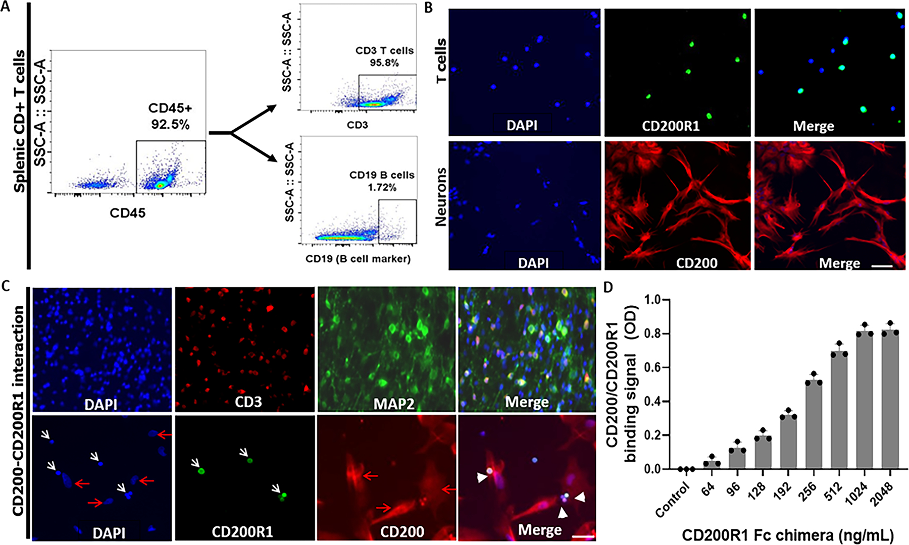
Background and purpose: CD200 (cluster of differentiation 200), a highly glycosylated protein primarily expressed on neurons in the central nervous system, binds with its receptor CD200R to form an endogenous inhibitory signal against immune responses. However, little is known about the effect of neuronal CD200 signaling in cerebral ischemia. The aim of this study was to investigate how neuronal CD200 signaling impacts poststroke inflammation and the ischemic injury.

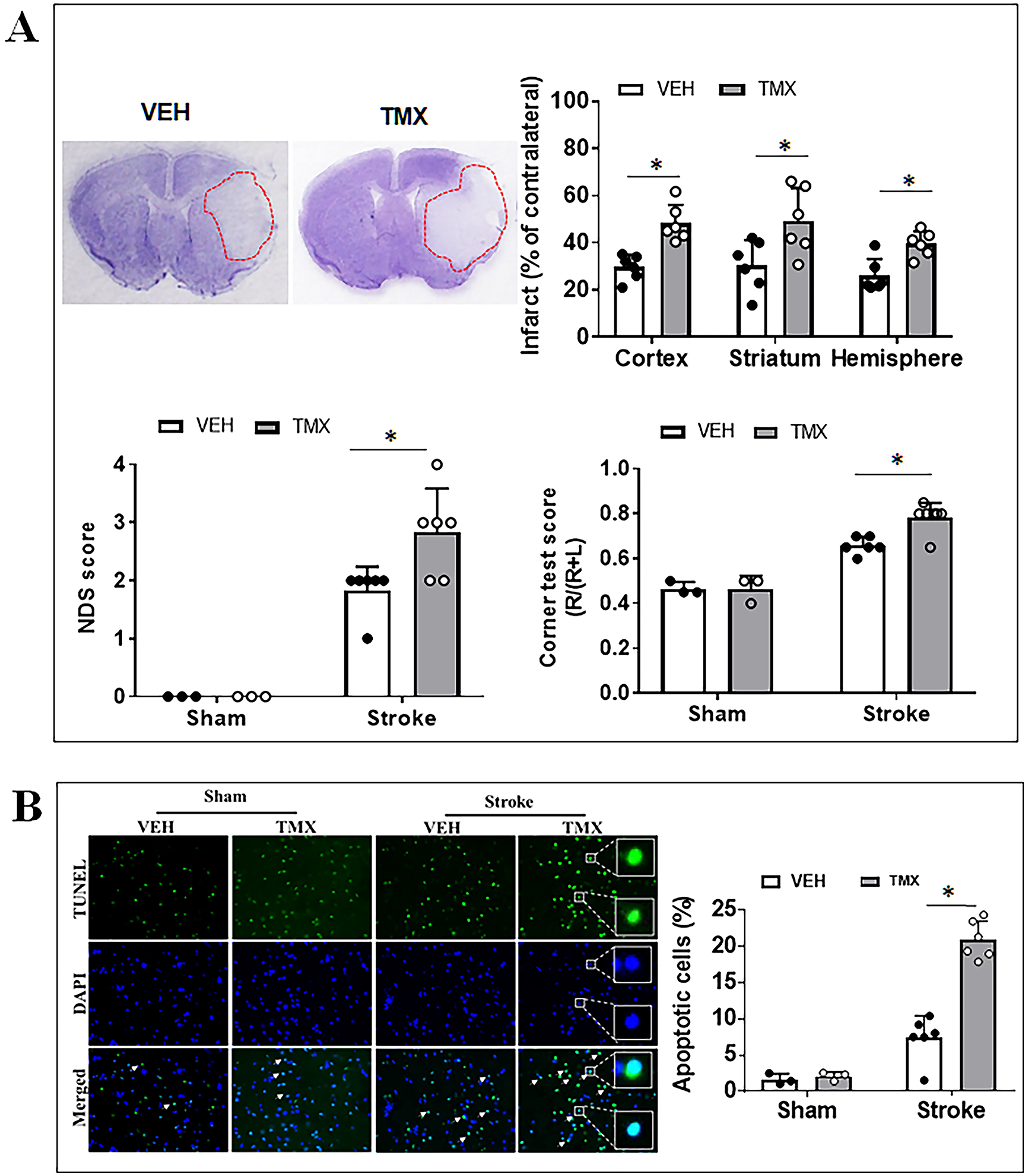
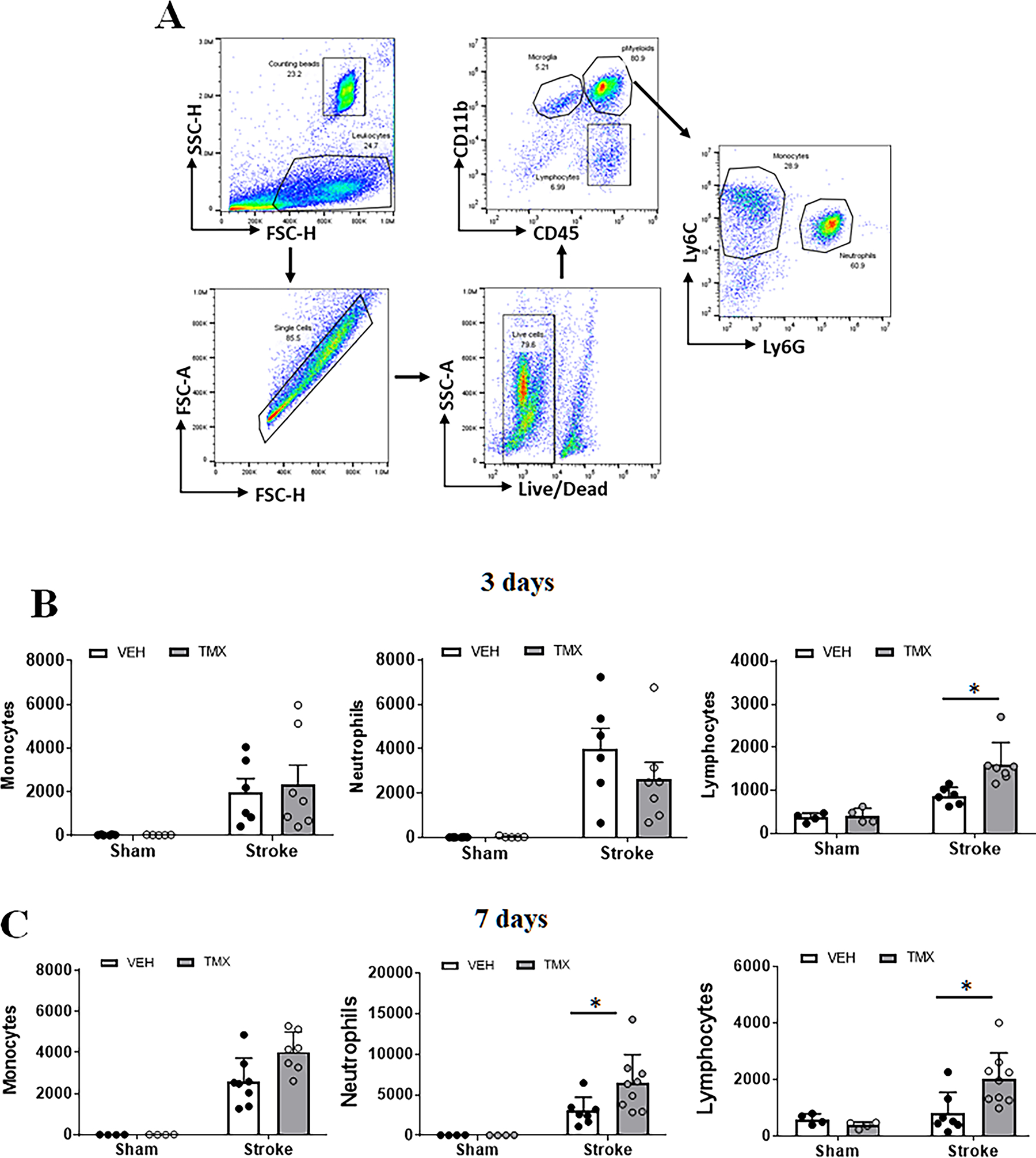
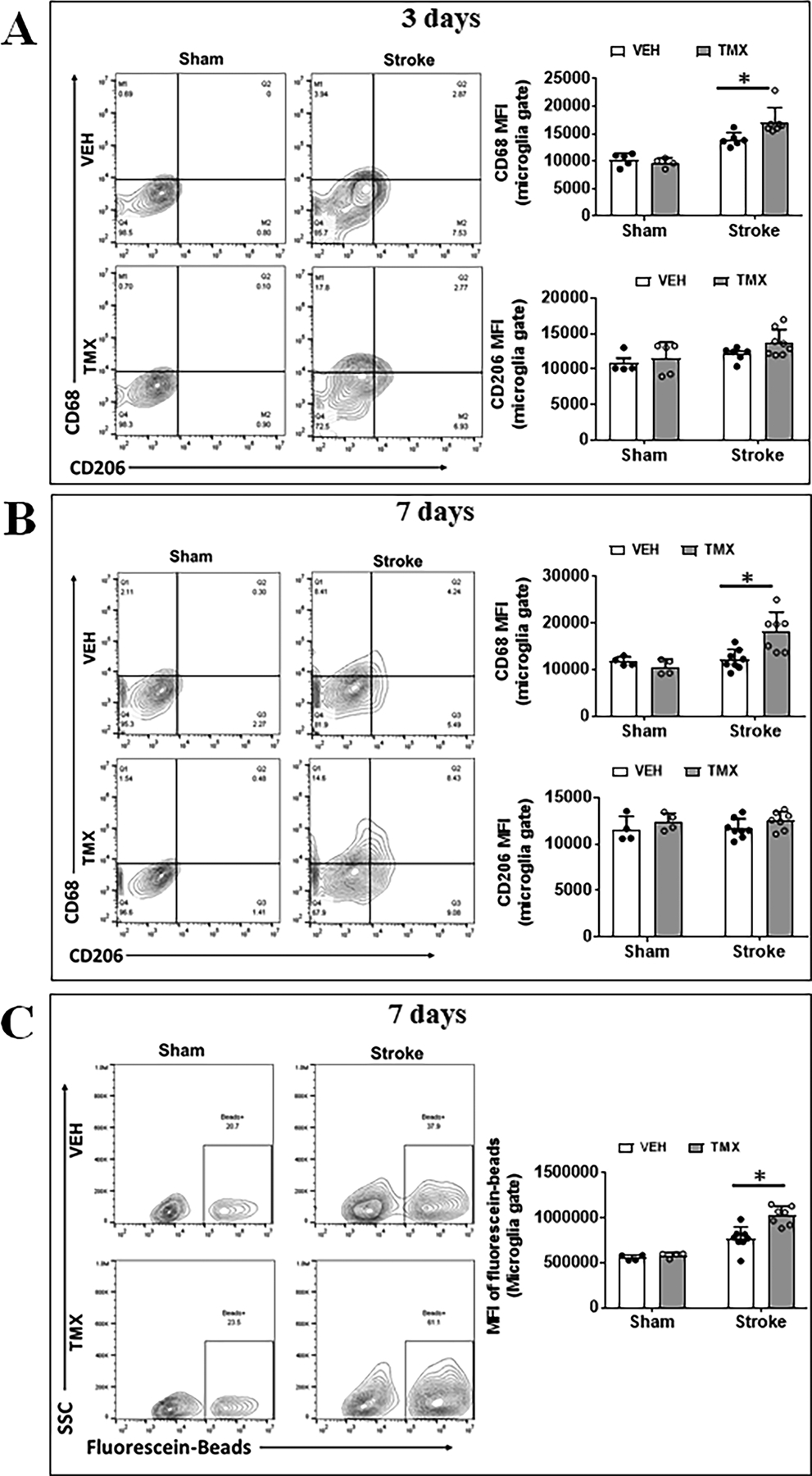
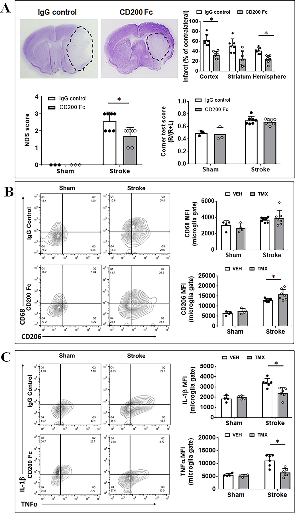
Methods: CD200 tma1lf/fl:Thy1CreER mice were treated with tamoxifen to induce conditional gene knockout (ICKO) of neuronal CD200. The mice were subjected to a 60-minute transient middle cerebral artery occlusion. Stroke outcomes, apoptotic cell death, immune cell infiltration, microglia activation, and other inflammatory profiles were evaluated at 3 and 7 days after stroke.

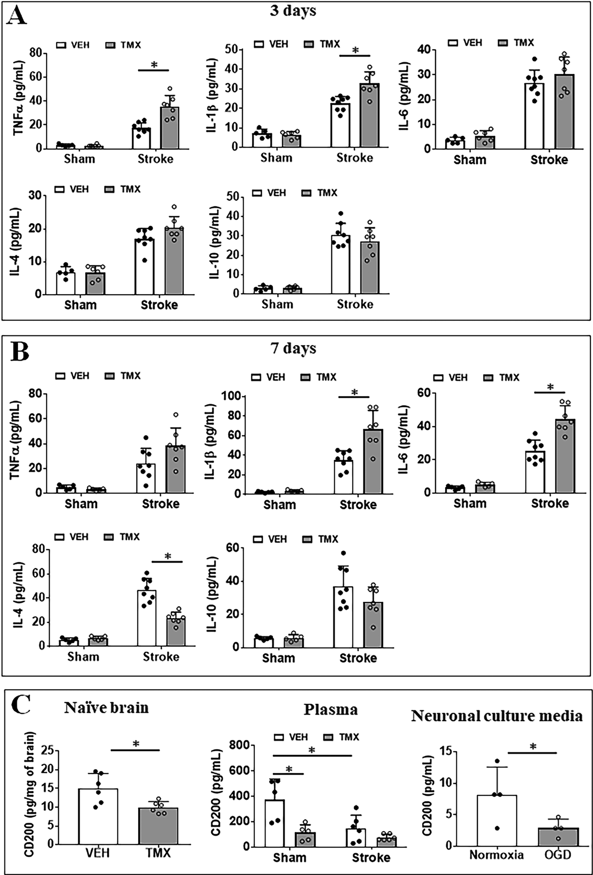
Results: Infarct volumes were significantly larger, and behavioral deficits more severe in ICKO versus control mice at 3 days after middle cerebral artery occlusion. Terminal deoxynucleotidyl transferase dUTP nick end labeling assay also revealed a significant increase in apoptotic neuronal death in CD200 ICKO mice. An enhancement in lymphocytic infiltration and microglial proinflammatory responses were revealed by flow cytometry at 3 and 7 days after stroke in ICKO mice, accompanied by an increased microglial phagocytosis activity. Plasma proinflammatory cytokine (TNFα [tumor necrosis factor alpha] and IL [interleukin]-1β) levels significantly increased at 3 days, and IL-1β/IL-6 levels increased at 7 days in ICKO versus control animals. ICKO led to significantly lower baseline level of CD200 both in brain and plasma.

Conclusions: Neuronal CD200 inhibits proinflammatory responses and is protective against stroke injury.

Keywords: immunoglobulin; inflammation; ischemic stroke; neurons; neuroprotection.


Conflict of interest statement

Disclosures

None of the authors have a conflict of interest relevant to this work.

Figures

Figure 1.
Figure 1.
Effects of neuronal CD200 signaling on stroke outcomes and neuronal apoptosis at 3 days after stroke. (A) Representative images of brain slices stained with cresyl violet (CV) and quantification of brain infarct volumes, NDS, and corner test scores in TMX vs. VEH mice. (B) TUNEL labelled cell apoptosis in peri-infarct area of TMX and VEH treated mice and the quantification of percent of TUNEL positive cells in ipsilateral hemispheres Each dot represents the average of % apoptotic cells from eight 20× fields/animal in the peri-infarct area. N=3 for sham and 6–7 for stroke per group; *P < 0.05.
Figure 2.
Figure 2.
Peripheral immune cell infiltration in ischemic brains at 3 and 7 days after stroke. (A) Representative dot plots depict the gating strategy used to differentiate the microglia (CD45intermediateCD11b+), peripheral myeloid cells (CD45highCD11b+) (pMyeloid), and lymphocytes (CD45highCD11b). In pMyeloid cells, monocytes are further gated as CD45highCD11b+Ly6C+Ly6G, and neutrophils as CD45highCD11b+Ly6G+ Ly6C. Absolute counts of infiltrating monocytes, neutrophils, and lymphocytes at 3d (B) and 7d (C) after stroke were shown. N=4–5 sham and 6–8 stroke animals per group; *P < 0.05.
Figure 3.
Figure 3.
Microglial activation after stroke in TMX and VEH treated mice. Representative FC plots of CD68/CD206 expressed on microglia at 3d (A) and 7d (B) after MCAO. Quantitative expression of CD68 and CD206 was measured as mean fluorescence intensity (MFI). (C) Representative plots illustrate the phagocytic activity of microglia at 7d after MCAO, which was quantified by MFI of bio-particles in microglia. N=6–8 stroke/4–5 sham animals per group; *P < 0.05.
Figure 4.
Figure 4.
Plasma cytokine levels after ischemic injury. Pro-inflammatory (TNFα, IL-1β and IL-6) and anti-inflammatory (IL-10 and IL-4) cytokine levels (pg/mL) were measured in the plasma of VEH and TMX treated sham/stroke mice at 3d (A) and 7d (B). N=5–6 sham and 6–7 stroke animals per group; *P < 0.05. (C) CD200 levels in naïve brains (left), in the plasma (middle) of TMX and VEH treated mice, and in neuronal culture medium after OGD (right). N=5–6 sham and 6–8 stroke animals per group; *P < 0.05. For neuronal culture medium, N=4 independent OGD experiments; *P < 0.05.
Figure 5.
Figure 5.
Stroke outcomes and flow cytometry in CD200 Fc treated mice. (A) Representative images of brain slices stained with cresyl violet (CV) and quantification of brain infarct volumes, NDS, and corner test scores in CD200Fc vs. IgG treated mice. N=3 for sham and 6–7 for stroke per group; *P < 0.05. (B) Microglial expression of cell membrane markers CD68/CD206 measured as mean fluorescence intensity (MFI). (C) Microglial expression of intracellular markers IL-1β/TNFα. For (B) and (C), n=4/sham group and n=6/stroke group; *P < 0.05.
Figure 6.
Figure 6.
Interaction of neuronal CD200 with lymphocytic CD200R1. (A) The purity of CD45+CD3+ T cells from spleens of C57BL/6 mice were confirmed by flow cytometry. (B) Representative ICC images showed splenic CD3+ T cells express CD200R1 (upper panels; green) and cultured primary neurons express CD200 (lower panels; red). (C) Neurons interact with CD3+ T cells. Upper panels, colocalization of CD3 (marker for T cells) and MAP2 (marker for neurons). Lower panels, colocalization of CD200 ligand on neurons and CD200R1 on T cells. White arrows indicate T cells; red arrows indicate neurons; white arrow heads indicate the interaction of CD200 with CD200R. Scale bar=100 μm. (D) ELISA binding assay of mouse biotinylated CD200Fc chimera protein with mouse CD200R1 chimera protein (captured by anti-CD200R1 antibody) immobilized on a Nunc MaxiSorp ELISA plate and in a concentration dependent manner (n = 4 assays per group). Control ELISA wells were without CD200R1 Fc chimera protein.

References

    1. Al Mamun A, Chauhan A, Yu H, Xu Y, Sharmeen R, Liu F. Interferon regulatory factor 4/5 signaling impacts on microglial activation after ischemic stroke in mice. Eur J Neurosci. 2018;47:140–149 - PMC - PubMed
    1. Al Mamun A, Chauhan A, Qi S, Ngwa C, Xu Y, Sharmeen R, Hazen AL, Li J, Aronowski JA, McCullough LD, et al. Microglial irf5-irf4 regulatory axis regulates neuroinflammation after cerebral ischemia and impacts stroke outcomes. Proc Natl Acad Sci U S A. 2020;117:1742–1752 - PMC - PubMed
    1. Szepesi Z, Manouchehrian O, Bachiller S, Deierborg T. Bidirectional microglia-neuron communication in health and disease. Frontiers in cellular neuroscience. 2018;12:323. - PMC - PubMed
    1. Tian L, Ma L, Kaarela T, Li Z. Neuroimmune crosstalk in the central nervous system and its significance for neurological diseases. Journal of neuroinflammation. 2012;9:155. - PMC - PubMed
    1. Shirley R, Ord EN, Work LM. Oxidative stress and the use of antioxidants in stroke. Antioxidants (Basel). 2014;3:472–501 - PMC - PubMed

Publication types

MeSH terms