

Computer-aided diagnosis with a convolutional neural network algorithm for automated detection of urinary tract stones on plain X-ray
- PMID: 34353306
- PMCID: PMC8340490
- DOI: 10.1186/s12894-021-00874-9
Computer-aided diagnosis with a convolutional neural network algorithm for automated detection of urinary tract stones on plain X-ray
Abstract
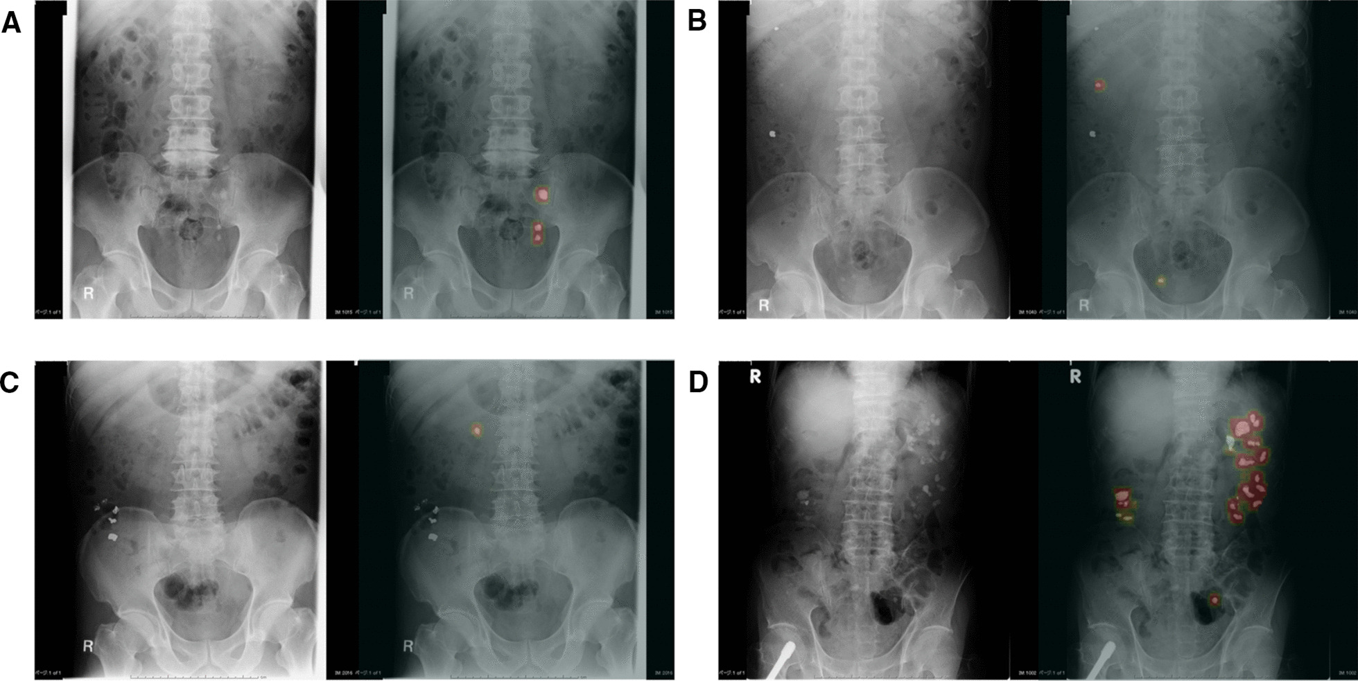
Background: Recent increased use of medical images induces further burden of their interpretation for physicians. A plain X-ray is a low-cost examination that has low-dose radiation exposure and high availability, although diagnosing urolithiasis using this method is not always easy. Since the advent of a convolutional neural network via deep learning in the 2000s, computer-aided diagnosis (CAD) has had a great impact on automatic image analysis in the urological field. The objective of our study was to develop a CAD system with deep learning architecture to detect urinary tract stones on a plain X-ray and to evaluate the model's accuracy.
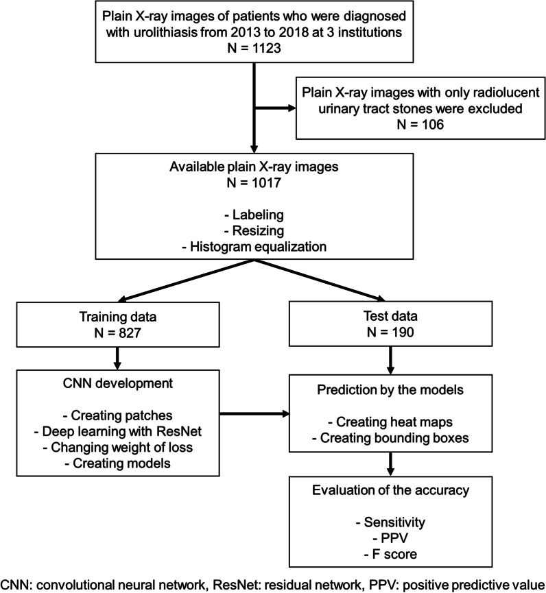
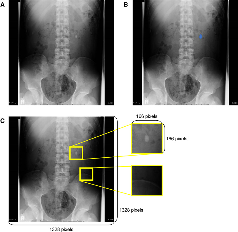
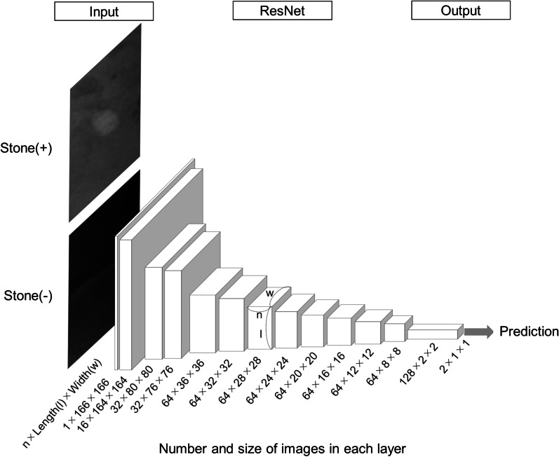
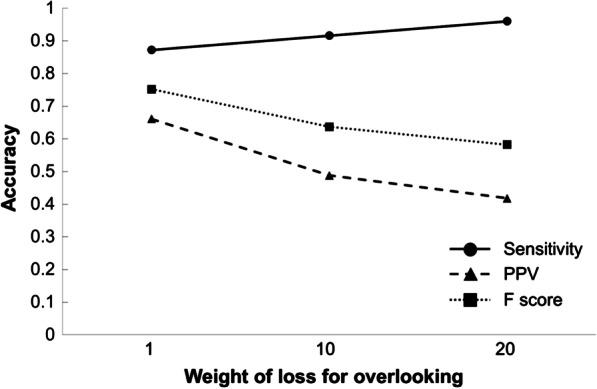
Methods: We collected plain X-ray images of 1017 patients with a radio-opaque upper urinary tract stone. X-ray images (n = 827 and 190) were used as the training and test data, respectively. We used a 17-layer Residual Network as a convolutional neural network architecture for patch-wise training. The training data were repeatedly used until the best model accuracy was achieved within 300 runs. The F score, which is a harmonic mean of the sensitivity and positive predictive value (PPV) and represents the balance of the accuracy, was measured to evaluate the model's accuracy.
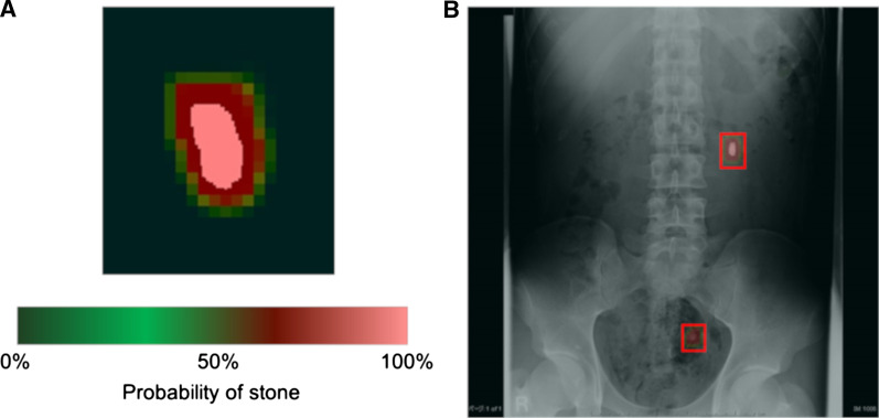
Results: Using deep learning, we developed a CAD model that needed 110 ms to provide an answer for each X-ray image. The best F score was 0.752, and the sensitivity and PPV were 0.872 and 0.662, respectively. When limited to a proximal ureter stone, the sensitivity and PPV were 0.925 and 0.876, respectively, and they were the lowest at mid-ureter.
Conclusion: CAD of a plain X-ray may be a promising method to detect radio-opaque urinary tract stones with satisfactory sensitivity although the PPV could still be improved. The CAD model detects urinary tract stones quickly and automatically and has the potential to become a helpful screening modality especially for primary care physicians for diagnosing urolithiasis. Further study using a higher volume of data would improve the diagnostic performance of CAD models to detect urinary tract stones on a plain X-ray.
Keywords: Artificial intelligence; Convolutional neural network; Deep learning; Urinary tract stone; Urolithiasis; X-ray.
© 2021. The Author(s).
Conflict of interest statement
The authors declare that they have no competing interests.
Figures
Similar articles
-
Deep Convolutional Neural Network-Based Computer-Aided Detection System for COVID-19 Using Multiple Lung Scans: Design and Implementation Study.J Med Internet Res. 2021 Apr 26;23(4):e27468. doi: 10.2196/27468. J Med Internet Res. 2021. PMID: 33848973 Free PMC article.
-
MABAL: a Novel Deep-Learning Architecture for Machine-Assisted Bone Age Labeling.J Digit Imaging. 2018 Aug;31(4):513-519. doi: 10.1007/s10278-018-0053-3. J Digit Imaging. 2018. PMID: 29404850 Free PMC article.
-
Computer aided detection of ureteral stones in thin slice computed tomography volumes using Convolutional Neural Networks.Comput Biol Med. 2018 Jun 1;97:153-160. doi: 10.1016/j.compbiomed.2018.04.021. Epub 2018 Apr 27. Comput Biol Med. 2018. PMID: 29730498
-
Can Artificial Intelligence Accurately Detect Urinary Stones? A Systematic Review.J Endourol. 2024 Aug;38(8):725-740. doi: 10.1089/end.2023.0717. Epub 2024 May 30. J Endourol. 2024. PMID: 38666692
-
Deep Learning Computer-Aided Diagnosis for Breast Lesion in Digital Mammogram.Adv Exp Med Biol. 2020;1213:59-72. doi: 10.1007/978-3-030-33128-3_4. Adv Exp Med Biol. 2020. PMID: 32030663 Review.
Cited by
-
Design and Validation of a Deep Learning Model for Renal Stone Detection and Segmentation on Kidney-Ureter-Bladder Images.Bioengineering (Basel). 2023 Aug 16;10(8):970. doi: 10.3390/bioengineering10080970. Bioengineering (Basel). 2023. PMID: 37627855 Free PMC article.
-
Identification of kidney stones in KUB X-ray images using VGG16 empowered with explainable artificial intelligence.Sci Rep. 2024 Mar 14;14(1):6173. doi: 10.1038/s41598-024-56478-4. Sci Rep. 2024. PMID: 38486010 Free PMC article.
-
Transforming urinary stone disease management by artificial intelligence-based methods: A comprehensive review.Asian J Urol. 2023 Jul;10(3):258-274. doi: 10.1016/j.ajur.2023.02.002. Epub 2023 May 2. Asian J Urol. 2023. PMID: 37538159 Free PMC article. Review.
-
Deep Learning Model for Computer-Aided Diagnosis of Urolithiasis Detection from Kidney-Ureter-Bladder Images.Bioengineering (Basel). 2022 Dec 16;9(12):811. doi: 10.3390/bioengineering9120811. Bioengineering (Basel). 2022. PMID: 36551017 Free PMC article.
-
Artificial intelligence in urolithiasis: a systematic review of utilization and effectiveness.World J Urol. 2024 Oct 17;42(1):579. doi: 10.1007/s00345-024-05268-8. World J Urol. 2024. PMID: 39417840
References
-
- Pfister SA, Deckart A, Laschke S, Dellas S, Otto U, Buitrago C, et al. Unenhanced helical computed tomography vs intravenous urography in patients with acute flank pain: accuracy and economic impact in a randomized prospective trial. Eur Radiol. 2003;13:2513–2520. doi: 10.1007/s00330-003-1937-1. - DOI - PubMed
MeSH terms
LinkOut - more resources
Full Text Sources
Miscellaneous
