

Emergency surgery for hemobilia due to hepatic artery pseudoaneurysm rupture complicated by Mirizzi syndrome type II: a case report
- PMID: 34353316
- PMCID: PMC8340468
- DOI: 10.1186/s12893-021-01314-z
Emergency surgery for hemobilia due to hepatic artery pseudoaneurysm rupture complicated by Mirizzi syndrome type II: a case report
Abstract
Background: Hemobilia refers to bleeding into the biliary tract. Hepatic artery pseudoaneurysm (HAP) rupture is an uncommon cause of hemobilia, and cases of HAP associated with Mirizzi syndrome are extremely rare. Although transarterial embolization is recommended as the first-line treatment for hemobilia, surgery is sometimes required.
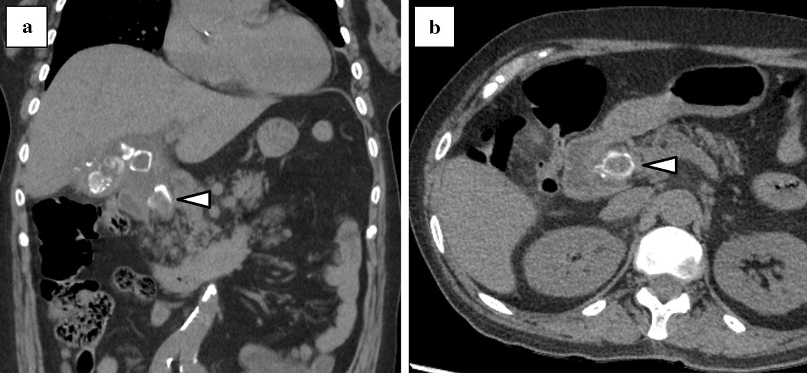
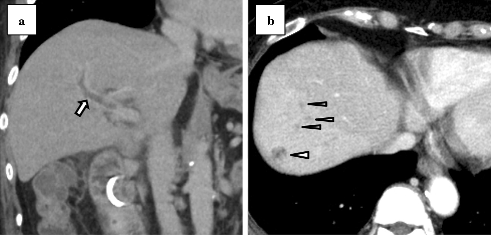
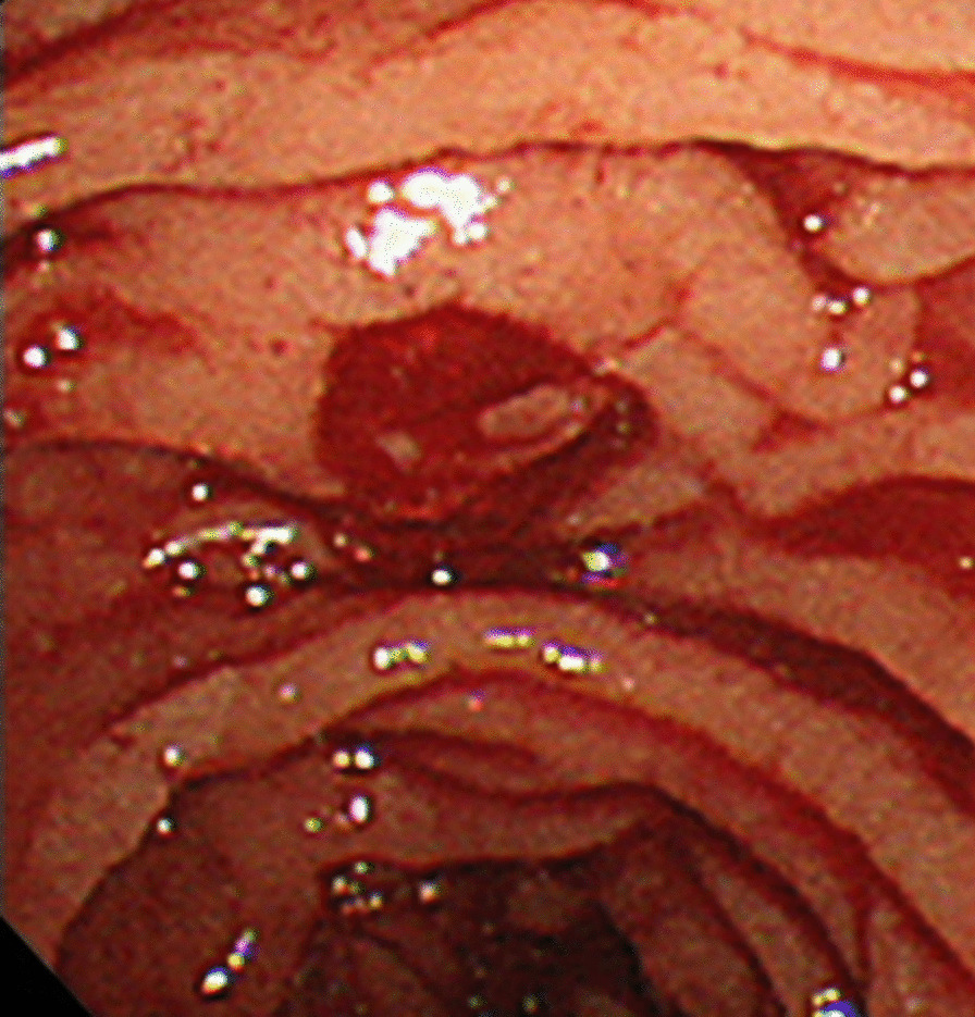
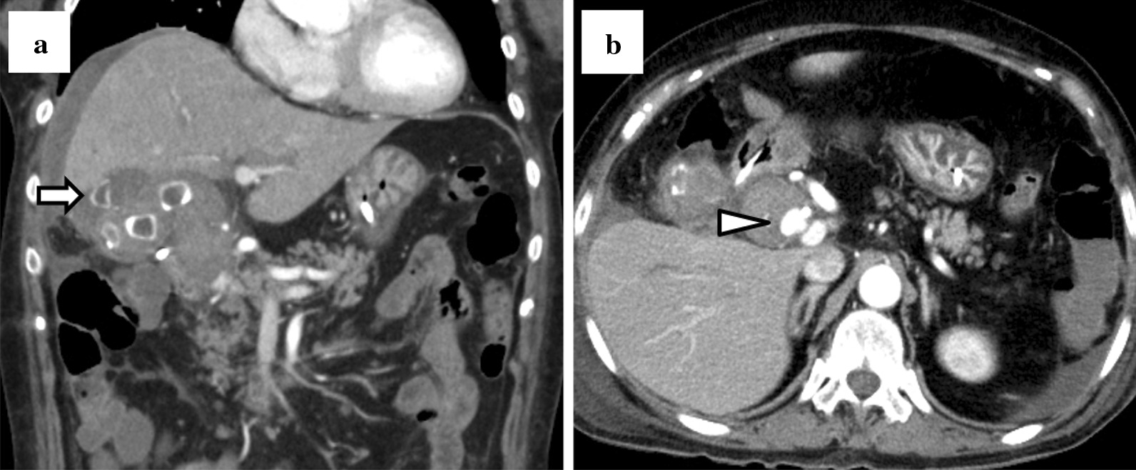
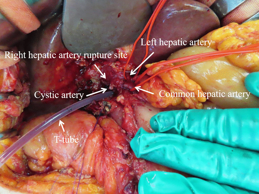
Case presentation: A 76-year-old woman was referred to our hospital with epigastric pain. She was febrile and had conjunctival icterus and epigastric tenderness. Laboratory tests revealed abnormal white blood cell count and liver function. An abdominal computed tomography (CT) revealed multiple calculi in the gallbladder, an incarcerated calculus in the cystic duct, and a slightly dilated common hepatic duct. Based on examination findings, she was diagnosed with Mirizzi syndrome type I, complicated by cholangitis. Intravenous antibiotics were administered, and we performed endoscopic retrograde cholangiopancreatography (ERCP) to place a drainage tube. The fever persisted; therefore, contrast-enhanced CT (CECT) was performed. This revealed portal vein thrombosis and hepatic abscesses; therefore, heparin infusion was administered. The following day, she complained of melena, and laboratory tests showed that she was anemic. ERCP was performed to change the drainage tube in the bile duct; however, bleeding from the papilla of Vater was observed. CECT demonstrated a right HAP with high-density fluid in the gallbladder and gallbladder perforation. Finally, she was diagnosed with hemobilia caused by HAP rupture, and emergency surgery was performed to secure hemostasis and control the infection. During laparotomy, we found that a right HAP had ruptured into the gallbladder. The gallbladder made a cholecystobiliary fistula, which indicated Mirizzi syndrome type II. Although we tried to repair the right hepatic artery, we later ligated it due to arterial wall vulnerability. Then, we performed subtotal cholecystectomy and inserted a T-tube into the common bile duct. There were no postoperative complications except for minor leakage from the T-tube insertion site. The patient was discharged after a total hospital stay of 7 weeks.
Conclusions: We experienced an extremely rare case of emergency definitive surgery for hemobilia due to HAP rupture complicated by Mirizzi syndrome type II. Surgery might be indicated when controlling underlying infections was required.
Keywords: Case report; Hemobilia; Hepatic artery; Mirizzi syndrome; Pseudoaneurysm; Surgery.
© 2021. The Author(s).
Conflict of interest statement
The authors declare that they have no competing interests.
Figures
References
Publication types
MeSH terms
LinkOut - more resources
Full Text Sources
