Multicenter Study
. 2021 Aug 5;16(1):349.
doi: 10.1186/s13023-021-01969-0.

Auditory cortex hypoperfusion: a metabolic hallmark in Beta Thalassemia

Affiliations
Multicenter Study

Auditory cortex hypoperfusion: a metabolic hallmark in Beta Thalassemia

Renzo Manara et al. Orphanet J Rare Dis. .

Abstract

Background: Sensorineural hearing loss in beta-thalassemia is common and it is generally associated with iron chelation therapy. However, data are scarce, especially on adult populations, and a possible involvement of the central auditory areas has not been investigated yet. We performed a multicenter cross-sectional audiological and single-center 3Tesla brain perfusion MRI study enrolling 77 transfusion-dependent/non transfusion-dependent adult patients and 56 healthy controls. Pure tone audiometry, demographics, clinical/laboratory and cognitive functioning data were recorded.

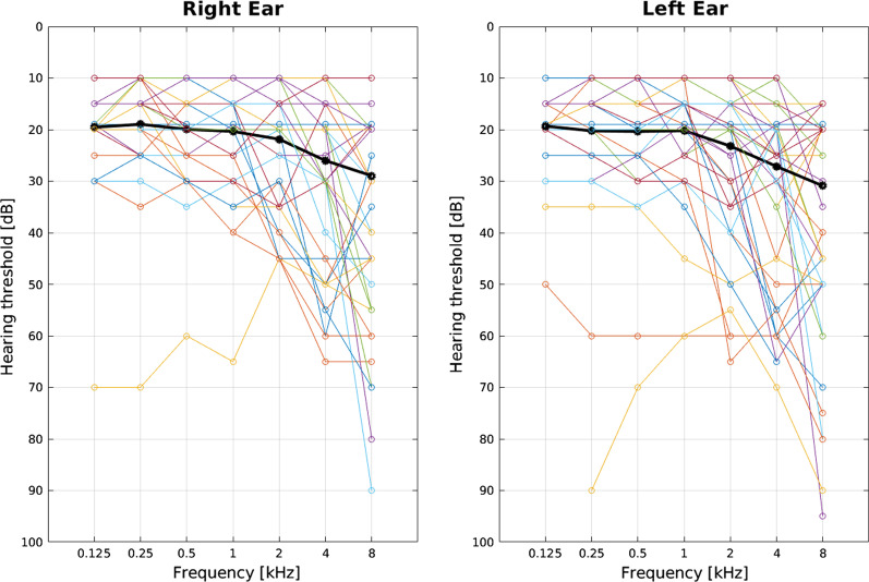
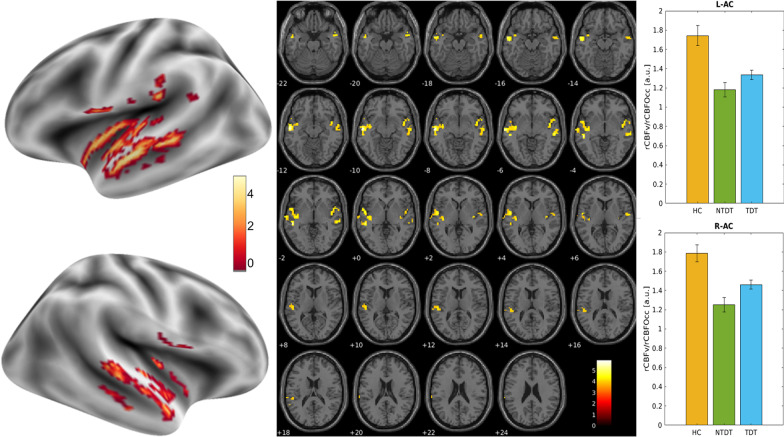
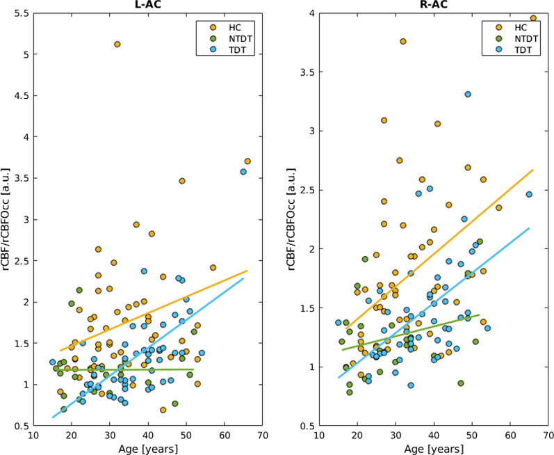
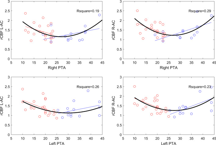
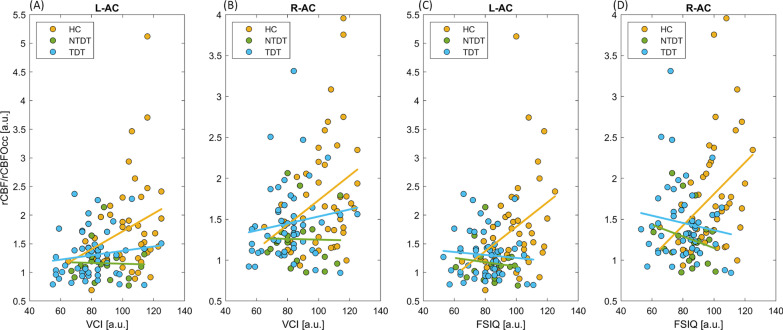
Results: Half of patients (52%) presented with high-frequency hearing deficit, with overt hypoacusia (Pure Tone Average (PTA) > 25 dB) in 35%, irrespective of iron chelation or clinical phenotype. Bilateral voxel clusters of significant relative hypoperfusion were found in the auditory cortex of beta-thalassemia patients, regardless of clinical phenotype. In controls and transfusion-dependent (but not in non-transfusion-dependent) patients, the relative auditory cortex perfusion values increased linearly with age (p < 0.04). Relative auditory cortex perfusion values showed a significant U-shaped correlation with PTA values among hearing loss patients, and a linear correlation with the full scale intelligence quotient (right side p = 0.01, left side p = 0.02) with its domain related to communication skills (right side p = 0.04, left side p = 0.07) in controls but not in beta-thalassemia patients. Audiometric test results did not correlate to cognitive test scores in any subgroup.

Conclusions: In conclusion, primary auditory cortex perfusion changes are a metabolic hallmark of adult beta-thalassemia, thus suggesting complex remodeling of the hearing function, that occurs regardless of chelation therapy and before clinically manifest hearing loss. The cognitive impact of perfusion changes is intriguing but requires further investigations.

Keywords: Brain; Hearing loss; Perfusion; Thalassemia; Transfusion medicine.


Conflict of interest statement

None of the authors have a relevant conflict of interest to disclose.

Figures

Fig. 1
Fig. 1
Audiometric thresholds for air conduction in the pure tone audiometry for all the patients (n = 71) and the two ears. Black lines indicate mean hearing loss across patients for each tested frequency
Fig. 2
Fig. 2
Left: T-maps of the statistical comparison between Healthy Controls and Thalassemic patients (TDT and NTDT) showed respectively on inflated brain surfaces and on the single subject T1w anatomical image. Right: Bar plot (and standard deviation bars) of the mean perfusion values across the three groups in the clusters of significant differences. Patients had significantly lower perfusion compared to Healthy Controls in both left (L-AC) and right (R-AC) auditory cortex. By subgroup analysis, the difference persisted significant between healthy controls and TDT; perfusion values did not differ between NTDT and TDT. rCBFv: relative cerebral blood flow values in the auditory cortex; rCBFOcc: relative cerebral blood flow in the occipital cortex
Fig. 3
Fig. 3
Relative left and right auditory cortex perfusion values according to age in healthy controls, Transfusion-Dependent Thalassemia (TDT) and Non-Transfusion-Dependent Thalassemia (NTDT) patients
Fig. 4
Fig. 4
Polynomial fitting of the second order between relative left and right auditory cortex perfusion values (rCBF L-AC and rCBF R-AC) and Pure Tone Average (PTA). Circles and lines indicate the subjects and the linear relationship between PTA and rCBF (red for hearing loss in at least one frequency (fHL), blue for hearing loss with abnormal Pure Tone Average (HL)); the curve in black is the second-order polynomial fitting (Robust fit bisquare) and the relative R-square value considering all the subjects together. Three subjects were excluded from this analysis because of outlier values of PTA, secondary to cholesteatoma in 1 and otosclerosis in 2
Fig. 5
Fig. 5
Relative left and right auditory cortex perfusion values according to Verbal Comprehension Index (VCI) (A, B) and Full Scale Intelligence Quotient (FSIQ) (C, D) in Healthy Controls (HC), Transfusion-Dependent Thalassemia (TDT) and Non-Transfusion-Dependent Thalassemia (NTDT) patients

References

    1. Borgna-Pignatti C, Marsella M. Iron chelation in thalassemia major. Clin Ther. 2015 doi: 10.1016/j.clinthera.2015.10.001. - DOI - PubMed
    1. Porter JB, Jaswon MS, Huehns ER, East CA, Hazell JWP. Desferrioxamine ototoxicity: evaluation of risk factors in thalassaemic patients and guidelines for safe dosage. Br J Haematol. 1989 doi: 10.1111/j.1365-2141.1989.tb07761.x. - DOI - PubMed
    1. Chonat S, Quinn CT. Current standards of care and long term outcomes for thalassemia and sickle cell disease. Adv Exp Med Biol. 2017;1013:59–87. doi: 10.1007/978-1-4939-7299-9_3. - DOI - PMC - PubMed
    1. Badfar G, Mansouri A, Shohani M, Karimi H, Khalighi Z, Rahmati S, et al. Hearing loss in Iranian thalassemia major patients treated with deferoxamine: a systematic review and meta-analysis. Caspian J Intern Med. 2017;8:239–249. - PMC - PubMed
    1. Osma U, Kurtoglu E, Eyigor H, Yilmaz MD, Aygener N. Sensorineural hearing loss in β-thalassemia patients treated with iron chelation. Ear Nose Throat J. 2015;94:481–485. doi: 10.1177/014556131509401206. - DOI - PubMed

Publication types

LinkOut - more resources