

Perivascular spaces are associated with tau pathophysiology and synaptic dysfunction in early Alzheimer's continuum
- PMID: 34353353
- PMCID: PMC8340485
- DOI: 10.1186/s13195-021-00878-5
Perivascular spaces are associated with tau pathophysiology and synaptic dysfunction in early Alzheimer's continuum
Abstract
Background: Perivascular spaces (PVS) have an important role in the elimination of metabolic waste from the brain. It has been hypothesized that the enlargement of PVS (ePVS) could be affected by pathophysiological mechanisms involved in Alzheimer's disease (AD), such as abnormal levels of CSF biomarkers. However, the relationship between ePVS and these pathophysiological mechanisms remains unknown.
Objective: We aimed to investigate the association between ePVS and CSF biomarkers of several pathophysiological mechanisms for AD. We hypothesized that ePVS will be associated to CSF biomarkers early in the AD continuum (i.e., amyloid positive cognitively unimpaired individuals). Besides, we explored associations between ePVS and demographic and cardiovascular risk factors.
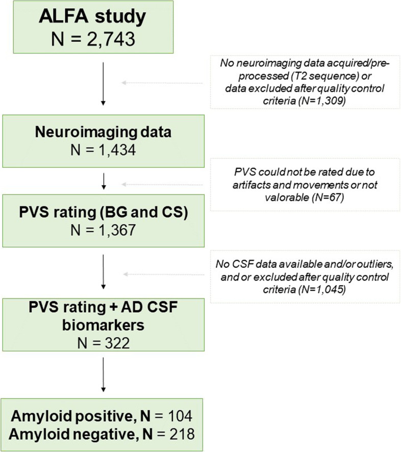
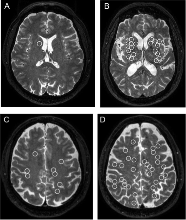
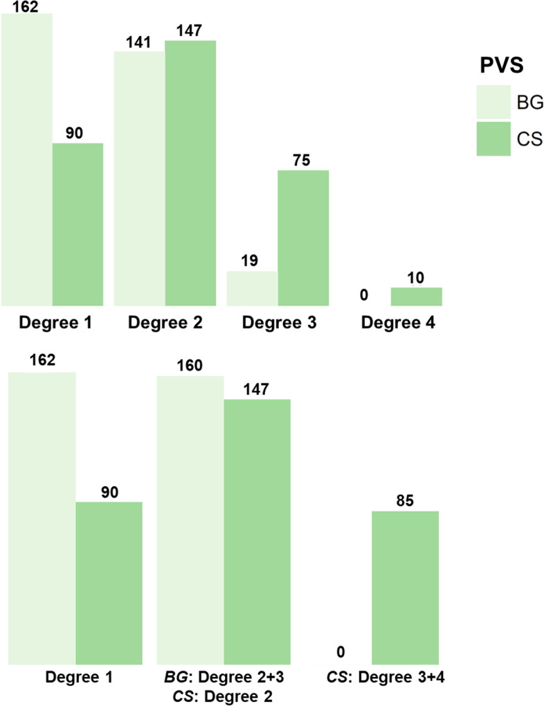
Methods: The study included 322 middle-aged cognitively unimpaired participants from the ALFA + study, many within the Alzheimer's continuum. NeuroToolKit and Elecsys® immunoassays were used to measure CSF Aβ42, Aβ40, p-tau and t-tau, NfL, neurogranin, TREM2, YKL40, GFAP, IL6, S100, and α-synuclein. PVS in the basal ganglia (BG) and centrum semiovale (CS) were assessed based on a validated 4-point visual rating scale. Odds ratios were calculated for associations of cardiovascular and AD risk factors with ePVS using logistic and multinomial models adjusted for relevant confounders. Models were stratified by Aβ status (positivity defined as Aβ42/40 < 0.071).
Results: The degree of PVS significantly increased with age in both, BG and CS regions independently of cardiovascular risk factors. Higher levels of p-tau, t-tau, and neurogranin were significantly associated with ePVS in the CS of Aβ positive individuals, after accounting for relevant confounders. No associations were detected in the BG neither in Aβ negative participants.
Conclusions: Our results support that ePVS in the CS are specifically associated with tau pathophysiology, neurodegeneration, and synaptic dysfunction in asymptomatic stages of the Alzheimer's continuum.
Keywords: Alzheimer’s disease; CSF biomarkers; MRI; Perivascular spaces; Tau pathophysiology; Virchow-Robin spaces.
© 2021. The Author(s).
Conflict of interest statement
GK is a full-time employee of Roche Diagnostics GmbH. The remaining authors declare that they have no conflict of interest. HZ has served at scientific advisory boards for Denali, Roche Diagnostics, Wave, Samumed, and CogRx; has given lectures in symposia sponsored by Fujirebio, Alzecure, and Biogen;and is a co-founder of Brain Biomarker Solutions in Gothenburg AB (BBS), which is a part of the GU Ventures Incubator Program. JLM has served/serves as a consultant or at advisory boards for the following for-profit companies or has given lectures in symposia sponsored by the following for-profit companies: Roche Diagnostics, Genentech, Novartis, Lundbeck, Oryzon, Biogen, Lilly, Janssen, Green Valley, MSD, Eisai, Alector, BioCross, GE Healthcare, ProMIS Neurosciences. KB has served as a consultant, at advisory boards, or at data monitoring committees for Abcam, Axon, Biogen, JOMDD/Shimadzu. Julius Clinical, Lilly, MagQu, Novartis, Roche Diagnostics, and Siemens Healthineers, and is a co-founder of Brain Biomarker Solutions in Gothenburg AB (BBS), which is a part of the GU Ventures Incubator Program. The other co-authors declare that they have no competing interests.
Figures
References
Publication types
MeSH terms
Substances
Grants and funding
LinkOut - more resources
Full Text Sources
Medical
Research Materials
Miscellaneous
