

Comment
doi: 10.3174/ajnr.A7234.
Epub 2021 Aug 5.
Reply
Affiliations
- PMID: 34353782
- PMCID: PMC8423047
- DOI: 10.3174/ajnr.A7234
Item in Clipboard
Comment
Reply
AJNR Am J Neuroradiol.
2021 Sep.
No abstract available
Figures
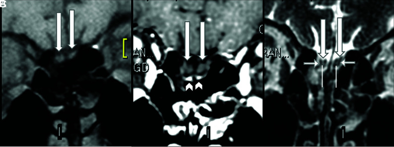
Normal olfactory bulbs and susceptibility artifacts seen on 1.5T MR imaging before the COVID-19 pandemic. The coronal precontrast fat-suppressed T1WI (A) and the postcontrast (B) and coronal FSE T2WI (C) demonstrate normal olfactory bulbs (long arrows). The olfactory bulbs are isointense to the cerebral cortex and normally hypointense on pre- (A) and postgadolinium sequences (B) and do not enhance. Susceptibility artifacts on the cribriform plate (B, arrowheads) are bilateral and symmetric below the olfactory bulbs and do not hinder the analysis. On thin-sliced coronal FSE T2WI, the normal olfactory bulbs show a “sandwich-like pattern,” which consists of a hyperintense central area (C, superior extremity of the vertical lines), similar to the cortical gray matter, and a hypointense periphery (C, short horizontal arrows), similar to the white matter that looks like the laminar layers of olfactory bulbs on histology.
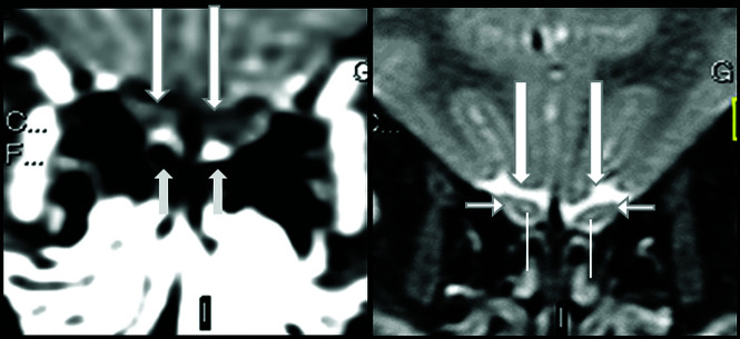
Normal olfactory bulbs and susceptibility artifacts seen on 1.5T MR imaging before the COVID-19 pandemic. The olfactory bulbus are normal, being hypointense, and do not enhance on thin-sliced coronal fat-suppressed postcontrast T1WI (A, long arrows). Susceptibility artifacts are bilateral and symmetric and can be recognized as hyperintensity located outside and adjacent to the inferior periphery of the normal olfactory bulbs, mainly at cribriform plate (A, short arrows). On coronal T2WI, the normal sandwich-like pattern is observed as the central portion of olfactory bulbs showing hyperintensity (B, superior extremity of the line), similar to that of gray matter, and the peripheral portion showing hypointensity, similar to that of white matter (B, short horizontal arrows).
Comment on
-
Anosmia in COVID-19 Associated with Injury to the Olfactory Bulbs Evident on MRI.AJNR Am J Neuroradiol. 2020 Sep;41(9):1703-1706. doi: 10.3174/ajnr.A6675. Epub 2020 Jun 25. AJNR Am J Neuroradiol. 2020. PMID: 32586960 Free PMC article.
-
Susceptibility Artifacts in the Anterior Cranial Fossa Mimicking Hemorrhage in Patients with Anosmia.AJNR Am J Neuroradiol. 2021 Sep;42(9):E64-E65. doi: 10.3174/ajnr.A7184. Epub 2021 Aug 5. AJNR Am J Neuroradiol. 2021. PMID: 34353783 Free PMC article. No abstract available.
References
Publication types
LinkOut - more resources
Full Text Sources