. 2021 Oct;100(4):372-387.
doi: 10.1124/molpharm.121.000336. Epub 2021 Aug 5.

Pharmacological Characterization of the Imipridone Anticancer Drug ONC201 Reveals a Negative Allosteric Mechanism of Action at the D2 Dopamine Receptor

Affiliations

Pharmacological Characterization of the Imipridone Anticancer Drug ONC201 Reveals a Negative Allosteric Mechanism of Action at the D2 Dopamine Receptor

R Benjamin Free et al. Mol Pharmacol. 2021 Oct.

Abstract

ONC201 is a first-in-class imipridone compound that is in clinical trials for the treatment of high-grade gliomas and other advanced cancers. Recent studies identified that ONC201 antagonizes D2-like dopamine receptors at therapeutically relevant concentrations. In the current study, characterization of ONC201 using radioligand binding and multiple functional assays revealed that it was a full antagonist of the D2 and D3 receptors (D2R and D3R) with low micromolar potencies, similar to its potency for antiproliferative effects. Curve-shift experiments using D2R-mediated β-arrestin recruitment and cAMP assays revealed that ONC201 exhibited a mixed form of antagonism. An operational model of allostery was used to analyze these data, which suggested that the predominant modulatory effect of ONC201 was on dopamine efficacy with little to no effect on dopamine affinity. To investigate how ONC201 binds to the D2R, we employed scanning mutagenesis coupled with a D2R-mediated calcium efflux assay. Eight residues were identified as being important for ONC201's functional antagonism of the D2R. Mutation of these residues followed by assessing ONC201 antagonism in multiple signaling assays highlighted specific residues involved in ONC201 binding. Together with computational modeling and simulation studies, our results suggest that ONC201 interacts with the D2R in a bitopic manner where the imipridone core of the molecule protrudes into the orthosteric binding site, but does not compete with dopamine, whereas a secondary phenyl ring engages an allosteric binding pocket that may be associated with negative modulation of receptor activity. SIGNIFICANCE STATEMENT: ONC201 is a novel antagonist of the D2 dopamine receptor with demonstrated efficacy in the treatment of various cancers, especially high-grade glioma. This study demonstrates that ONC201 antagonizes the D2 receptor with novel bitopic and negative allosteric mechanisms of action, which may explain its high selectivity and some of its clinical anticancer properties that are distinct from other D2 receptor antagonists widely used for the treatment of schizophrenia and other neuropsychiatric disorders.


Figures

Fig. 1.
Fig. 1.
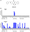
ONC201 shows high D2-like receptor selectivity. (A) Structure of the imipridone ONC201. (B) Screening panels demonstrating antagonism by ONC201 (top) or haloperidol (bottom) of agonist-stimulated β-arrestin recruitment to various GPCRs. The GPCR screening study was performed by DiscoverX, Inc. (Fremont, CA). Briefly, cells were preincubated with 10 μM of either ONC201 or haloperidol followed by an EC80 concentration of agonist for each indicated receptor (dopamine was the agonist used for dopamine receptors). β-arrestin recruitment was detected through the addition of DiscoverX PathHunter detection reagent cocktail. Microplates were read with a PerkinElmer EnvisionTM instrument for chemiluminescent signal detection. Assay results, run in duplicate, are presented as the mean percent inhibition of the indicated GPCR for each compound tested. Compound activity was analyzed using the CBIS data analysis suite (ChemInnovation, CA) and expressed as a percentage of the antagonism observed, as adapted from (Prabhu et al., 2018). Note that in (A), the y-axis does not extend to 100%. For a full description of the DiscoverX gpcrMAX assay panel see: http://www.DiscoverX.com.
Fig. 2.
Fig. 2.
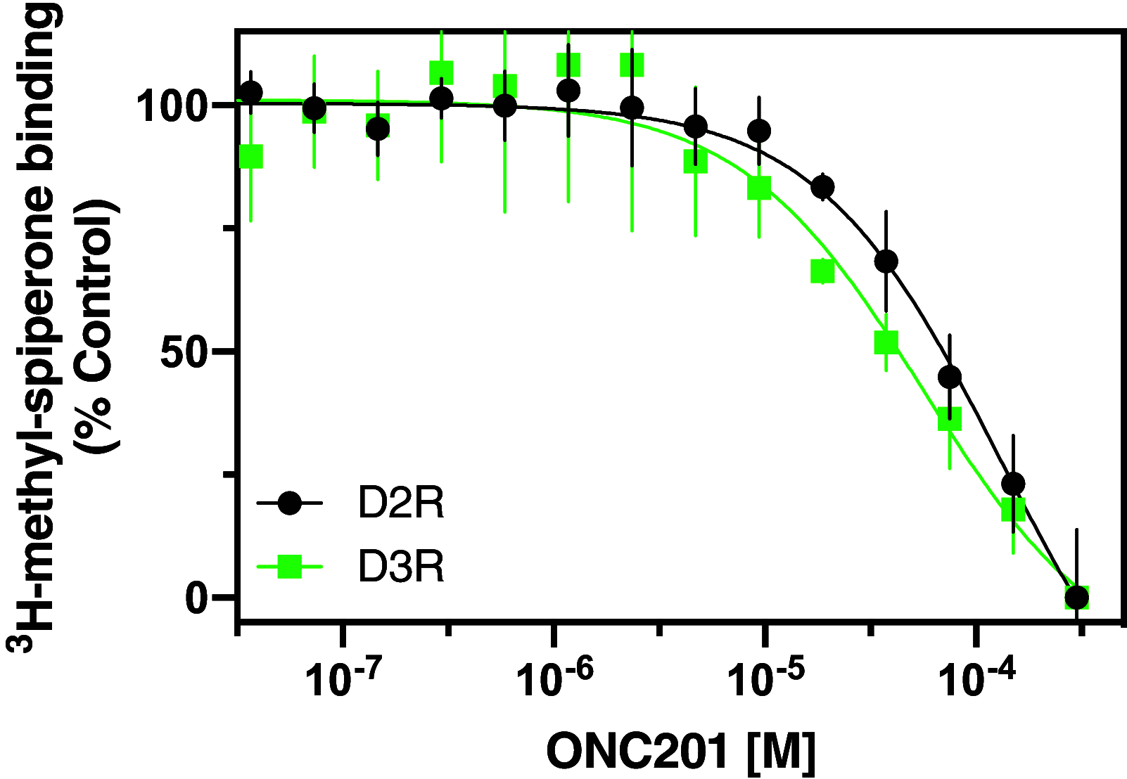
Radioligand binding competition assays to determine ONC201 affinity for the D2R and D3R. Radioligand binding assays using [3H]methylspiperone were performed as described in the Materials and Methods. Briefly, membranes from CHO cells stably expressing either the D2R or D3R were harvested and incubated with the indicated concentrations of ONC201 and 0.5 nM [3H]methylspiperone. The data are expressed as percentage of the control specific binding and represent mean ± S.D. values from three independent experiments performed in triplicate. Mean ONC201 Ki values [95% C.I.] for each receptor were calculated from the IC50 values using the Cheng-Prusoff equation (Cheng and Prusoff, 1973) and found to be 65.6 μM [44–102] for the D2R and 27.9 μM [9.2–99] for the D3R.
Fig. 3
Fig. 3
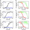
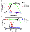
Pharmacological activity of ONC201 on D2-like dopamine receptors. β-arrestin recruitment assays were performed as described in the Materials and Methods. Data are expressed as a percentage of the maximum dopamine response in each assay and represent the mean ± S.D. values from 3 experiments each performed in triplicate. EC50 or IC50 values are expressed as means (95% CI). ONC201 did not produce any measurable agonist response for any of the tested receptors. (A) Dopamine stimulated β-arrestin recruitment to the D2R with an EC50 of 58 nM [40–86]. (B) β-arrestin recruitment to the D2R was stimulated with an EC80 concentration of dopamine (1 μM) and coincubated with the indicated concentrations of either sulpiride or ONC201 resulting in IC50 values of 48 nM [35–68] and 11 μM [5.6–21], respectively. (C) Dopamine stimulated β-arrestin recruitment to the D3R with an EC50 of 3 nM [2–4.7]. (D) β-arrestin recruitment to the D3R was stimulated with an EC80 concentration of dopamine (30 nM) and coincubated with the indicated concentrations of either spiperone or ONC201 resulting in IC50 values of 5.3 nM [3.8–7.5] and 21 μM [9.6–51], respectively. (E) Dopamine stimulated β-arrestin recruitment to the D4R with an EC50 of 100 nM [47–218]. (F) β-arrestin recruitment to the D4R was stimulated with an EC80 concentration of dopamine (1 μM) and coincubated with the indicated concentrations of either spiperone or ONC201 resulting in an IC50 value of 1.6 nM [0.6–4.5] for spiperone, whereas incomplete inhibition was observed for ONC201 (IC50 > 100 μM).
Fig. 4.
Fig. 4.
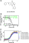
ONC201 isomer [4,3-d] is inactive at the D2R. (A) Structure of ONC201 isomer [4,3-d]. (B) β-arrestin recruitment assays were performed as described in the Materials and Methods. Data are expressed as a percentage of the maximum dopamine response and represent the mean ± S.D. values from 3 experiments, each performed in triplicate. Cells were incubated with an EC80 concentration of dopamine (1 μM) in the presence of increasing concentrations of sulpiride or the ONC201 isomer [4,3-d]. Sulpiride antagonized the D2R-mediated dopamine response with an IC50 of 48 nM [35–68] [mean (95% CI)], while the ONC201 isomer [4,3-d] was unable to block the response. (C) Dopamine-mediated β-arrestin recruitment assays were conducted by stimulating the D2R with the indicated concentrations of dopamine with or without various concentrations of the ONC201 isomer [4,3-d]. The EC50 of dopamine was not significantly affected by any concentration of the ONC201 isomer [4,3-d].
Fig. 5.
Fig. 5.
Functional antagonism of D2R- and D4R-mediated inhibition of forskolin-stimulated cAMP accumulation. CHO cells stably expressing either the D2R (A) or D4R (B) were incubated with 10 μM of forskolin plus the indicated drug combinations, and the resulting cAMP levels were determined using the LANCE assay as described in the Materials and Methods. Data are expressed as a percentage of the inhibition observed with dopamine alone (% control) and are expressed as the mean ± S.D. values from at least 3 experiments each performed in triplicate. EC50 or IC50 values are expressed as means (95% CI). (A) ONC201 failed to show any measurable agonist response. Dopamine dose-dependently inhibited cAMP accumulation with an EC50 value of 2.3 nM [1.5–3.6]. For antagonist-mode assays, inhibition of cAMP accumulation by the D2R was promoted with an EC80 concentration of dopamine (10 nM) and coincubated with the indicated concentrations of either sulpiride or ONC201 resulting in IC50 values of 1.5 nM [1.3–1.7] and 9.3 μM [5.7–15], respectively. (B) ONC201 failed to show any measurable agonist response. Dopamine dose-dependently inhibited cAMP accumulation with an EC50 value of 0.6 nM [0.3–1.2]. For antagonist-mode assays, inhibition of cAMP accumulation by the D4R was promoted with an EC80 concentration of dopamine (10 nM) and coincubated with the indicated concentrations of either spiperone or ONC201, resulting in an IC50 value of 9.8 nM [7.9–12] for spiperone, whereas incomplete inhibition was observed for ONC201 (IC50 > 100 μM).
Fig. 6.
Fig. 6.
Functional effects of ONC201 on D2R-mediated activation of Rho and G12. HEK293 cells were transiently cotransfected with the D2R and either the Rho or G12 BRET biosensors, and dopamine-stimulated BRET responses were measured as described in the Materials and Methods. Data are expressed as a percentage of the maximum dopamine response (% control) in each assay and represent the mean ± S.D. values from 3 experiments each performed in triplicate. EC50 or IC50 values are expressed as mean values (95% CI). (A) Dopamine dose-dependently stimulated Rho activation with an EC50 of 185 nM [97–385]. Concurrent treatment of the cells with 200 nM YM-254890 (+YM) or overnight treatment with 100 ng/ml +PTX resulted in dopamine EC50 values of 430 nM [162–986] and 207 nM [99–447], respectively, which were not significantly different from control values. (B) Rho activation (BRET) was stimulated with an EC80 concentration of dopamine (1 μM) and treated with the indicated concentrations of sulpiride or ONC201 resulting in IC50 values of 63 nM [17–198] and 9.4 μM [3.3–35], respectively. (C) Dopamine dose-dependently stimulated G12 activation with an EC50 of 310 nM [216–442], whereas ONC201 had no stimulatory effect. (D) G12 activation (BRET) was stimulated with an EC80 concentration of dopamine (1 μM) and treated with the indicated concentrations of sulpiride or ONC201 resulting in IC50 values of 18 nM [12–27] and 33 μM [20–55], respectively.
Fig. 7.
Fig. 7.
Curve-shift assays indicate that ONC201 behaves in an allosteric manner with dopamine at the D2R. D2R-mediated β-arrestin recruitment assays (A) or cAMP inhibition assays (LANCE) (B) were conducted by stimulating the receptor with the indicated concentrations of dopamine with or without various concentrations of ONC201, as described in the Materials and Methods. For the cAMP assay, the cells were preincubated with 10 μM forskolin to stimulate cAMP production. Data are expressed as a percentage of the maximum dopamine response seen in the absence of ONC201 (% control). Data points represent the mean ± S.D. of at least three independent experiments each performed in triplicate. The curves represent the best global fit of grouped data as determined using an operational model of allosterism (Leach et al., 2007), as described in the Materials and Methods, with the corresponding functional parameters being summarized in Table 1.
Fig. 8.
Fig. 8.
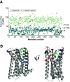
Alanine scanning mutagenesis identifies amino acids important for ONC201 antagonism of D2R signaling. HEK293T cells were transiently transfected with the wild-type (WT) D2R or D2R mutants and a chimeric Gα16 subunit. After 22 hours, calcium flux experiments were performed as described in the Materials and Methods. (A) Ca2+ flux was measured in response to 1 nM dopamine for each of the 442 D2R clones shown in Supplemental Fig. 1E and expressed as a percentage of the wild-type D2R response (light green symbols). For each D2R clone in the alanine-scan library, Ca2+ flux was also measured in response to 1 nM dopamine in the presence of 100 μM ONC201 and expressed as a percentage of the dopamine-stimulated response for the wild-type D2R (cyan and red symbols). The data represent average values from three experiments. Mutant clones (red symbols) were considered to be critical for ONC201 inhibition if they demonstrated Ca2+ flux values greater than 2 S.D.s above the average flux value (AV + 2 S.D.) observed in the presence of 100 μM ONC201. These eight residues (red symbols) are listed in Table 2. (B) The eight residues identified in (A) are depicted in the risperidone-D2R crystal structure [(Wang et al., 2018), PDB 6CM4]. Four colors depict the distinct geographic regions of these eight residues.
Fig. 9.
Fig. 9.
D2R mutants that affect ONC201 antagonism are not impaired in dopamine stimulation or sulpiride antagonism of G protein-mediated signaling. (A) HEK293 cells were transfected with the untagged wild-type D2R, or the indicated D2R mutants, along with Gαo1-RLuc8, Gγ2-mVenus, and Gβ1. After 48 hours, dopamine-stimulation of Go activation was assessed using a BRET assay as described in the Materials and Methods. The dopamine EC50 values for each D2R construct are shown in Table 3. (B) The cells were transfected as in (A) followed by stimulation with an EC80 concentration of dopamine (7 nM) and increasing concentrations of the orthosteric antagonist sulpiride. BRET ratio values were determined and the sulpiride IC50 values for each D2R construct are shown in Table 3. (C) HEK293 cells were transfected with the untagged wild-type D2R, or the indicated D2R mutants, along with the CAMYEL cAMP BRET biosensor. After 48 hours, the cells were incubated with 10 μM forskolin to stimulate cAMP accumulation, plus the indicated concentrations of dopamine. BRET ratio values were determined as described in the Materials and Methods with the dopamine EC50 values shown in Table 3. (D) The cells were transfected and treated as in (C) followed by stimulation with an EC80 concentration of dopamine (25 nM) and increasing concentrations of sulpiride. BRET ratio values were determined, and the sulpiride IC50 values for each D2R construct are shown in Table 3. In each panel, the data are expressed as a percentage of the wild-type D2R response. Data points represent the mean ± S.D. of at least three independent experiments each performed in triplicate.
Fig. 10.
Fig. 10.
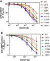
ONC201 antagonism of D2R-mediated G protein-mediated signaling is severely impaired in select D2R point mutants. (A) Go BRET activation assays were performed as described in Fig. 9 and the Materials and Methods. Cells were incubated with an EC80 concentration of dopamine (7 nM) and increasing concentrations of ONC201. BRET ratios were determined, and the data are expressed as a percentage of the wild-type D2R response. The mean IC50 values for each D2R construct are shown in Table 3. (B) CAMYEL BRET cAMP inhibition assays were performed as described in Fig. 9 and the Materials and Methods. The cells were incubated with an EC80 concentration of dopamine (25 nM) and increasing concentrations of ONC201. BRET ratios were determined, and the data are expressed as a percentage of the maximum wild-type D2R response. The mean IC50 values for each D2R construct are shown in Table 3. Data points represent the mean ± S.D. of at least three independent experiments each performed in triplicate.
Fig. 11.
Fig. 11.
The binding pose of ONC201 in the inactive state of the D2R overlaps with that of spiperone. Molecular docking was performed as described in the Methods using a model (see Supplement for PDB file) of the D2R in an inactive state based on a crystal structure of the D2R in complex with the antagonist spiperone (Im et al., 2020) (PDB: 7DFP). The superposition of the spiperone (colored in cyan) and ONC201 (colored in orange) poses are shown both from a side view (A) and an extracellular view (B) of the receptor’s TM regions. Receptor residues within the orthosteric pocket (D114) and allosteric pocket (V91 and E95) are designated.
Fig. 12.
Fig. 12.
The two possible ONC201 binding poses in the extracellular vestibule of the D2R in the active state. Molecular docking was performed as described in the Methods using an active state structure of the D2R (Yin et al., 2020) (PDP: 6VMS). Poses 1 (A) and 2 (B) of ONC201 (colored in orange) in the extracellular vestibule are shown with dopamine (colored in yellow) bound to the orthosteric binding site (see Supplement for PDB files of poses 1 and 2). Orthosteric residues D114, S193, and S197 form direct interactions with dopamine. For each pose, MD frames were extracted every 60 ns for the last 300 ns of their corresponding MD trajectories.

References

    1. Allen JE, Crowder RN, El-Deiry WS (2015) First-in-class small molecule ONC201 induces DR5 and cell death in tumor but not normal cells to provide a wide therapeutic index as an anti-cancer agent. PLoS One 10:e0143082. - PMC - PubMed
    1. Allen JEKrigsfeld GMayes PAPatel LDicker DTPatel ASDolloff NGMessaris EScata KAWang W, et al. (2013) Dual inactivation of Akt and ERK by TIC10 signals Foxo3a nuclear translocation, TRAIL gene induction, and potent antitumor effects. Sci Transl Med 5:171ra17. - PMC - PubMed
    1. Arang N, Gutkind JS (2020) G protein-coupled receptors and heterotrimeric G proteins as cancer drivers. FEBS Lett 594:4201–4232. - PMC - PubMed
    1. Ballesteros JA, Weinstein H (1995) Integrated methods for the construction of three-dimensional models and computational probing of structure-function relations in G protein-coupled receptors. Methods in Neuroscience 25:366–428.
    1. Beaulieu JM, Gainetdinov RR (2011) The physiology, signaling, and pharmacology of dopamine receptors. Pharmacol Rev 63:182–217. - PubMed

Publication types

MeSH terms

LinkOut - more resources