. 2021 Aug 6;373(6555):700-704.
doi: 10.1126/science.abe6821.

Maturation of the matrix and viral membrane of HIV-1

Affiliations

Maturation of the matrix and viral membrane of HIV-1

Kun Qu et al. Science. .

Abstract

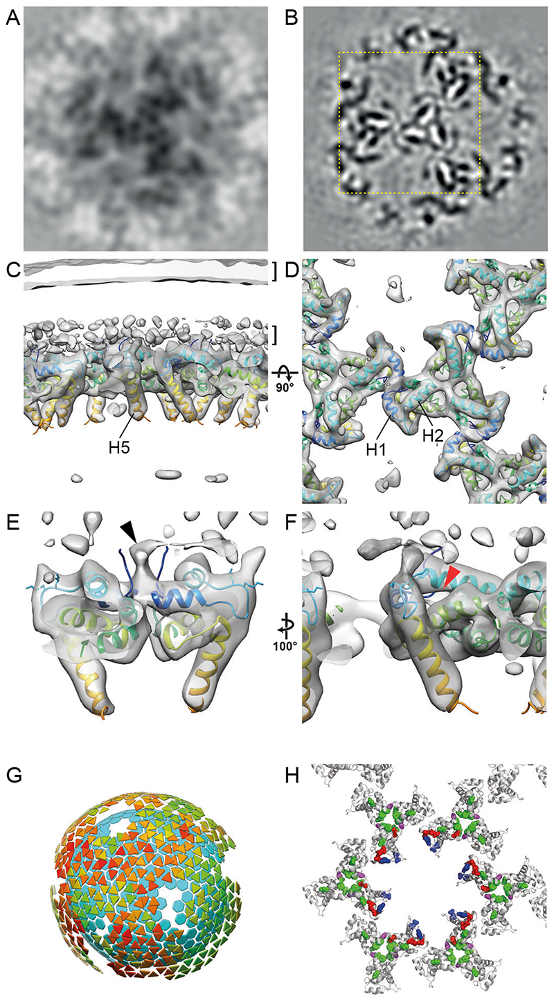
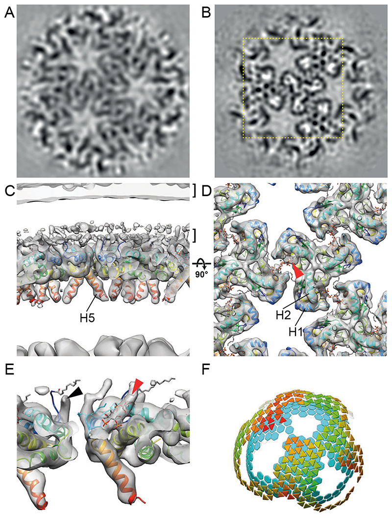
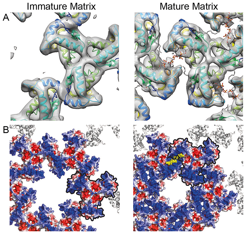
Gag, the primary structural protein of HIV-1, is recruited to the plasma membrane for virus assembly by its matrix (MA) domain. Gag is subsequently cleaved into its component domains, causing structural maturation to repurpose the virion for cell entry. We determined the structure and arrangement of MA within immature and mature HIV-1 through cryo-electron tomography. We found that MA rearranges between two different hexameric lattices upon maturation. In mature HIV-1, a lipid extends out of the membrane to bind with a pocket in MA. Our data suggest that proteolytic maturation of HIV-1 not only assembles the viral capsid surrounding the genome but also repurposes the membrane-bound MA lattice for an entry or postentry function and results in the partial removal of up to 2500 lipids from the viral membrane.


Conflict of interest statement

Competing interests:

Authors declare no competing interests.

Figures

Fig. 1
Fig. 1. Tomograms of immature and mature HIV-1 particles.
(A) Top: Schematic of the Gag protein domain architecture with cleavage sites marked as vertical black lines. The D25N mutation (red X) inactivates the viral PR. Bottom: Computational slices through a representative tomogram of immature HIV-1 (cHIV PR-) particles. Density is black. The striated Gag layer is distinctly observed in immature particles (black arrowheads). (B) As in (A) for cleaved, mature HIV-1 (cHIV) particles. Distinct conical CA cores are observed (white arrowheads). (C) Slices grazing the inner surface of the membrane at the top of the particles in (B) revealing a regular MA lattice. Gold fiducials are removed from the images. Scale bar: 100 nm.
Fig. 2
Fig. 2. The immature HIV-1 matrix structure.
(A, B) Slices through reconstructions of the immature HIV-1 MA lattice in cHIV PR- (A) and HIV-1NL4-3 PR- (B). The boxed region in (B) is shown at a higher magnification in (D). Density is black. (C, D) Isosurface views of the cryo-ET reconstruction (grey) for immature HIV-1NL4-3 PR- MA lattice cut perpendicular to the membrane (C) or viewed from the top towards the virus center (D). The two layers of density corresponding to the lipid headgroup layers are indicated by brackets. The structure of monomeric MA determined by NMR (PDBID: 2H3Q (18); colored blue to red from N to C terminus) was fitted as a rigid body into the density. Helices 1, 2 and 5 are marked. (E) As in (C), enlarged and cut to reveal density corresponding to the N-terminal residues (black arrowhead). (F) As in (E) but rotated. The red arrowhead indicates the unoccupied PI(4,5)P2 binding pocket. (G) Lattice map derived from subtomogram averaging for the immature HIV-1NL4-3 PR- MA lattice. Lattice maps illustrate positions and orientations of MA trimers as triangles colored on a scale from red (lower cross correlation to average structure) to green (higher cross correlation to average structure). The positions of CA hexamers are indicated as cyan hexagons to illustrate the relationship between MA and CA layers. (H) Immature MA lattice is shown as grey ribbons and residues whose mutation has been reported to modulate Env incorporation are shown as colored spheres. Mutations at residues L12, L30 and L74 (red), E16 and E98 (blue), and T69 (purple) impair Env incorporation (, , –33). Mutations in V34, F43, Q62 and S66 (green) can rescue Env incorporation defects (11, 28, 31, 43), except those caused by mutations at T69 (purple) (28). E16 and E98 (blue) face the hole in the MA lattice in the immature virion.
Fig. 3
Fig. 3. The mature HIV-1 matrix structure.
(A, B) Slices through reconstructions of the mature HIV-1 MA lattice in cHIV (A) and cHIV MA-SP1 (B). The boxed region in (B) is shown at a higher magnification in (D). (C, D) An isosurface view of the cryo-ET reconstruction for mature cHIV MA-SP1 MA lattice fitted with the structure of monomeric MA, cut perpendicular to the membrane (C) or viewed from the top towards the virus center (D). The two layers of density corresponding to the lipid headgroup layers are indicated by brackets. The structure of monomeric MA determined by NMR (PDBID: 2H3Q (18); colored blue to red from N to C terminus) was fitted as a rigid body into the density. Helices 1, 2 and 5 are marked. Density is observed in the PI(4,5)P2 binding site (red arrowhead), and the structure of PI(4,5)P2 as resolved bound to MA by NMR (PDBID: 2H3V (18)), is shown as a stick model. (E) As in (C), enlarged and cut to reveal density corresponding to the N-terminal residues (black arrowhead), and PI(4,5)P2 (red arrowhead). (F) As in Fig. 2G, lattice map for the mature cHIV MA-SP1 MA lattice and the underlying CA lattice.
Fig. 4
Fig. 4. Comparison of the immature and mature HIV-1 MA lattices.
(A) Immature and mature MA lattices are shown with one trimer aligned. In the immature lattice, contact with neighbouring trimers is mediated by N-terminal regions and the PI(4,5)P2 binding site is empty. In the mature lattice, contact with neighbouring trimers is mediated by the region surrounding the occupied PI(4,5)P2 binding site. (B) Electrostatic surface potential maps of the hexameric lattice of immature and mature MA trimers. The red (-5 kT/e) and blue (+5 kT/e) colours represent negatively and positively charged electric potentials. PI(4,5)P2 is shown in yellow. The negatively-charged lipid headgroups are surrounded by positively-charged residues. One MA trimer aligned as in (A) is outlined in black. The potential of the surface of MA facing the holes at the hexamer positions in the lattice changes from positive to neutral/negative during maturation. The hole becomes smaller.

Comment in

  • Maturation of HIV-1.
    Hikichi Y, Freed EO. Hikichi Y, et al. Science. 2021 Aug 6;373(6555):621-622. doi: 10.1126/science.abj9075. Science. 2021. PMID: 34353938 No abstract available.

References

    1. Sundquist WI, Kräusslich H-G. HIV-1 assembly, budding, and maturation. Cold Spring Harbor Perspectives in Medicine. 2012;2:a006924. - PMC - PubMed
    1. Freed EO. HIV-1 assembly, release and maturation. Nature Reviews Microbiology. 2015;13:484–496. 2015 13:8. - PMC - PubMed
    1. Mattei S, Schur FK, Briggs JAG. Retrovirus maturation — an extraordinary structural transformation. Current Opinion in Virology. 2016;18:27–35. - PubMed
    1. Pornillos O, Ganser-Pornillos BK. Maturation of retroviruses. Current Opinion in Virology. 2019;36:47–55. - PMC - PubMed
    1. Murakami T, Ablan S, Freed EO, Tanaka Y. Regulation of human immunodeficiency virus type 1 Env-mediated membrane fusion by viral protease activity. Journal of Virology. 2004;78:1026–1031. - PMC - PubMed

Publication types

MeSH terms

Substances