. 2021 Aug 5;11(1):15943.
doi: 10.1038/s41598-021-95660-w.

Fluorescence/photoacoustic imaging-guided nanomaterials for highly efficient cancer theragnostic agent

Affiliations

Fluorescence/photoacoustic imaging-guided nanomaterials for highly efficient cancer theragnostic agent

Vu Hoang Minh Doan et al. Sci Rep. .

Erratum in

Abstract

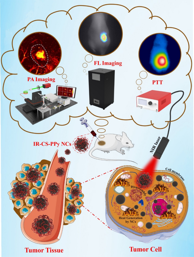
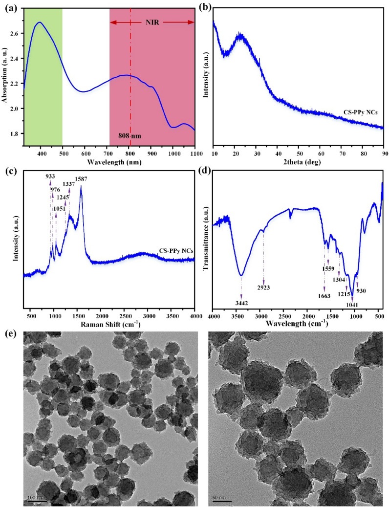
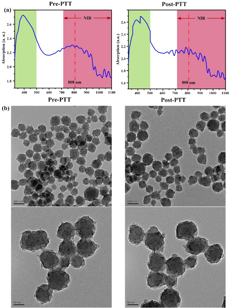
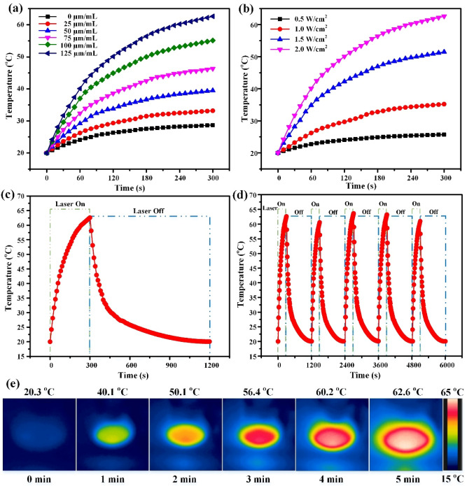
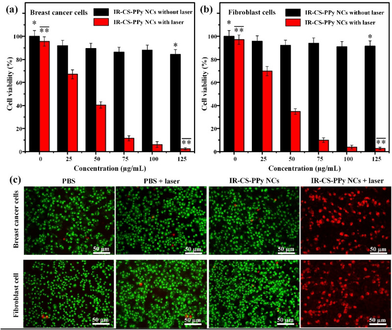
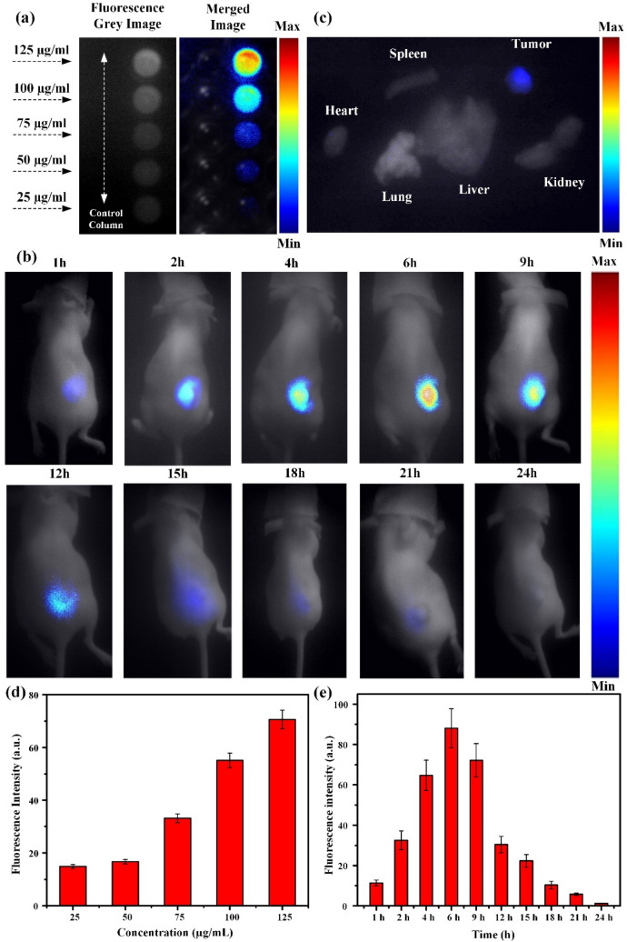
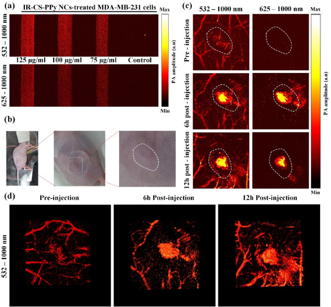
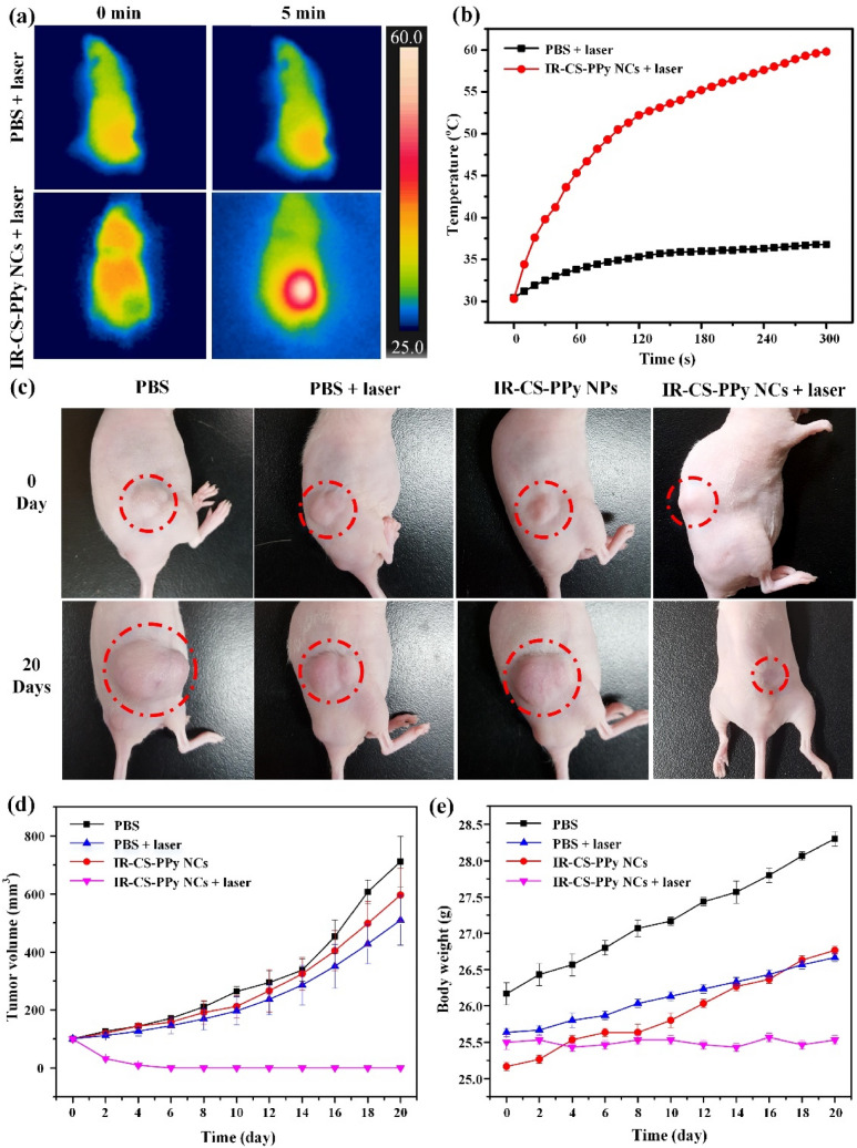
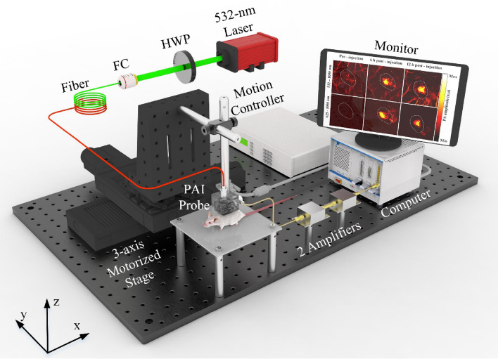
Imaging modalities combined with a multimodal nanocomposite contrast agent hold great potential for significant contributions in the biomedical field. Among modern imaging techniques, photoacoustic (PA) and fluorescence (FL) imaging gained much attention due to their non-invasive feature and the mutually supportive characteristic in terms of spatial resolution, penetration depth, imaging sensitivity, and speed. In this present study, we synthesized IR783 conjugated chitosan-polypyrrole nanocomposites (IR-CS-PPy NCs) as a theragnostic agent used for FL/PA dual-modal imaging. A customized FL and photoacoustic imaging system was constructed to perform required imaging experiments and create high-contrast images. The proposed nanocomposites were confirmed to have great biosafety, essentially a near-infrared (NIR) absorbance property with enhanced photostability. The in vitro photothermal results indicate the high-efficiency MDA-MB-231 breast cancer cell ablation ability of IR-CS-PPy NCs under 808 nm NIR laser irradiation. The in vivo PTT study revealed the complete destruction of the tumor tissues with IR-CS-PPy NCs without further recurrence. The in vitro and in vivo results suggest that the demonstrated nanocomposites, together with the proposed imaging systems could be an effective theragnostic agent for imaging-guided cancer treatment.


Conflict of interest statement

The authors declare no competing interests.

Figures

Figure 1
Figure 1
Schematic representation of IR-CS–PPy NCs for dual-channel fluorescence/photoacoustic imaging-guided photothermal therapy. The scheme was created using Servier Medical Art (http://smart.servier.com/).
Figure 2
Figure 2
Characterization of CS–PPy NCs (a) UV–Vis–NIR absorbance spectrum CS–PPy NCs dispersion in water. (b) XRD patterns of CS–PPy NCs. (c) Raman spectrum of CS–PPy NCs. (d) FTIR spectrum of CS–PPy NCs. (e) FE-TEM image of CS–PPy NCs.
Figure 3
Figure 3
Characterization of IR-CS–PPy NCs (a) UV–Vis spectrum of IR-CS–PPy NCs (125 μg/mL) before and after PTT. (b) FE-TEM images of IR-CS–PPy NCs (125 μg/mL) before and after PTT.
Figure 4
Figure 4
(a) The temperature elevation in an aqueous solution of different IR-CS–PPy NCs concentrations (0, 25, 50, 75, 100, 125 μg/mL) as a function of irradiation time by 808 nm laser at 2 W/cm2 of laser power densities. (b) The temperature elevation of IR-CS–PPy NCs aqueous solution at a concentration of 125 μg/mL under 808-nm laser irradiation at different power densities (0.5 W/cm2, 1.0 W/cm2, 1.5 W/cm2, and 2.0 W/cm2) for 5 min. (c) The photothermal response of IR-CS–PPy NCs (125 μg/mL) aqueous solution exposed to an 808-nm laser source at 2 W/cm2 for 300 s and then the laser was shut off for about 900 s. (d) Temperature profiles of IR-CS–PPy NCs (125 μg/mL) aqueous solution for five on/off cycles. (e) The corresponding NIR thermographic images of the well containing IR-CS–PPy NCs (125 μg/mL) during 300 s of laser irradiation (2 W/cm2).
Figure 5
Figure 5
The cell viability of (a) MDA-MB-231 breast cancer and (b) L929 human normal fibroblast cells treated with IR-CS–PPy NCs with and without NIR laser (2 W/cm2, 5 min). (b) The cell viability of L929 cells treated with IR-CS–PPy NCs with and without NIR laser (2 W/cm2, 5 min). (c) AO/PI staining of MDA-MB-231 cells and L929 cells treated with PBS, PBS + laser (2 W/cm2, 5 min), 125 µg/mL IR-CS–PPy NCs, and 125 µg/mL IR-CS–PPy NCs + laser (2 W/cm2, 5 min). Data are shown as the mean ± standard deviation (n = 3). (*Significant p < 0.05).
Figure 6
Figure 6
(a) In vitro fluorescence imaging of control cells and MDA-MB-231 cell treated with different concentrations of IR-CS–PPy NCs (25, 50, 75, 100, and 125 μg/mL) (b) In vivo fluorescence imaging of MDA-MB-231 tumor-bearing nude mice at different time points after local injection of IR-CS–PPy NCs. (c) Ex vivo fluorescence imaging of the organs including, the heart, liver, lung, kidney, and spleen, and tumors excised from the mice treated with IR-CS–PPy NCs at 24 h post local injection. (d) The mean fluorescence intensities of different concentrations of IR-CS–PPy NCs were measured, displaying the highest fluorescence intensity at 125 μg/mL. (e) The mean fluorescence intensities of tumors were quantified at different time points, showing a peak value after 6 h injection of IR-CS–PPy NCs. Data are shown as the mean ± standard deviation (n = 3). Min = 0, and Max = 100.
Figure 7
Figure 7
(a) In vitro PAI of MDA-MB-231 cells incubated with various concentrations of IR-CS–PPy NCs using PAI system with 532–1000 nm wavelength and 625–1000 nm wavelength. (b) Representative digital photographs of MDA-MB-231 tumor-bearing nude mice for in vivo PAI. The white dash lines indicate the tumor area. (c) In vivo PAI of tumor tissues in MDA-MB-231 tumor-bearing nude mice at 0, 6, and 12 h after injection of IR-CS–PPy NCs (100 μL) using PAI system with 532–1000 nm wavelength, and 625–1000 nm wavelength. (d) Representative 3D PA images of tumor tissues MDA-MB-231 tumor-bearing nude mice at 0, 6, and 12 h after injection of IR-CS–PPy NCs using PAI system with 532–1000 nm wavelength.
Figure 8
Figure 8
(a) NIR thermographic images of tumor-bearing mice with intratumoral injection of PBS and IR-CS–PPy NCs with 808-nm NIR laser irradiation at 2.0 W/cm2 for 5 min. (b) Temperature change of tumor-bearing mice after intratumoral injection of with IR-CS–PPy NCs with 808-nm NIR laser irradiation at 2.0 W/cm2 for 5 min. (c) The digital photographs of tumor-bearing mice taken at day 0 before treatment and 20 days after treatment. (d) Tumor volume growth curves of different groups of mice after different treatments. Data presented as mean ± standard deviation. (n = 3). (e) The body weight after different treatments indicated in 20 days. Data are shown as the mean ± standard deviation (n = 3).
Figure 9
Figure 9
The proposed fluorescence imaging system (a) Schematic diagram of fluorescence system and the controller printed circuit board (PCB) (b) 3D design of controller PCB (c) 3D design of fluorescence system.
Figure 10
Figure 10
Schematic of the proposed photoacoustic imaging system. HWP half-wave plate, FC fiber coupler.

References

    1. Agabeigi R, Rasta SH, Rahmati-Yamchi M, Salehi R, Alizadeh E. Novel chemo-photothermal therapy in breast cancer using methotrexate-loaded folic acid conjugated Au@SiO2 Nanoparticles. Nanoscale Res. Lett. 2020 doi: 10.1186/s11671-020-3295-1. - DOI - PMC - PubMed
    1. Azamjah N, Soltan-Zadeh Y, Zayeri F. Global trend of breast cancer mortality rate: A 25-year study. Asian Pac. J. Cancer Prev. 2019 doi: 10.31557/APJCP.2019.20.7.2015. - DOI - PMC - PubMed
    1. Feng Y, Spezia M, Huang S, Yuan C, Zeng Z, Zhang L, Ji X, Liu W, Huang B, Luo W, Liu B, Lei Y, Du S, Vuppalapati A, Luu HH, Haydon RC, He TC, Ren G. Breast cancer development and progression: Risk factors, cancer stem cells, signaling pathways, genomics, and molecular pathogenesis. Genes Dis. 2018;5(2):77–106. doi: 10.1016/j.gendis.2018.05.001. - DOI - PMC - PubMed
    1. Jeffe DB, Pérez M, Cole EF, Liu Y, Schootman M. The effects of surgery type and chemotherapy on early-stage breast cancer patients’ quality of life over 2-year follow-up. Ann. Surg. Oncol. 2016;23(3):735–743. doi: 10.1245/s10434-015-4926-0. - DOI - PMC - PubMed
    1. Ratajczak MZ, Jadczyk T, Schneider G, Kakar SS, Kucia M. Induction of a tumor-metastasis-receptive microenvironment as an unwanted and underestimated side effect of treatment by chemotherapy or radiotherapy. J. Ovarian Res. 2013 doi: 10.1186/1757-2215-6-95. - DOI - PMC - PubMed

Publication types