Review
. 2021 Sep;24(9):1198-1209.
doi: 10.1038/s41593-021-00904-7. Epub 2021 Aug 5.

Revisiting the neurovascular unit

Affiliations
Review

Revisiting the neurovascular unit

Samantha Schaeffer et al. Nat Neurosci. 2021 Sep.

Abstract

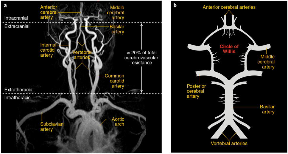
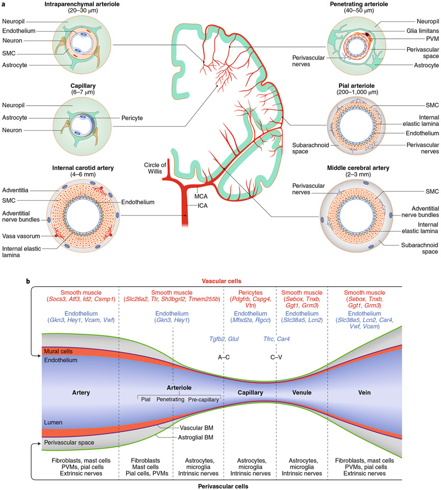
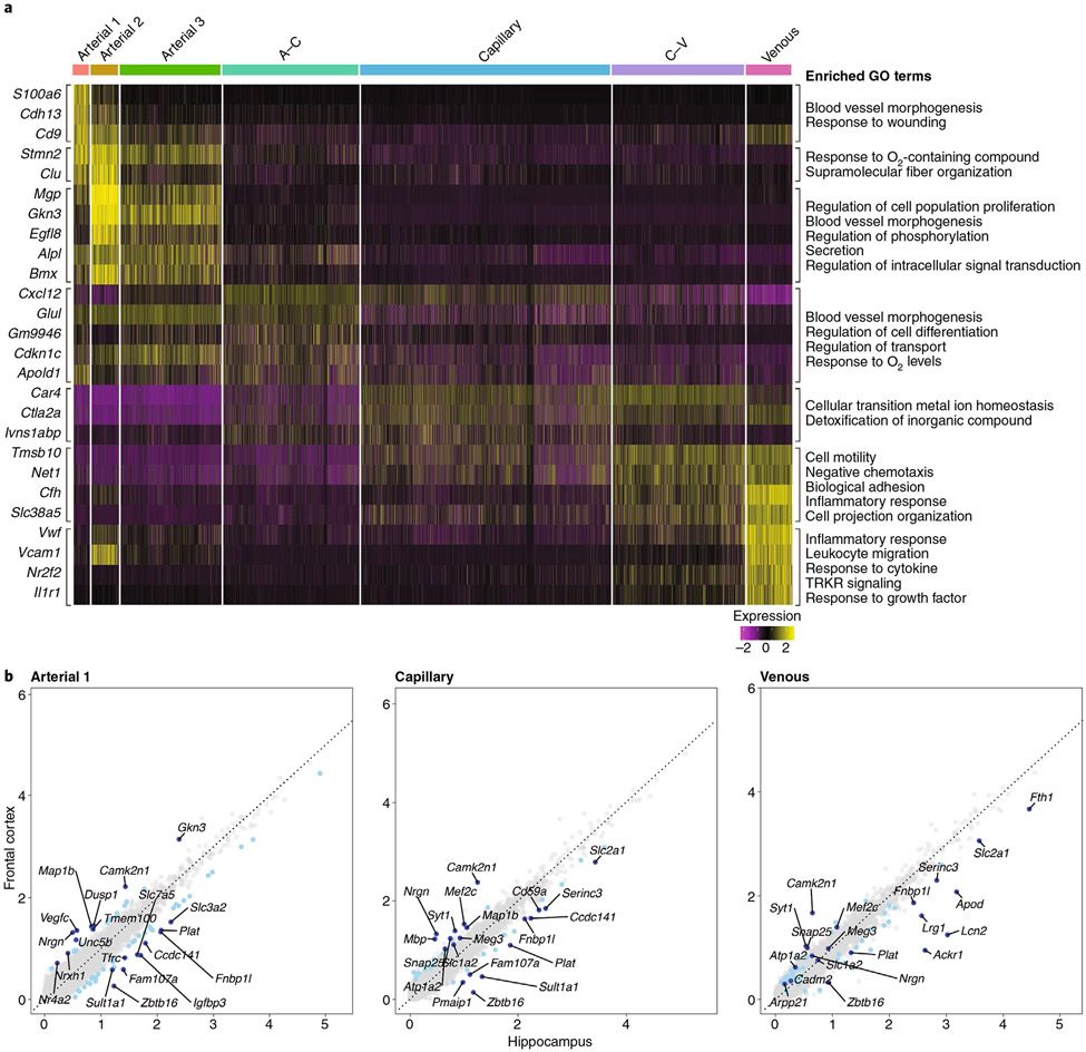
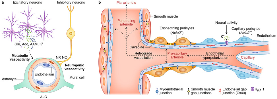
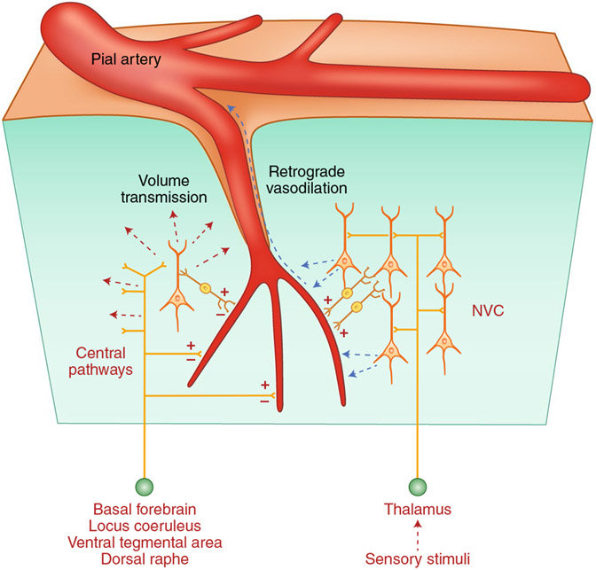
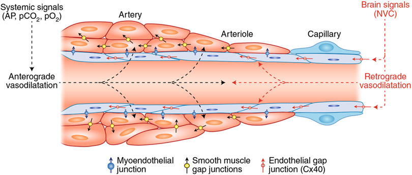
The brain is supplied by an elaborate vascular network that originates extracranially and reaches deep into the brain. The concept of the neurovascular unit provides a useful framework to investigate how neuronal signals regulate nearby microvessels to support the metabolic needs of the brain, but it does not consider the role of larger cerebral arteries and systemic vasoactive signals. Furthermore, the recently emerged molecular heterogeneity of cerebrovascular cells indicates that there is no prototypical neurovascular unit replicated at all levels of the vascular network. Here, we examine the cellular and molecular diversity of the cerebrovascular tree and the relative contribution of systemic and brain-intrinsic factors to neurovascular function. Evidence supports the concept of a 'neurovascular complex' composed of segmentally diverse functional modules that implement coordinated vascular responses to central and peripheral signals to maintain homeostasis of the brain. This concept has major implications for neurovascular regulation in health and disease and for brain imaging.


Figures

Fig. 1 ∣
Fig. 1 ∣. Anatomy of the large vessels supplying the brain.
a, Common carotid arteries arise from large intrathoracic arteries and give rise to the internal carotid arteries that enter the skull and merge into the circle of Willis. Vertebral arteries run along the cervical vertebrae and enter the skull and join to form the basilar artery, which merges into the circle of Willis. On the basis of AP gradient measurements, extracranial arteries are responsible for ~20% of the total cerebrovascular resistance, which suggests that they contribute to the regulation of cerebral perfusion. b, Schematic representation of the circle of Willis and its major branches. Image provided by A. Gupta.
Fig. 2 ∣
Fig. 2 ∣. Segmental heterogeneity of cerebral arteries and diversity of vascular and perivascular cells.
a, The internal carotid artery has a thick layer of SMCs surrounded by nerves arising from cranial autonomic ganglia (extrinsic innervation) embedded in perivascular connective tissue (adventitia). The internal elastic lamina separates SMCs from the endothelial cell monolayer. In the middle cerebral artery (MCA) and pial arteriolar branches, the SMC layer becomes progressively thinner, and a perivascular nerve plexus surrounds the vascular wall. Penetrating arterioles dive into the substance of the brain surrounded by a perivascular space where perivascular macrophages (PVMs) and other cells reside. As the vessel becomes smaller (intraparenchymal arterioles), the vascular basement membrane fuses with the glial basement membrane and perivascular nerves are replaced by nerve terminals from interneurons or subcortical pathways (intrinsic innervation). In capillaries, SMCs are replaced by pericytes. Vascular diameters indicated under the vascular segments refer to the human cerebral circulation. Venous SMCs are morphologically, functionally and molecularly distinct from arterial SMCs. b, Each segment of the cerebrovascular tree is characterized by diverse vascular and perivascular cells. The vascular and astroglial membranes delimit the perivascular space, which disappears when these membranes fuse together. Pial arterioles give rise to penetrating arterioles, the first-order branch of which is defined as precapillary arterioles. For mural and endothelial cells, genes enriched in each vascular segment are also indicated. For SMCs, we used the database from ref. , in which segmental assignment was validated by in situ hybridization. For endothelial cells, we used ref. , in which the segmental assignment was predicted in silico. A–C represents marker endothelial genes at the arteriolar–capillary transition and C–V at the capillary–venular transition. BM, basement membrane; ICA, internal carotid artery.
Fig. 3 ∣
Fig. 3 ∣. Endothelial expression heatmap and scatter plot of differentially expressed genes in the neocortex and the hippocampus.
a, Analysis of single-cell RNA-seq data from the Saunders database of whole-brain endothelial cells. The heatmap shows scaled, log-normalized expression of the top discriminative genes per endothelial cell cluster (left) identified using the SEURAT toolkit. The color bar (top) denotes assignment for endothelial cells ordered by position in the vascular tree according to validated markers,. On the right, the GO terms that define the biological process in which the differentially expressed genes may be involved are presented. GO terms were derived from the biological process subset of MSigDB’s v7.1 GO gene sets (C5) for Mus musculus. All significant differentially expressed genes were used for analysis, and resulting pathways with a false discovery rate q-value of <0.05 are presented (see Supplementary Methods for details). b, Scatter plot of differentially expressed genes in neocortical and hippocampal endothelial cells. Comparative analysis of endothelial genes (gray dots) scaled, log-normalized expression in the frontal cortex and the hippocampus mined from the Saunders database. Differentially expressed genes between the frontal cortex and the hippocampus are indicated in light blue, with the top ten most regulated genes indicated in dark blue. The GO terms referring to these differentially expressed genes are presented in Supplementary Table 1 (see Supplementary Methods for details).
Fig. 4 ∣
Fig. 4 ∣. Local and remote vascular components of NVC.
a, Excitatory neurons have a powerful effect on local neural activity and energy metabolism, and may drive local vascular changes through neurotransmitters, vasoactive ions, as well as by-products of neural activity such as adenosine (Ado) and arachidonic acid metabolites (AAMs). Astrocytes may also participate in this process. Interneurons, which have little impact on local neural activity, may signal blood vessels through direct neurovascular connections releasing neuropeptides (NPs) and NO. Glu, glutamate. b, The local vascular response elicited by overall neural activity leads to hyperpolarization of endothelial cells through KIR2.1 channels. Whether mesh and thin-strand (capillary) pericytes (ACTA2-negative) participate in this process remains unclear. Hyperpolarization is propagated retrogradely through inter-endothelial gap junctions and is transmitted to contractile mural cells, ACTA2-containing ensheathing pericytes and SMCs, likely via myoendothelial junctions and KIR channels. In turn, mural cell hyperpolarization suppresses voltage-gated Ca2+ channel activity, resulting in intracellular Ca2+ depletion, relaxation of the contractile apparatus and vasodilatation. The relaxation is then transmitted to adjacent mural cells through intercellular gap junctions, leading to retrograde vasodilation, which eventually reaches pial arterioles at the brain surface.
Fig. 5 ∣
Fig. 5 ∣. Sources and targets of brain intrinsic vasoactive signals.
Central pathways arising from the basal forebrain and brainstem nuclei contact cerebral blood vessels directly or through interposed interneurons (intrinsic innervation) and can either increase (+) or decrease (−) CBF diffusely. Neurotransmitters released from these pathways may also affect more distant vessels through volume transmission. Somatosensory or visual stimuli originating from the thalamus produce localized increases in blood flow by activating local neurons, which in turn release vasoactive agents (NVC; see Fig. 4 for details).
Fig. 6 ∣
Fig. 6 ∣. Central and peripheral vasoactive signals regulating CBF.
Vasoactive signals arising from the periphery (systemic signals) exert anterograde effects on all segments of the cerebrovascular tree by acting mainly on SMCs (anterograde vasodilatation). Brain-intrinsic vasoactive signals evoked by NVC and, possibly, central pathways arise within the substance of the brain and propagate retrogradely to microvascular SMCs and beyond via inter-endothelial junctions (retrograde vasodilatation). During activities of daily living, in which major changes in AP and blood gases occur, maintenance of the perfusion and homeostatic balance of the brain depends on the dynamic and coordinated interaction of these vasoactive signals engaging all segments of the neurovascular complex.

References

    1. Iadecola C The neurovascular unit coming of age: a journey through neurovascular coupling in health and disease. Neuron 96, 17–42 (2017). - PMC - PubMed
    1. Alves de Lima K, Rustenhoven J & Kipnis J Meningeal immunity and its function in maintenance of the central nervous system in health and disease. Annu. Rev. Immunol 38, 597–620 (2020). - PubMed
    1. Koizumi T, Kerkhofs D, Mizuno T, Steinbusch HWM & Foulquier S Vessel-associated immune cells in cerebrovascular diseases: from perivascular macrophages to vessel-associated microglia. Front. Neurosci 13, 1291 (2019). - PMC - PubMed
    1. Faraco G et al. Dietary salt promotes cognitive impairment through tau phosphorylation. Nature 574, 686–690 (2019). - PMC - PubMed
    1. Paredes I, Himmels P & Ruiz de Almodovar C Neurovascular communication during CNS development. Dev. Cell 45, 10–32 (2018). - PubMed

Publication types