. 2020 Mar 24;30(13):1905544.
doi: 10.1002/adfm.201905544. Epub 2020 Feb 13.

A Wirelessly Controlled Smart Bandage with 3D-Printed Miniaturized Needle Arrays

Affiliations

A Wirelessly Controlled Smart Bandage with 3D-Printed Miniaturized Needle Arrays

Hossein Derakhshandeh et al. Adv Funct Mater. .

Abstract

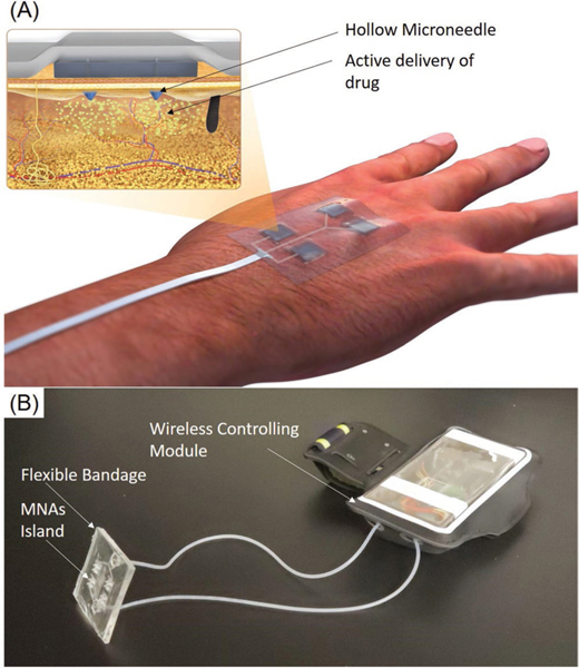
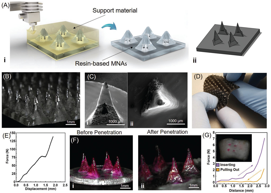
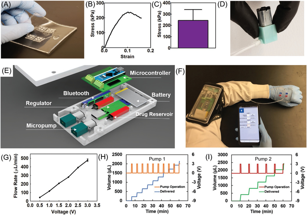
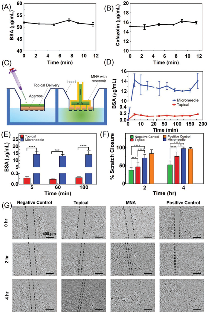
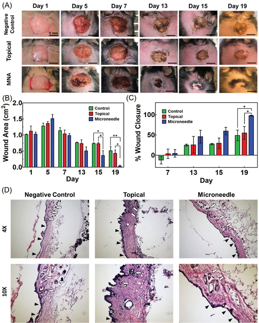
Chronic wounds are one of the most devastating complications of diabetes and are the leading cause of nontraumatic limb amputation. Despite the progress in identifying factors and promising in vitro results for the treatment of chronic wounds, their clinical translation is limited. Given the range of disruptive processes necessary for wound healing, different pharmacological agents are needed at different stages of tissue regeneration. This requires the development of wearable devices that can deliver agents to critical layers of the wound bed in a minimally invasive fashion. Here, for the first time, a programmable platform is engineered that is capable of actively delivering a variety of drugs with independent temporal profiles through miniaturized needles into deeper layers of the wound bed. The delivery of vascular endothelial growth factor (VEGF) through the miniaturized needle arrays demonstrates that, in addition to the selection of suitable therapeutics, the delivery method and their spatial distribution within the wound bed is equally important. Administration of VEGF to chronic dermal wounds of diabetic mice using the programmable platform shows a significant increase in wound closure, re-epithelialization, angiogenesis, and hair growth when compared to standard topical delivery of therapeutics.

Keywords: 3D-printed needles; active drug delivery; chronic wounds; smart bandages.


Conflict of interest statement

Conflict of Interest The authors declare no conflict of interest.

Figures

Figure 1.
Figure 1.
The wirelessly controlled smart bandage equipped with MNAs for delivery of therapeutics. A) Schematic of the engineered bandage and its operation. B) A representative photograph of the bandages for the delivery of multiple drugs. The system had two modules, a wearable bandage with integrated MNAs connected to the controlling module, which can communicate wirelessly with a smartphone in order to control the drug delivery rate.
Figure 2.
Figure 2.
Fabrication and characterization of 3D-printed MNAs. A) Schematic of 3D-printing MNAs showing i) the fabrication process and ii) a representative computer model view of the designed hollow MNAs. B) Microscopic image of sample MNAs. C) SEM images of i) front view and ii) top view of a single hollow needle. D) Multimaterial printing of MNAs for creating rigid needles on a flexible base. E) The characterization of the mechanical properties of MNA islands (four needles) (n = 4). F) Microscopic view of MNAs i) before and ii) after pig skin penetration test (n = 4). G) Characterization of the insertion and pull out force of the MNA islands applied to pig skin and image of pig skin targeted with painted MNAs (inset).
Figure 3.
Figure 3.
Characterization of microcontroller and wireless software. A) Photograph of a typical fabricated flexible bandage with bonded hollow MNAs on a PDMS-made bandage. B) A representative stress-strain curve of a peel-off test for assessment of the bonding strength of resin to PDMS. C) The bonding strength of resin and PDMS substrates (n = 6). D) Photograph of the micropump used for the drug delivery system. E) Schematic view of the microcontroller and its components. F) Example of the integrated system operation on the human body. G) Calibration plot of the micropump flow rate as a function of applied voltage (n = 3). H,I) The cumulative delivered solution using two independent pumps on a single bandage subjected to different periodic functions of applied voltage calibrated to produce a flow rate of 200 μL every 6 min for pump 1 and 400 μL every 10 min for pump 2 (n = 3 for each pump).
Figure 4.
Figure 4.
Characterizing the drug release and its effect on cellular cultures. A,B) The concentration of BSA and cefazolin in solutions perfused through the engineered bandage and MNAs over time. The results suggest an insignificant change in concentration (n = 3 for each solution). C) Schematic of the two-compartment in vitro model used for simulating chronic wounds covered by a crust and necrotic tissue used for comparing the topical and MNA-based drug delivery. D) The cumulative concentration of the BSA in the bottom chamber representing the wound bed after the administration of 20 μg mL−1 solution of BSA through 2 mm thick agarose gel (3% w/v) within a cell culture insert (n = 3 for each group). E) The cumulative drug concentration after 5, 60, and 180 min postdrug administration (***P < 0.001, ****P < 0.0001). F,G) Scratch assay on the culture of HUVECs receiving the following treatments: 1) 50 ng mL−1 of VEGF in the culture medium (positive control), 2) no VEGF (negative control), 3) equivalent to 50 ng mL−1 delivered topically, and 4) equivalent to 50 ng mL−1 delivered using the MNAs (n = 5 for each group) (***P < 0.001, ****P < 0.0001). Representative micrographs are shown in (G).
Figure 5.
Figure 5.
Animal studies for assessment of the effectiveness of VEGF delivery through MNAs on diabetic wound healing. Full thickness wounds (1 cm × 1 cm) were formed on the dorsum of diabetic mice. A) Representative images showing wound healing progression in three mice groups (control (no VEGF) (n = 4), topically applied VEGF (n = 4), and MNA-based VEGF delivery (n = 5)) over 19 days. B,C) Significant wound closure (95%) was observed in MNA-based VEGF delivery group while no treatment and topical delivery groups showed an average healing rate of 40% and 50%, respectively (*P < 0.05, **P < 0.01). Wound closure rate was calculated as the ratio (percentage) of the open wound area at each measured time point divided by the area of the wound at time 0 (day 1). D) H&E staining for characterization of granulation tissue and neovascularization as well as hair growth in the skin in all three study groups is shown in two different magnifications. The ulcers with an underlying bed of granulation tissue are denoted with black arrowheads and hair growth is denoted with white arrows.

References

    1. Frykberg RG, Banks J, Adv. Wound Care 2015, 4, 560. - PMC - PubMed
    1. Werdin F, Tenenhaus M, Rennekampff H-OJTL, Lancet 2008, 372, 1860; - PubMed
    2. Lindholm C, Searle R, Int. Wound J 2016, 13, 5. - PMC - PubMed
    1. Sen CK, Gordillo GM, Roy S, Kirsner R, Lambert L, Hunt TK, Gottrup F, Gurtner GC, Longaker MT, Wound Repair Regener. 2009, 17, 763; - PMC - PubMed
    2. Fife CE, Carter MJ, Wounds 2012, 24, 10. - PubMed
    1. Guo S, DiPietro LA, J. Dent. Res 2010, 89, 219; - PMC - PubMed
    2. Saghazadeh S, Rinoldi C, Schot M, Kashaf SS, Sharifi F, Jalilian E, Nuutila K, Giatsidis G, Mostafalu P, Derakhshandeh H, Yue K, Swieszkowski W, Memic A, Tamayol A, Khademhosseini A, Adv. Drug Delivery Rev 2018, 127, 138; - PMC - PubMed
    3. Guo J, Dardik A, Fang K, Huang R, Gu Y, Stem Cell Res. Ther 2017, 8, 228. - PMC - PubMed
    1. Demidova-Rice TN, Hamblin MR, Herman IM, Adv. Skin Wound Care 2012, 25, 304; - PMC - PubMed
    2. Percival, McCarty SM, Lipsky B, Adv. Wound Care 2015, 4, 373. - PMC - PubMed

LinkOut - more resources