. 2021 Jul 20:15:665757.
doi: 10.3389/fnins.2021.665757. eCollection 2021.

Injection of Anti-proBDNF Attenuates Hippocampal-Dependent Learning and Memory Dysfunction in Mice With Sepsis-Associated Encephalopathy

Affiliations

Injection of Anti-proBDNF Attenuates Hippocampal-Dependent Learning and Memory Dysfunction in Mice With Sepsis-Associated Encephalopathy

Yan-Hui Cui et al. Front Neurosci. .

Abstract

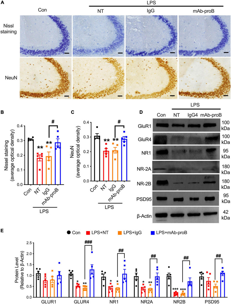
Sepsis-associated encephalopathy (SAE) is a risk factor for cognitive and memory dysfunction; however, the mechanism remains unclear. Brain-derived neurotrophic factor (BDNF) was reported to have a positive effect on cognition and emotion regulation, but the study of its precursor, proBDNF, has been limited. This study aimed to elucidate the effects and associated mechanisms of hippocampal proBDNF in a lipopolysaccharide (LPS)-induced SAE mouse model. In this study, we found that the mice exhibited cognitive dysfunction on day 7 after LPS injection. The expression of proBDNF and its receptor, p75 NTR , was also increased in the hippocampus, while the levels of BDNF and its receptor, TrkB, were decreased. A co-localization study showed that proBDNF and p75 NTR were mainly co-localized with neurons. Furthermore, LPS treatment reduced the expression of NeuN, Nissl bodies, GluR4, NR1, NR2A, and NR2B in the hippocampus of SAE mice. Furthermore, an intrahippocampal or intraperitoneal injection of anti-proBDNF antibody was able to ameliorate LPS-induced cognitive dysfunction and restore the expression of NeuN, Nissl bodies, GluR4, NR1, NR2A, NR2B, and PSD95. These results indicated that treatment with brain delivery by an intrahippocampal and systemic injection of mAb-proBDNF may represent a potential therapeutic strategy for treating patients with SAE.

Keywords: cognition and memory dysfunction; hippocampus; p75NTR; proBDNF; sepsis associated encephalopathy.


Conflict of interest statement

The authors declare that the research was conducted in the absence of any commercial or financial relationships that could be construed as a potential conflict of interest.

Figures

FIGURE 1
FIGURE 1
Behavioral tests after the establishment of the sepsis-associated encephalopathy mouse model. (A) Timeline of behavioral tests after administration of lipopolysaccharide. (B,C) The total distance and the time spent in the center field of the open field test (n = 9/group). (D,E) The recognition index of short-term or long-term memory tasks in the novel object recognition test (n = 9/group). (F,G) The percentage of time mice stay in the novel arm in the short-term or long-term memory task in the Y maze test (n = 9/group). Data are expressed as mean ± SEM. **p < 0.01, ***p < 0.001.
FIGURE 2
FIGURE 2
The expression pattern of proBDNF, BDNF, and their receptors in sepsis-associated encephalopathy mice. (A–D) The expression level of BDNF, TrkB, sortilin, and p75NTR at different time points after the lipopolysaccharide injection (n = 4/group). (E–K) Immunoblot analysis of different time points and the quantitative results of the expression of proBDNF, p75NTR, sortilin, BDNF, and TrkB (n = 4/group). Data are expressed as mean ± SEM. *p < 0.05, **p < 0.01, ***p < 0.001, and ****p < 0.0001.
FIGURE 3
FIGURE 3
proBDNF and its receptor p75NTR are mainly expressed in hippocampal neurons. (A,B) proBDNF accumulated in hippocampal neurons (upper scale bar = 100 μm, lower scale bar = 50 μm, n = 4/group). (C,D) proBDNF and its receptor p75NTR mainly co-existed in the neuron (scale bar = 50 μm, n = 4/group). Data are expressed as mean ± SEM. *p < 0.05.
FIGURE 4
FIGURE 4
Intrahippocampal microinjection of mAb-proB improved the sepsis-associated encephalopathy-induced cognitive impairment. (A) The behavioral intervention flowchart of the experiment. (B) The efficiency of neutralizing mAb-proB in hippocampus tissue. Scale bar = 100 μm. (C,D) The total traveled distance and percentage of time mice spent in the center of the field via open field test (n = 9/group). (E,F) The recognition index of short/long-term memory tasks of the novel object recognition test (n = 9/group). (G,H) The percentage of the time that mice stay in the novel arm of the short/long-term memory tasks in the Y maze test (n = 9/group). Data are expressed as mean ± SEM. *p < 0.05, **p < 0.01, ****p < 0.0001 vs. sham group; #p < 0.05, ##p < 0.01, LPS + IgG vs. LPS + mAb-proB.
FIGURE 5
FIGURE 5
Systemic delivery of neutralizing mAb-proB partially reversed the sepsis-associated encephalopathy-induced cognitive impairment. (A) The behavior schedule of the experiment. (B,C) The total distance traveled and percentage of time during which mice stay in the center of the field (n = 9–10/group). (D,E) The recognition index of the short/long-term memory tasks of the novel object recognition test (n = 10/group). (F,G) The percentage of time that mice stay in the novel arm of the short/long-term memory tasks in the Y maze test (n = 10/group). Data are expressed as mean ± SEM. **p < 0.01, ***p < 0.001, ****p < 0.0001 vs. control; #p < 0.05, ##p < 0.01, LPS + IgG vs. LPS + mAb-proB.
FIGURE 6
FIGURE 6
MAb-proB upregulated the expression of neuronal cells and synapse-associated proteins in sepsis-associated encephalopathy mice. (A–C) Representative images and quantitation of Nissl bodies and NeuN-positive neuronal cells (scale bar = 50 μm, n = 4/group). (D,E) Immunoblot analysis of GluR1, GluR4, NR1, NR-2A, NR-2B, and PTSD95 (n = 5/group). Data are expressed as mean ± SEM. *p < 0.05, **p < 0.01, ***p < 0.001 vs. control; #p < 0.05, ##p < 0.01, ###p < 0.001, LPS + IgG vs. LPS + mAb-proB.

References

    1. Abramova A. Y., Pertsov S. S., Kozlov A. Y., Nikenina E. V., Kalinichenko L. S., Dudnik E. N., et al. (2013). Cytokine levels in rat blood and brain structures after administration of lipopolysaccharide. Bull. Exp. Biol. Med. 155 417–420. 10.1007/s10517-013-2166-5 - DOI - PubMed
    1. Alonso M., Vianna M. R., Depino A. M., Mello e Souza T., Pereira P., Szapiro G., et al. (2002). BDNF-triggered events in the rat hippocampus are required for both short- and long-term memory formation. Hippocampus 12 551–560. 10.1002/hipo.10035 - DOI - PubMed
    1. An L., Li X., Tang C., Xu N., Sun W. (2018). Hippocampal proBDNF facilitates place learning strategy associated with neural activity in rats. Brain Struct. Funct. 223 4099–4113. 10.1007/s00429-018-1742-x - DOI - PubMed
    1. Angelucci F., Brene S., Mathe A. A. (2005). BDNF in schizophrenia, depression and corresponding animal models. Mol. Psychiatry 10 345–352. 10.1038/sj.mp.4001637 - DOI - PubMed
    1. Antunes M., Biala G. (2012). The novel object recognition memory: neurobiology, test procedure, and its modifications. Cogn. Process. 13 93–110. 10.1007/s10339-011-0430-z - DOI - PMC - PubMed