. 2021 Jul 20:12:683911.
doi: 10.3389/fimmu.2021.683911. eCollection 2021.

Insight Into Polysaccharides From Panax ginseng C. A. Meyer in Improving Intestinal Inflammation: Modulating Intestinal Microbiota and Autophagy

Affiliations

Insight Into Polysaccharides From Panax ginseng C. A. Meyer in Improving Intestinal Inflammation: Modulating Intestinal Microbiota and Autophagy

Dandan Wang et al. Front Immunol. .

Abstract

Polysaccharides from Panax ginseng C. A. Meyer (P. ginseng) are the main active component of P. ginseng and exhibit significant intestinal anti-inflammatory activity. However, the therapeutic mechanism of the ginseng polysaccharide is unclear, and this hinders the application for medicine or functional food. In this study, a polysaccharide was isolated from P. ginseng (GP). The primary structure and morphology of the GP were studied by HPLC, FT-IR spectroscopy, and scanning electron microscopy (SEM). Further, its intestinal anti-inflammatory activity and its mechanism of function were evaluated in experimental systems using DSS-induced rats, fecal microbiota transplantation (FMT), and LPS-stimulated HT-29 cells. Results showed that GP modulated the structure of gut microbiota and restored mTOR-dependent autophagic dysfunction. Consequently, active autophagy suppressed inflammation through the inhibition of NF-κB, oxidative stress, and the release of cytokines. Therefore, our research provides a rationale for future investigations into the relationship between microbiota and autophagy and revealed the therapeutic potential of GP for inflammatory bowel disease.

Keywords: Panax ginseng C. A. Meyer; autophagy; gut microbiota; intestinal inflammation; polysaccharide.


Conflict of interest statement

The authors declare that the research was conducted in the absence of any commercial or financial relationships that could be construed as a potential conflict of interest.

Figures

Figure 1
Figure 1
Description and analysis of GP (A) Summarized extraction procedure. (B) HPLC of monosaccharide PMP derivatization. 1. PMP. 2. Mannose. 3. Rhamnose. 4. Glucuronic acid. 5. Galacturonic acid. 6. Glucose. 7. Galactose. 8. Arabinose. 9. Fucose. (C) Infrared spectroscopy. (D) Morphology by SEM. (E) Molecular weight distribution.
Figure 2
Figure 2
GP alleviated the intestinal inflammation in rats with DSS induced colitis. (A) Experimental operation process. (B) H&E stained sections of the differently treated rats as described above. (C, D) The effects of GP on Rats’ basal body weight and Spleen weight. (E–G) Changes of cytokines in different groups in vivo. All data shown are representative of 3 independent experiments. Bars in graphs represent mean ± SD, #P < 0.05, ##P < 0.01 VS Control group; *P < 0.05, **P < 0.01 VS DSS group.
Figure 3
Figure 3
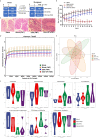
Effects of GP on gut microbiota. (A) Rarefaction curves of OTU quantity. (B) Heatmap at the level of phylumgenus. (C) Heatmap at the level of genus. (D) A phylogenetictree of OTUs by GraPhlAn visualization. (E) PCA analysis. (F) LPS in colon contents. Bars in graphs represent mean ± SD, ##P < 0.01 VS Control group; **P < 0.01 VS DSS group.
Figure 4
Figure 4
GP inhibits inflammation via TLR4 pathway and activates mTOR dependent autophagy in DSS-induced colitis in vivo. (A, C) Immunofluorescence for TLR4 and p62 was performed on colon sections. (B, D) The expressions of TLR4 and p62 were calculated relative to DAPI staining from three independent experiments. (E) The expression of autophagy-related and inflammation-related proteins measured by western blot. (F–K) The expressions of proteins were quantified by the ratio of phosphorylated protein/total protein and total amount protein/GAPDH. All data shown are representative of 3 independent experiments. Bars in graphs repr Activated TLR4 directly triggers the phosphorylation of mTOR. With the increase of TLR4 in the DSS group, the phosphorylated mTOR was upregulated followed by the dysfunction of autophagy. To investigate whether autophagy was impaired in DSS-induced intestinal inflammation, the expression of autophagy-related proteins (including LC3B, p62, and phosphorylated mTOR) were evaluated by immunofluorescence and Western blot. Compared with the control group, the expression of p62, a cargo protein degraded inside autolysosomes, was upregulated after DSS stimulation, indicating the inhibition of autophagy. Defective autophagy was further confirmed by the decrease in LC3BII/LC3BI when compared with the control group. Autophagy was recovered to a normal level after treatment with GP, indicated by decreased p62 and increased LC3BII/LC3BI levels. These results demonstrated that GP relieved intestinal inflammation by promoting mTOR-dependent autophagy and blocking the inflammatory cascade. Bars in graphs represent mean ± SD, #P < 0.05, ##P < 0.01 VS Control group; *P < 0.05 VS DSS group.
Figure 5
Figure 5
Spearman correlation analysis of the bacterial genera with highest abundance and phenotypes. *P < 0.05, **P < wfi 20.01.
Figure 6
Figure 6
Transplanting of fecal microbiome intervened by GP improves intestinal inflammation. (A) The experimental protocol for FMT. (B) Changes of body weight (n=10). (C) Histopathological changes after HE staining. (D) Rarefaction curves of OUT quantity. (E) PCA analysis. (F) The gut microbiota composition among experimental groups at phylum/genus level.
Figure 7
Figure 7
Transplanting of fecal microbiome intervened by GP improves intestinal inflammation. (A) Immunofluorescence for p-NF-κB was performed on colon sections. (B) The expression of autophagy-related and inflammation-related proteins measured by western blot. (C, D) The expressions of proteins were quantified by the ratio of phosphorylated protein/total protein and total amount protein/GAPDH. All data shown are representative of 3 independent experiments. Bars in graphs represent mean ± SD, #P < 0.05, ##P < 0.01 VS Control group; *P < 0.05, **P < 0.01 VS DSS group, n.s., no significant differences.
Figure 8
Figure 8
Blocking upstream TLR4-MAPK pathway activated autophagy and quenched inflammation. (A) Changes of cytokines contents in different groups in LPS-induced HT-29 cells. (B) The changes of autophagy-related and inflammation-related proteins measured by western blot. (C–H) The expressions of proteins were quantified by the ratio of phosphorylated protein/total protein and total amount protein/GAPDH. All data shown are representative of 3 independent experiments. Bars in graphs represent mean ± SD, *P < 0.05, **P < 0.01. #P < 0.05, ##P < 0.01 VS Control group.
Figure 9
Figure 9
Schematic representation of the proposed mechanism of GP on ameliorating intestinal inflammation.

References

    1. Rooks MG, Garrett WS. Gut Microbiota, Metabolites and Host Immunity. Nat Rev Immunol (2016) 16(6):341–52. 10.1038/nri.2016.42 - DOI - PMC - PubMed
    1. Barker N. Adult Intestinal Stem Cells: Critical Drivers of Epithelial Homeostasis and Regeneration. Nat Rev Mol Cell Biol (2014) 15(1):19–33. 10.1038/nrm3721 - DOI - PubMed
    1. Miller YI, Choi SH, Wiesner P, Bae YS. The SYK Side of TLR4: Signalling Mechanisms in Response to LPS and Minimally Oxidized LDL. Br J Pharmacol (2012) 167(5):990–9. 10.1111/j.1476-5381.2012.02097.x - DOI - PMC - PubMed
    1. Rogers M, Moore R, Cohen J. The Relationship Between Faecal Endotoxin and Faecal Microflora of the C57BL Mouse. J Hygiene (1985) 95(2):397–402. 10.1017/S0022172400062823 - DOI - PMC - PubMed
    1. Di Lorenzo F, Kubik Ł., Oblak A, Lorè NI, Cigana C, Lanzetta R, et al. . Activation of Human Toll-Like Receptor 4 (TLR4)·Myeloid Differentiation Factor 2 (MD-2) by Hypoacylated Lipopolysaccharide From a Clinical Isolate of Burkholderia Cenocepacia. J Biol Chem (2015) 290(35):21305–19. 10.1074/jbc.M115.649087 - DOI - PMC - PubMed

Publication types

MeSH terms