. 2021 Jul 20:12:616761.
doi: 10.3389/fgene.2021.616761. eCollection 2021.

Variants in LAMC3 Causes Occipital Cortical Malformation

Affiliations

Variants in LAMC3 Causes Occipital Cortical Malformation

Xiaohang Qian et al. Front Genet. .

Abstract

Occipital cortical malformation (OCCM) is a disease caused by malformations of cortical development characterized by polymicrogyria and pachygyria of the occipital lobes and childhood-onset seizures. The recessive or complex heterozygous variants of the LAMC3 gene are identified as the cause of OCCM. In the present study, we identified novel complex heterozygous variants (c.470G > A and c.4030 + 1G > A) of the LAMC3 gene in a Chinese female with childhood-onset seizures. Cranial magnetic resonance imaging was normal. Functional experiments confirmed that both variant sites caused premature truncation of the laminin γ3 chain. Bioinformatics analysis predicted 10 genes interacted with LAMC3 with an interaction score of 0.4 (P value = 1.0e-16). The proteins encoded by these genes were mainly located in the basement membrane and extracellular matrix component. Furthermore, the biological processes and molecular functions from gene ontology analysis indicated that laminin γ3 chain and related proteins played an important role in structural support and cellular processes through protein-containing complex binding and signaling receptor binding. KEGG pathway enrichment predicted that the LAMC3 gene variant was most likely to participate in the occurrence and development of OCCM through extracellular matrix receptor interaction and PI3K-Akt signaling pathway.

Keywords: ECM-receptor interaction; Lamc3; PI3K-Akt signaling pathway; childhood-onset seizures; occipital cortical malformation.


Conflict of interest statement

The authors declare that the research was conducted in the absence of any commercial or financial relationships that could be construed as a potential conflict of interest.

Figures

FIGURE 1
FIGURE 1
Clinical and genetic characterizations. (A) Cranial MRI showed normal brain structure without abnormal signal and lateral ventricular dilation. (B) The pedigree of this family shows only the patient had epilepsy, and her parents and two younger siblings were both unaffected. (C) Direct polymerase chain reaction sequencing revealed a heterozygous variant c.4030 + 1G > A on exon 24 of the father’s LAMC3 gene, and a heterozygous variant c.470G > A on exon 2 of the mother’s LAMC3 gene.
FIGURE 2
FIGURE 2
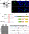
The functional analysis of complex heterozygous variants in LAMC3 gene. (A) The LAMC3 WT group expressed a normal protein with a molecular weight of 198 kDd and a truncated protein with a molecular weight of 47 kDd in LAMC3 variant group (c.470G > A). (B) The cellular localization of LAMC3 in WT and variant group was in the cytoplasm without significant morphological difference. (C) Sanger sequence of minigene in in WT (above) and variant (below) group (c.4030 + 1G > A). (D) The schematic diagrams showed the minigene comprising intron 23, exon 24, and intron 24 of the WT or variant (c.4030 + 1G > A). The variant c.4030 + 1G > A affected the normal splicing of LAMC3 mRNA, resulting in 4 bp base (ATAC) retention at the left of intron 24, “*” represents variant location. (E) Agarose gel electrophoresis of RT-PCR fragments showed a similar length of the product between the WT and variant group. (F) The Sanger sequences showed a 4 bp base (ATAC) retention at the left of intron 24 in the variant group.
FIGURE 3
FIGURE 3
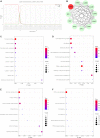
The bioinformatics analysis of LAMC3 gene. (A) The first 19 amino acids of the laminin γ3 chain were predicted as a signal peptide. (B) The protein-protein interaction network showed 10 genes (DAG1, LAMA1, LAMA3, LAMA5, LAMB1, LAMB2, LAMB3, NID1, NID2, and NTN4) that have potential interactions with LAMC3. (C) The cellular component of gene ontology showed LAMC3 and its interacting genes were mainly located in the basement membrane, extracellular matrix organization, laminin complex. (D) The biological process of gene ontology indicated these genes were mainly associated with extracellular matrix organization, cell adhesion, cellular component organization, and anatomical structure morphogenesis. (E) The molecular function of these genes were primarily involved in structural molecule activity, protein-containing complex binding, signaling receptor binding, and laminin-1 binding. (F) The KEGG pathway enrichment analysis showed eight potential signal pathways of these genes, including ECM-receptor interaction, amoebiasis, small cell lung cancer, toxoplasmosis, focal adhesion, human papillomavirus infection, PI3K-Akt signaling pathway, and pathways in cancer.

References

    1. Afawi Z., Oliver K. L., Kivity S., Mazarib A., Blatt I., Neufeld M. Y., et al. (2016). Multiplex families with epilepsy: success of clinical and molecular genetic characterization. Neurology 86 713–722. 10.1212/WNL.0000000000002404 - DOI - PMC - PubMed
    1. Barak T., Kwan K. Y., Louvi A., Demirbilek V., Saygı S., Tüysüz B., et al. (2011). Recessive LAMC3 mutations cause malformations of occipital cortical development. Nat. Genet. 43 590–594. 10.1038/ng.836 - DOI - PMC - PubMed
    1. Barkovich A. J., Guerrini R., Kuzniecky R. I., Jackson G. D., Dobyns W. B. (2012). A developmental and genetic classification for malformations of cortical development: update 2012. Brain 135 1348–1369. 10.1093/brain/aws019 - DOI - PMC - PubMed
    1. Benova B., Jacques T. S. (2019). Genotype-phenotype correlations in focal malformations of cortical development: a pathway to integrated pathological diagnosis in epilepsy surgery. Brain Pathol. 29 473–484. 10.1111/bpa.12686 - DOI - PMC - PubMed
    1. Bonnans C., Chou J., Werb Z. (2014). Remodelling the extracellular matrix in development and disease. Nat. Rev. Mol. Cell Biol. 15 786–801. 10.1038/nrm3904 - DOI - PMC - PubMed

LinkOut - more resources