. 2021 Jul 27:2021:9929461.
doi: 10.1155/2021/9929461. eCollection 2021.

Development of a Human Intestinal Organoid Model for In Vitro Studies on Gut Inflammation and Fibrosis

Affiliations

Development of a Human Intestinal Organoid Model for In Vitro Studies on Gut Inflammation and Fibrosis

Leonidas Kandilogiannakis et al. Stem Cells Int. .

Abstract

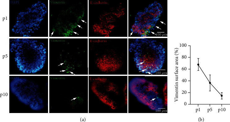
Inflammatory Bowel Diseases (IBDs) are characterized by chronic intestinal inflammation and fibrosis, the latter being the predominant denominator for long-term complications. Epithelial and mesenchymal 2D cultures are highly utilized in vitro models for the preclinical evaluation of anti-inflammatory and antifibrotic therapies. More recently, human intestinal organoids (HIOs), a new 3D in vitro model derived from pluripotent stem cells, have the advantage to closely resemble the architecture of the intestinal mucosa. However, the appropriate timing for the study of inflammatory and fibrotic responses, during HIO development, has not been adequately investigated. We developed HIOs from the human embryonic stem cell line, H1, and examined the expression of mesenchymal markers during their maturation process. We also investigated the effect of inflammatory stimuli on the expression of fibrotic and immunological mediators. Serial evaluation of the expression of mesenchymal and extracellular matrix (ECM) markers revealed that HIOs have an adequately developed mesenchymal component, which gradually declines through culture passages. Specifically, CD90, collagen type I, collagen type III, and fibronectin were highly expressed in early passages but gradually diminished in late passages. The proinflammatory cytokines IL-1α and TNF-α induced the mRNA expression of fibronectin, collagen types I and III, tissue factor (TF), and alpha-smooth muscle actin (α-SMA) primarily in early passages. Similarly, HIOs elicited strong mRNA and protein mesenchymal (CXCL10) and epithelial (CXCL1, CCL2, CXCL8, and CCL20) chemokine responses in early but not late passages. In contrast, the epithelial tight junction components, CLDN1 and JAMA, responded to inflammatory stimulation independently of the culture passage. Our findings indicate that this HIO model contains a functional mesenchymal component, during early passages, and underline the significance of the mesenchymal cells' fitness in inflammatory and fibrotic responses. Therefore, we propose that this model is suitable for the study of epithelial-mesenchymal interactions in early passages when the mesenchymal component is active.


Conflict of interest statement

The authors have no conflict to declare.

Figures

Figure 1
Figure 1
Development and characterization of HIOs. (a) Developmental stages of HIO formation. (b, c) HIOs stained against Desmin and E-cadherin, indicating fibroblast and epithelial cell populations, respectively. (d) HIOs stained positive for EpCam, cytokeratin, and CDX2, indicating intestinal epithelial cells. (e) MUC2-positive goblet cells and Chromogranin A-positive endocrine epithelial cells found in HIOs, surrounded by Villin-expressing epithelial cells. (f) HIOs stained positive for either KLF5 or SOX9, indicating the existence of intestinal epithelial stem cell niches that support the already-differentiated E-cadherin- and Villin-expressing epithelial cells. Representative 40x snapshots are shown in (a) and 28.8x immunofluorescence images in (b–f). (b–f) Images were obtained using a light sheet microscope. (c–f) Images are selected z-slices from the HIO total volume. (b) A 3D volume of an organoid.
Figure 2
Figure 2
Mesenchymal evolution along HIO passaging. HIOs stained for the epithelial marker, E-cadherin, and the mesenchymal marker, vimentin, in three different passages (a). Vimentin-positive staining area is shown to be reduced through subsequent passages, suggesting that the mesenchymal component is gradually decreased (b). Representative 40x immunofluorescence snapshots are shown.
Figure 3
Figure 3
Expression of mesenchymal markers and ECM components during passaging. The mRNA levels of CD90, collagen types I and III, and fibronectin were gradually reduced after passage 2 (a–d), with the exception of α-SMA, which showed a tendency to increase during passages 4-6, but later decreased to basal levels (e). ND: nondetectable. All experiments were performed in triplicate. The gene expression of each studied gene was normalized against GAPDH gene expression in the same sample using the 2-ΔΔCt method. Passage 1 expression levels were set as a reference point, and expression levels in later passages were compared to that. Data are presented as the mean ± standard error of the mean (SEM).
Figure 4
Figure 4
HIOs exhibit fibrotic responses to inflammatory cytokines. IL-1α and TNF-α (2C) induced the mRNA expression of α-SMA in passage 4 (a), fibronectin in passages 4 and 8 (b), TF in passage 4 (c), collagen type I in passages 2 and 4 (d), and collagen type III in passages 2, 4, and 6 (e). Concentrations of cytokines used: IL-1α 5 ng/ml, TNF-α 50 ng/ml. All experiments were performed in triplicate. The gene expression of each studied gene was normalized against GAPDH gene expression in the same sample using the 2-ΔΔCt method. In every passage, expression levels of treated organoids were normalized against those of the control ones. Data are presented as the mean ± standard error of the mean (SEM).
Figure 5
Figure 5
HIOs exhibit mesenchymal and epithelial chemokine mRNA responses to inflammatory cytokines. IL-1α and TNF-α (2C) induced the mRNA expression of CXCL10 in passage 2 (a), CXCL11 in passage 2 (b), CXCL1 in passages 2 and 4 (c), CXCL8 in passages 2, 4, and 6 (d), CCL2 in passages 2 and 4 (e), and CCL20 in passages 2 and 4 (f). Concentrations of cytokines used: IL-1α 5 ng/ml, TNF-α 50 ng/ml. All experiments were performed in triplicate. The gene expression of each studied gene was normalized against GAPDH gene expression in the same sample using the 2-ΔΔCt method. In every passage, expression levels of treated organoids were normalized against those of the control ones. Data are presented as the mean ± standard error of the mean (SEM).
Figure 6
Figure 6
HIOs exhibit mesenchymal and epithelial chemokine protein responses to inflammatory cytokines. IL-1α and TNF-α (2C) 24 h and 48 h stimulation induced the protein expression of CXCL1 (b), CXCL8 (c), CCL2 (d), and CCL20 (e) in passages 2 and 4, while CXCL10 was induced only in passage 2 (a). CCL20 was also induced in passage 6, after 48 h stimulation. Concentrations of cytokines used: IL-1α 5 ng/ml, TNF-α 50 ng/ml. All experiments were performed in triplicate. Data are presented as the mean ± standard error of the mean (SEM).
Figure 7
Figure 7
Epithelial responses to proinflammatory cytokines decrease during passaging. IL-1α and TNF-α (2C) induced the mRNA expression of OCLN in passage 4 (a), CLDN1 in passages 2 and 8 (b), ZO1 in passage 4 (c), and JAMA in passages 2, 8, and 10 (d). Concentrations of cytokines used: IL-1α 5 ng/ml, TNF-α 50 ng/ml. All experiments were performed in triplicate. The gene expression of each studied gene was normalized against GAPDH gene expression in the same sample using the 2-ΔΔCt method. In every passage, expression levels of treated organoids were normalized against those of the control ones. Data are presented as the mean ± standard error of the mean (SEM).

References

    1. Chang J. T. Pathophysiology of inflammatory bowel diseases. The New England journal of medicine. 2020;383(27):2652–2664. doi: 10.1056/NEJMra2002697. - DOI - PubMed
    1. West N. R., Investigators O. I. B. D. C., Hegazy A. N., et al. Oncostatin M drives intestinal inflammation and predicts response to tumor necrosis factor-neutralizing therapy in patients with inflammatory bowel disease. Nature Medicine. 2017;23(5):579–589. doi: 10.1038/nm.4307. - DOI - PMC - PubMed
    1. Parikh K., Antanaviciute A., Fawkner-Corbett D., et al. Colonic epithelial cell diversity in health and inflammatory bowel disease. Nature. 2019;567(7746):49–55. doi: 10.1038/s41586-019-0992-y. - DOI - PubMed
    1. Kinchen J., Chen H. H., Parikh K., et al. Structural remodeling of the human colonic mesenchyme in inflammatory bowel disease. Cell. 2018;175(2):372–386.e17. doi: 10.1016/j.cell.2018.08.067. - DOI - PMC - PubMed
    1. Duijvestein M., Battat R., Vande Casteele N., et al. Novel therapies and treatment strategies for patients with inflammatory bowel disease. Current treatment options in gastroenterology. 2018;16(1):129–146. doi: 10.1007/s11938-018-0175-1. - DOI - PubMed

LinkOut - more resources