. 2021 Jun 20;10(7):e1743-e1749.
doi: 10.1016/j.eats.2021.03.016. eCollection 2021 Jul.

Biologic Tuberoplasty With an Acellular Dermal Allograft for Massive Rotator Cuff Tears

Affiliations

Biologic Tuberoplasty With an Acellular Dermal Allograft for Massive Rotator Cuff Tears

Raffy Mirzayan et al. Arthrosc Tech. .

Abstract

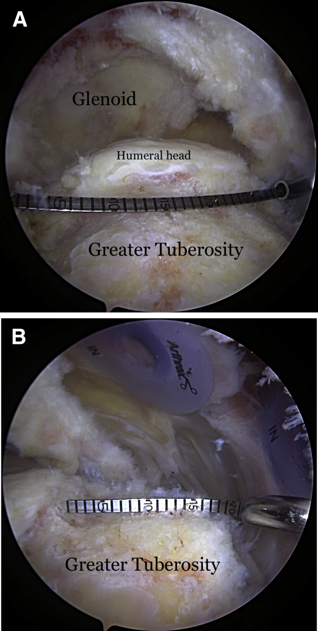
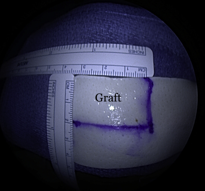
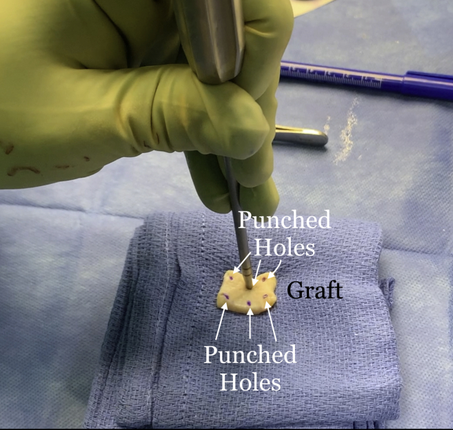
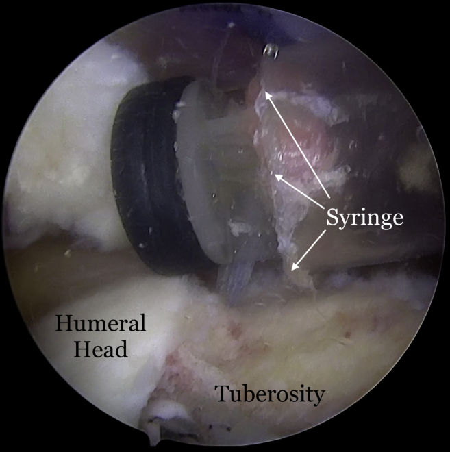
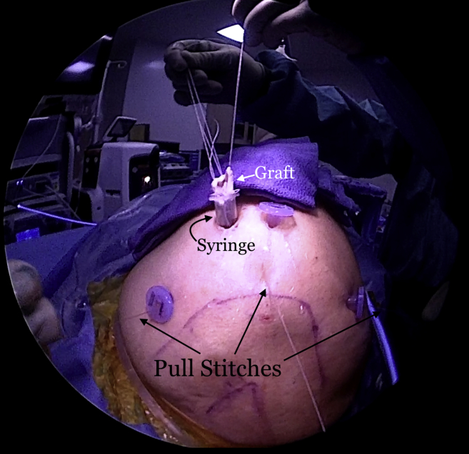
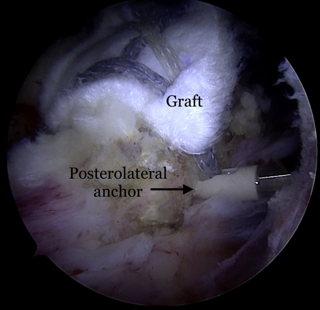
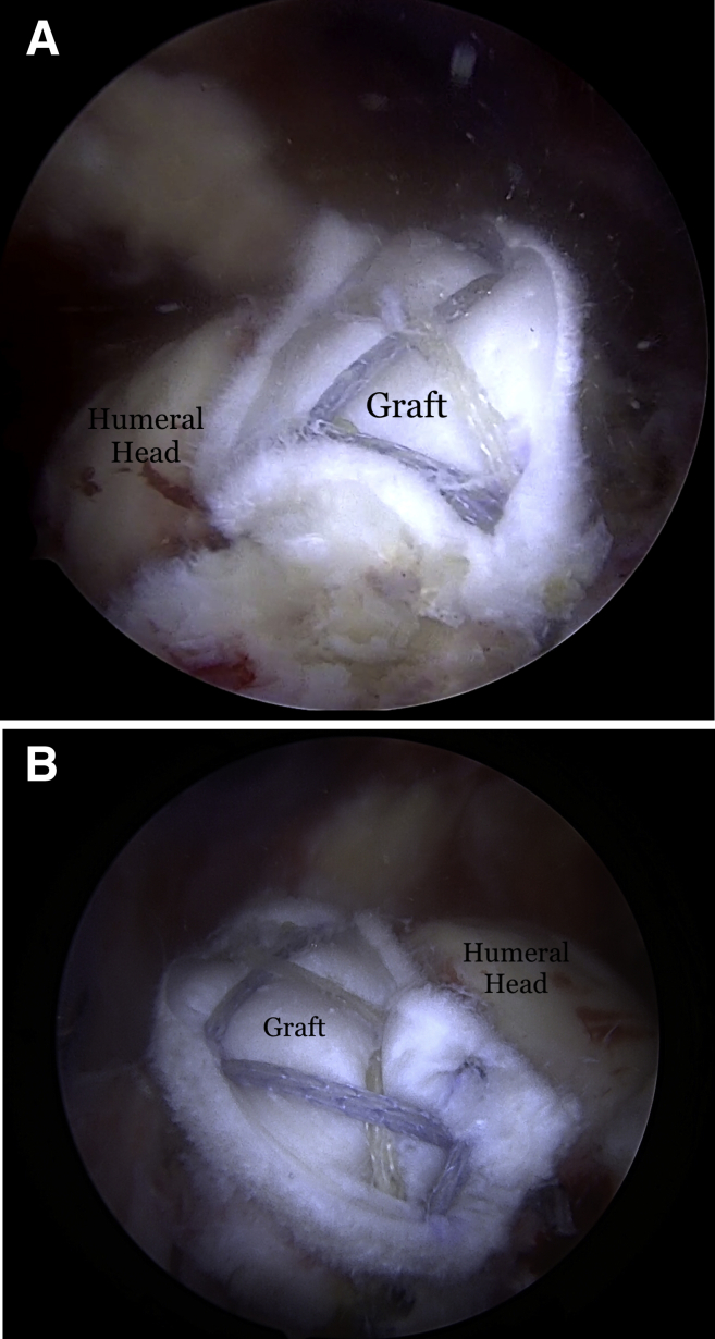
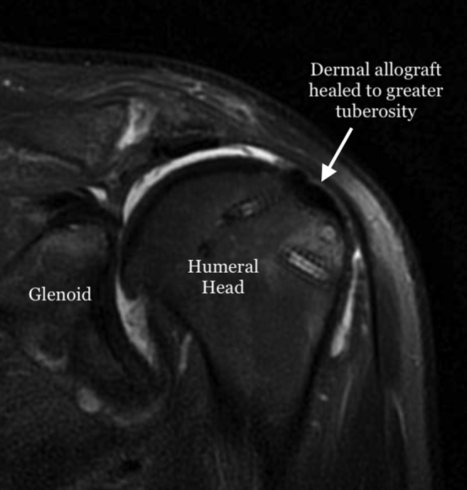
We present the technique of biologic tuberoplasty, where an acellular dermal allograft is used to cover the tuberosity in patients with massive irreparable cuff tears to prevent bone-to-bone contact between the tuberosity and acromion when active elevation is attempted. This technique can be performed in patients with massive rotator cuff tears who are low-demand and have significant medical comorbidities, poor bone quality, or who are not candidates for a reverse shoulder arthroplasty or who cannot tolerate a lengthy rehabilitation protocol. It is less time-consuming than a superior capsule reconstruction and more bone-sparing than traditional tuberoplasty. Patients with massive rotator cuff tears involving the supraspinatus and a portion of the infraspinatus lose their force couple, leading to superior humeral head migration with abutment of the tuberosity against the acromion upon deltoid activation. The center of rotation moves superiorly with deltoid contraction, developing an acromiohumeral articulation. This results in bone-to-bone contact between the acromion and humerus, leading to pain and acetabularization of the acromion over time. Coverage of the tuberosity with the acellular dermal allograft acts as an interpositional tissue and prevents bone-to-bone contact between the tuberosity and acromion.


Figures

Fig 1
Fig 1
(A) View from lateral portal of a right shoulder with the patient in lateral decubitus position demonstrating the measurement of the length of the tuberosity. (B) View from posterior portal of a right shoulder demonstrating the measurement of the width of the tuberosity.
Fig 2
Fig 2
A 30-mm × 20-mm piece of the graft is obtained from a 3-mm thick ArthroFlex 301 (LifeNet Health, Virginia Beach, VA) dermal allograft.
Fig 3
Fig 3
Holes are punched in the graft to allow for easier suture/FiberTape passage.
Fig 4
Fig 4
View from posterior portal of a right shoulder with the patient in lateral decubitus position demonstrating the insertion of a 10-cc syringe with its tip cut off (white arrows).
Fig 5
Fig 5
View from posterior portal of a right shoulder with the patient in lateral decubitus position. The 2 FiberTapes, the repair stitch, and the loop end of the shuttle stitch from each anchor are taken out of the syringe, leaving behind the pull end of the shuttle stitch.
Fig 6
Fig 6
Exterior view of a right shoulder with the patient in the lateral decubitus position. The sutures from each anchor are kept separate and clamped to the syringe for suture management.
Fig 7
Fig 7
Exterior view of a right shoulder with the patient in the lateral decubitus position. The repair stitch is passed in a mattress fashion at the edge of the graft. A towel is placed on the patient's arm near the syringe cannula to pass the sutures and FiberTapes through the graft.
Fig 8
Fig 8
Exterior view of a right shoulder with the patient in the lateral decubitus position. The final suture passage and graft preparation before insertion into the subacromial space are shown here. The mattress sutures at the medial edge are passed with the repair stitch from each anchor. The FiberTapes are passed through the pre-punched holes on the medial edge of the graft and then passed back down through the graft in an expanded speedbridge configuration. This allows better control of the lateral portion of the graft when the lateral row anchors are being inserted.
Fig 9
Fig 9
Exterior view of a right shoulder with the patient in the lateral decubitus position. The pull stitches are pulled sequentially, pulling the graft into the subacromial space through the syringe.
Fig 10
Fig 10
Right shoulder viewed from the posterior portal with the patient in the lateral decubitus position demonstrating the insertion of the posterolateral anchor placement.
Fig 11
Fig 11
(A) Final graft placement and fixation seen in the right shoulder viewed from the posterior portal with the patient in the lateral decubitus position and (B) viewed from the anterior portal.
Fig 12
Fig 12
T2-weighted fat-saturated magnetic resonance imaging scan of a left shoulder 3 months after surgery demonstrating healing and incorporation of the graft to the greater tuberosity.

References

    1. Burkhart S.S. Fluoroscopic comparison of kinematic patterns in massive rotator cuff tears. A suspension bridge model. Clin Orthop Relat Res. 1992:144–152. - PubMed
    1. Ecklund K.J., Lee T.Q., Tibone J., Gupta R. Rotator cuff tear arthropathy. J Am Acad Orthop Surg. 2007;15:340–349. - PubMed
    1. Green A. Chronic massive rotator cuff tears: Evaluation and management. J Am Acad Orthop Surg. 2003;11:321–331. - PubMed
    1. Mihata T., McGarry M.H., Pirolo J.M., Kinoshita M., Lee T.Q. Superior capsule reconstruction to restore superior stability in irreparable rotator cuff tears: A biomechanical cadaveric study. Am J Sports Med. 2012;40:2248–2255. - PubMed
    1. Lobao MH, Melvani RT, Abbasi P, Parks BG, Murthi AM. Dermal allograft superior capsule reconstruction biomechanics and kinematics [published online January 19, 2021]. J Shoulder Elbow Surg.https://doi.org/10.1016/j.jse.2020.11.024. - DOI - PubMed

LinkOut - more resources