. 2020 Sep 29:10:565950.
doi: 10.3389/fonc.2020.565950. eCollection 2020.

Prognostic Value and Potential Immunoregulatory Role of SCARF1 in Hepatocellular Carcinoma

Affiliations

Prognostic Value and Potential Immunoregulatory Role of SCARF1 in Hepatocellular Carcinoma

Daniel A Patten et al. Front Oncol. .

Abstract

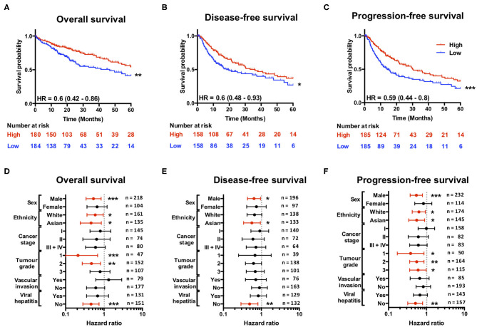
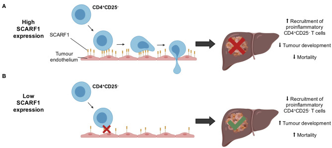
Scavenger receptor class F member 1 (SCARF1) is thought to play an important role in the selective recruitment of CD4+ T cells to liver sinusoidal endothelial cells during chronic liver disease. However, the contribution of SCARF1 to hepatocellular carcinoma (HCC) is currently unknown. We utilized publically-available RNA-sequencing data from The Cancer Genome Atlas (TGCA) to explore SCARF1 expression in HCC and correlated it with a number of clinicopathological features. Flow adhesion assays were used to determine the role of SCARF1 in CD4+ T cell subset recruitment. SCARF1 expression was downregulated in HCC tumor tissues, compared to non-tumoral tissues, and loss of SCARF1 expression was associated with poorly differentiated/aggressive tumors. Additionally, higher SCARF1 expression in HCC tumor tissues was highly prognostic of better overall, disease-free and progression-free survival. SCARF1 within HCC was largely associated with tumor endothelial cells and adhesion studies suggested that it played a role in the specific recruitment of proinflammatory CD4+ T cells (CD4+CD25-) to HCC tumor tissues. Endothelial SCARF1 expression in tumor biopsies may provide critical prognostic information. Additionally, SCARF1 may also be a novel endothelial target that could help re-programme the microenvironment of HCC by promoting effector T cell tumor infiltration.

Keywords: leukocyte recruitment; liver cancer; scavenger receptor; tumor endothelial cells; tumor microenviroment.


Figures

Figure 1
Figure 1
SCARF1 gene and protein expression is downregulated in HCC tumors. (A) Regulation of scavenger receptor gene expression in HCC tumors (blue; n = 371), compared to non-tumoral tissues (red; n = 162). *, **, *** and **** are representative of statistical significance as measured by the Kruskal–Wallis test, where p ≤ 0.05, p ≤ 0.01, p ≤ 0.005, and p ≤ 0.001, respectively. RSEM = RNA-Seq by Expectation Maximization. (B) Comparison of SCARF1 gene expression in non-tumoral (NT) tissues with HCC tumor tissues (T). ****Indicates statistical significance as measured by Mann–Whitney U-test, where p ≤ 0.001 (C). Representative images of SCARF1 immunohistochemical staining (brown) in HCC tumor (Right panels) and matched distal, non-tumorous (Left panels) tissues from the same patient. Scale bar = 400 μm; zoomed image scale bar = 100 μm. (D) Surface area quantification of immunohistochemical staining in matched HCC tumor (T) and non-tumorous (NT) tissues was performed by via threshold analysis using ImageJ software. **Indicates statistical significance as measured by a paired T-test, where p ≤ 0.01. n = 5, with the average of 5 random high-power fields of view taken per section. Data in (A,B) was generated from the TGCA dataset using the University of California Santa Cruz (UCSC) Xena tool (https://xenabrowser.net/).
Figure 2
Figure 2
More advanced and aggressive tumors exhibit lower SCARF1 expression. (A) SCARF1 expression in HCC tumor tissues of different histological grade. * and ** are representative of statistical significance as measured by the Kruskal–Wallis test, where p ≤ 0.05 and p ≤ 0.01, respectively. (B) SCARF1 expression in HCC tumor tissues from the four cancer stages. **indicates statistical significance as measured by the Kruskal–Wallis test, where p ≤ 0.01. SCARF1 expression correlated to tumor aggression parameters (C). Aneuploidy score (n =355) and (D) Buffa Hypoxia score (n = 361). Data in this Figure was generated from the TGCA dataset using the cBioPortal website (https://www.cbioportal.org/) (accessed 25th Feb 2020).
Figure 3
Figure 3
SCARF1 expression is predictive of survival in HCC. (A) Overall survival, (B) Disease-free survival, and (C) Progression-free survival of HCC patients separated into two groups (“High” and “Low” expression) via the median expression of SCARF1. *, ** and *** indicate statistical significance where p ≤ 0.05, p ≤ 0.01, or p ≤ 0.005, respectively. HR = hazard ratio. Forest plots of (D) Overall survival, (E) Disease-free survival, and (F) Progression-free survival in relation to various clinicopathological features of HCC patients. Data is displayed as hazard ratio with 95% confidence intervals. (D–F) Red plots highlight clinicopathological parameters in which statistical significance was achieved. *, ** and *** indicate statistical significance where p ≤ 0.05, p ≤ 0.01 or p ≤ 0.005, respectively. Data in this Figure was generated with use of KM Plotter (http://kmplot.com/analysis/).
Figure 4
Figure 4
SCARF1 is expressed in HCC tumor endothelial cells. (A) Correlation of scavenger receptor gene expression with tumor-associated cell-specific markers. This analysis was performed via the cBioPortal website (https://www.cbioportal.org/) (accessed 10th August 2020). n = 358. The heatmap was generated with use of the Heatmapper website (http://www.heatmapper.ca/). The black box highlights the expression profile of SCARF1. (B) Representative image of dual color immunofluorescent staining of SCARF1 (green) and CD31 (red) within HCC tumor sinusoids. Scale bar = 40 μm. White dashed line delineates site of intensity measurements. (C) Intensity measurements of immunofluorescent staining shown in (B).
Figure 5
Figure 5
SCARF1 potentially mediates the recruitment of proinflammatory CD4+ T cells to HCC tumors. (A) Correlation of SCARF1 expression with CD4 expression in HCC tumor tissues. n = 358. (B) Correlation of SCARF1 expression with the extent of CD4+ T cell infiltration in HCC tumor tissues. n = 358. (C) Schematic representation of a flow-based adhesion assay with primary human lymphocytes flowed across primary human LSEC. (D) Quantification of percentage of adherent CD4+ T cell subsets [regulatory (CD4+CD25+) and effector (CD4+CD25)] in the presence of SCARF1 blocking antibody or isotype-matched control (Control) antibody. **** indicates statistical significance as measured by a paired T-test, where p ≤ 0.001. n = 3 independent experiments with different LSEC and lymphocyte donors, with 12 fields of view taken from each. Data in (B) was generated with use of Tumor IMmune Estimation Resource (TIMER; https://cistrome.shinyapps.io/timer/; accessed 12th May 2020). Image in (C) created with BioRender.com.
Figure 6
Figure 6
Schematic representation of potential mechanism of action of SCARF1 in HCC tumors. (A) In the presence of high expression of SCARF1 in tumor endothelial cells, proinflammatory CD4+CD25 T cells are recruited to HCC tumors, resulting in decreased tumor development and, ultimately, decreased mortality. (B) In low expression of SCARF1 in tumor endothelial cells, recruitment of CD4+CD25 T cells lower, resulting in increased tumor development and mortality. Image created with BioRender.com.

References

    1. Bertuccio P, Turati F, Carioli G, Rodriguez T, La Vecchia C, Malvezzi M, et al. . Global trends and predictions in hepatocellular carcinoma mortality. J Hepatol. (2017) 67:302–9. 10.1016/j.jhep.2017.03.011 - DOI - PubMed
    1. Debes JD, Carrera E, Mattos AZ, Prieto JE, Boonstra A. Hepatocellular carcinoma, a unique tumor with a lack of biomarkers. Ann Hepatol. (2019) 18:786–7. 10.1016/j.aohep.2019.07.009 - DOI - PubMed
    1. O'Rourke JM, Sagar VM, Shah T, Shetty S. Carcinogenesis on the background of liver fibrosis: implications for the management of hepatocellular cancer. World J Gastroenterol. (2018) 24:4436–47. 10.3748/wjg.v24.i39.4436 - DOI - PMC - PubMed
    1. Zhang JP, Yan J, Xu J, Pang XH, Chen MS, Li L, et al. . Increased intratumoral IL-17-producing cells correlate with poor survival in hepatocellular carcinoma patients. J Hepatol. (2009) 50:980–9. 10.1016/j.jhep.2008.12.033 - DOI - PubMed
    1. Yao W, He JC, Yang Y, Wang JM, Qian YW, Yang T, et al. . The prognostic value of tumor-infiltrating lymphocytes in hepatocellular carcinoma: a systematic review and meta-analysis. Sci Rep. (2017) 7:7525. 10.1038/s41598-017-08128-1 - DOI - PMC - PubMed