Review
. 2021 Jun 29;24(8):102798.
doi: 10.1016/j.isci.2021.102798. eCollection 2021 Aug 20.

Engineering with keratin: A functional material and a source of bioinspiration

Affiliations
Review

Engineering with keratin: A functional material and a source of bioinspiration

Benjamin S Lazarus et al. iScience. .

Abstract

Keratin is a highly multifunctional biopolymer serving various roles in nature due to its diverse material properties, wide spectrum of structural designs, and impressive performance. Keratin-based materials are mechanically robust, thermally insulating, lightweight, capable of undergoing reversible adhesion through van der Waals forces, and exhibit structural coloration and hydrophobic surfaces. Thus, they have become templates for bioinspired designs and have even been applied as a functional material for biomedical applications and environmentally sustainable fiber-reinforced composites. This review aims to highlight keratin's remarkable capabilities as a biological component, a source of design inspiration, and an engineering material. We conclude with future directions for the exploration of keratinous materials.

Keywords: Biomaterials; Engineering materials; Materials design.


Conflict of interest statement

The authors declare no competing interests.

Figures

None
Graphical abstract
Figure 1
Figure 1
Comparison between the atomic scale and sub-nanoscale of α- and β-keratin Both α- and β-keratin, composed of amino acids, are similar at the atomic scale. The secondary protein structures are distinct for α (helix)- and β (sheet)-keratin at the sub-nanoscale. The subsequent polypeptide chains both form dimers which assemble into protofilaments and finally intermediate filaments. At the scale of IFs, both structures converge despite the differences in their diameters.
Figure 2
Figure 2
Once α and β keratin form IFs, their general structure converges before splitting at larger length scales The IFs embed in an amorphous matrix which then forms macrofibrils. These macrofibrils fill dead pancake-shaped keratinocyte cells, which stack on top of each other forming lamellae. From there, the structure of each keratinous system diverges to fulfill its specific function better. On the micro-, meso-, and macroscale, a vast range of designs and configurations are formed from the keratinous building blocks.
Figure 3
Figure 3
Intercellular suture structures are present on the surface of many keratinocytes (A) Human hair. Reproduced with permission (Yu et al., 2017a). Copyright 2017, Elsevier. (B) Pangolin scales. Reproduced with permission (Wang et al., 2016c). Copyright 2016, Elsevier.
Figure 4
Figure 4
Mechanical properties of keratin and keratinous materials (A) Idealized stress-strain curve of α-keratin showing three distinct regions. This is a representative curve and does not take into account factors like viscoelasticity or structural deformation mechanisms. Still, it does highlight the plateau yield region and the range of these three phases of deformation. Reproduced with permission (McKittrick et al., 2012). Copyright 2012, Springer. (B) Spring and dashpot configuration of the two-phase model that is used to incorporate the hydration-induced viscoelasticity of the amorphous matrix. (C) Tensile stress-strain curves of bird feathers and claws test at different humidities at a strain rate of 0.11 min−1. Adapted with permission (Taylor et al., 2004). Copyright 2004, Springer. (D) Effect of strain rate on biopolymers' strength (whale baleen, hair, pangolin) and the synthetic polymer PMMA. Reproduced with permission (Wang et al., 2019). Copyright 2018, Wiley.
Figure 5
Figure 5
Full period (one rotation, corresponding to -CCNCCNCCNCC-) for α-helix (0.52 nm) and corresponding distance for β-pleated sheet (1.2 nm) The stretched β configuration with the same chain (-CCNCCNCCNCC-) has a length of 1.39 nm. The formation of pleats reduces the length to 1.2 nm. The theoretical strain corresponding to full transformation is equal to 1.34; this is seldom achieved in real cases. Reproduced with permission (Yu et al., 2017a). Copyright 2017, Elsevier.
Figure 6
Figure 6
Ashby diagram demonstrating toughness vs. modulus for different biological material Reproduced with permission (Wang et al., 2016b). Copyright 2016, Elsevier.
Figure 7
Figure 7
Hydration-induced mechanical reversiblilty is a common trait amongst keratinous systems Reversible deformation of the feather shaft induced by hydration; top: restraightening of a deformed feather with hydration and recovery of its initial shape; bottom: sequence of events as the IF-amorphous matrix composite is first deformed and then hydrated. Adapted with permission (Quan et al., 2021). Copyright 2021, Nature.
Figure 8
Figure 8
Keratin provides many functions in nature In the following section, bioinspired designs based on keratinous systems will be broken down into the classifications shown in this figure.
Figure 9
Figure 9
Horse hooves have been a great source of inspiration for tough material designs with fracture control properties (A) A schematic of the horse hoof's micro- and meso-structure showing reinforced tubules embedded in layers of pancake-shaped cells. These cells are filled with IFs. Reproduced with permission (Kasapi and Gosline, 1999). Copyright 1999, Company of Biologists. (B) Schematic showing different epoxy arrangements infiltrated PLA samples inspired by the hoof's layered structure. (C) Crack propagation through flat layered samples before peak stress (top left), at peak stress (bottom left), during failure (top right), and after failure (bottom left). (D) Failure pattern of zigzag layered samples. (E) Schematic showing how cracks interact with a jagged layered structure. (F) Force-extension curve (left) and energy absorption-extension curve (right) of samples with layered structures of different angles. Reproduced with permission (Rice and Tan, 2019). Copyright 2019, Elsevier Ltd.
Figure 10
Figure 10
Tubular structures in hooves have attracted significant attention for bioinspired designs (A) Schematic of different tubular arrangements modeled after the hoof with tubules (yellow) represented as hexagonal prisms (left). Graph of normalized KIC for each model (middle), and representative images of the damage zone for each model after testing (right). Reproduced with permission (Wang et al., 2020a). Copyright 2020, Elsevier B.V. (B) Schematic of different models with increasing complexity culminating in double-phase tubules embedded in a layered structure (top). Images (middle) and optical micrographs (bottom) of the different samples after drop tower tests where the impact energy was 100KJ/m2. Open Access (Huang, 2018).
Figure 11
Figure 11
Designs containing structures inspired by the hoof wall have been fabricated to create materials with improved crashworthiness The top two rows of images show the naturally occurring horse hoof, while the bottom row shows designs of increasing complexity that incorporate the tubular and lamellar microstructure of the keratinous hoof sheath. Reproduced with permission (Ma et al., 2020). Copyright 2020, Elsevier Ltd.
Figure 12
Figure 12
Bighorn sheep horns can endure tremendous impacts and have been the muse for several impact-resistant bioinspired designs (A) The horn's structure (top) with SEM images of its tubular and layered structure. Schematics and images of bioinspired designs with unreinforced tubules embedded in a layered configuration. The layers relative to the tubules' orientation are the opposite of the hooves while the orientation of the tubules to the impact direction is also reversed. (B) Stress-strain curves of the horn and bioinspired samples in different orientations. (C) Images of failure mechanisms of bioinspired samples when compressed in different orientations with respect to print direction. Open Access (Huang, 2018).
Figure 13
Figure 13
Bighorn sheep horns absorb tremendous impacts in nature, so researchers envision helmets inspired by the horn's microstructure (A) Visualization of the hierarchical structure with an emphasis on the microstructure of the bighorn sheep horn. (B) Conception of a helmet with a gradient in tubular porosity between the interior and exterior. (C) Cross section of the protective tubular region showing a variation in tubule size through the helmet's thickness. Reprinted with permission (Kassar et al., 2016).
Figure 14
Figure 14
Whale baleen is a part of the filter-feeding apparatus of baleen whales and is able to withstand high stresses and impacts from fish that get sucked into the whale's mouth. Bioinspired models have shown that the structure of the baleen helps endow it with admirable properties (A) Image of a cross section of whale baleen showing the tubule layer sandwiched between a solid shell of keratin. (B) Stress-strain curves of the baleen in each orientation showing significant differences in response based on loading direction. Stress-strain curves of the bioinspired models, indicating the design's superiority with all of the features incorporated in tandem in model iv. Reproduced with permission (Wang et al., 2019). Copyright 2018, WILEY-VCH Verlag GmbH & Co. KGaA.
Figure 15
Figure 15
Polar bears can survive in some of the harshest environments on earth, largely due to their warm fur. Bioinspired models based on porous hairs have been fabricated to harness the remarkable thermal properties exhibited by polar bear hair (A) SEM images of polar bear hair radial (left) and longitudinal (right) cross sections. (B) Design set up for freeze spinning system used to fabricate bioinspired polar bear hairs fibers. (C) SEM images of bioinspired hair cross sections fabricated at different temperatures. (D) Plot of average pore size vs. fiber strength in the bioinspired fibers. (E) Plot of difference in heat between the top of fibers and bottom of fibers with varying average pore size when placed on a heated stage over a range of temperatures (−20°C - 80°C). Reproduced with permission (Cui et al., 2018). Copyright 2018, WILEY-VCH Verlag GmbH & Co. KGaA.
Figure 16
Figure 16
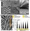
Progression of bioinspired designs based on the attachment mechanism found in the feather vane (A) SEM micrograph of the feather vane showing a branched network of barbs, barbules, and hooklets. (B) First hook and groove-inspired sample. (C) Modified hook and groove structure with a closer match in stiffness to the actual feather vane. (D) Advanced replication of the feather vane to incorporate membrane flaps for directional permeability. (E) The first groove-only unidirectional sliding structure. (F) Two-dimensional sliding structure, which shows textile-like behavior. (G) Cubic sliding structure which provides tailored stiffness in three dimensions. Adapted with permission (Sullivan et al., 2019). Copyright 2019, Elsevier.
Figure 17
Figure 17
Geckos use van der Waals forces generated by densely packed setal arrays on the feet to climb even the sheerest surfaces. Many researchers have attempted to replicate this structure to create reversible, dry adhesives (A) SEM image of the branched gecko setal array. The inset image shows the split-fiber endings with tilted, spatula-shaped tips (Rong et al., 2014). (B) SEM image of synthetic gecko-inspired adhesive composed of polymer micropillars with densely packed carbon nanotubes glued to the end. Open Access (Rong et al., 2014). Copyright 2013, the authors. (C) SEM image of bioinspired, tilted micropillars composed of polyurethane that mimic the gecko setae's directional gripping strength. Reproduced with permission (Murphy et al., 2009a). Copyright 2009, Wiley-VCH Verlag GmbH & Co. KGaA. (D) SEM images of three hierarchical tiers of mushroom-shaped pillars composed of polyurethane that mimic the hierarchical branched structure found in the gecko pad. Reproduced with permission (Murphy et al., 2009b). Copyright 2009, American Chemical Society.
Figure 18
Figure 18
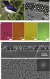
Structural color found in avian feathers and bioinspired analogs (A) Violet-backed starling and TEM micrograph of the multi-layered structure of hollo melanosomes and a thin film of keratin. (B) Structural color produced by SMNPs. (C) Micrograph detailing the arrangement of SMNPs as a thin film. Adapted with permission (Xiao et al., 2015). Copyright 2015, American Chemical Society.
Figure 19
Figure 19
The multiscale surface roughness and fine nanoscaled grooves on feathers help them repel water (A) Schematic showing how the hamuli on penguin feathers trap air beneath water droplets creating an air cushion and minimizing the amount of material in contact with the water. (B) Bioinspired polyamide nanofiber membrane fabricated via asymmetric electrode electrospinning. (C) Chart of contact angle and adhesive force versus location on the polyamide membrane highlighting the effect of fiber density. Reproduced with permission (Wang et al., 2016d, 2016e). Copyright 2016, American Chemical Society (D) SEM image of cotton fiber with precipitated chitosan nanoribbons on the surface inspired by duck feathers. (E) SEM image of polyester fibers with precipitated chitosan “nanoflowers” on the surface. Reproduced with permission (Liu et al., 2008). Copyright 2008, IOP Publishing Ltd.
Figure 20
Figure 20
Much like the gecko pad, the outer layer of skin on the gecko has hydrophobic, self-cleaning properties due to its rough mesostructure, which researchers have attempted to replicate (A) SEM images of natural gecko skin (i-iii) alongside SEM images of biomimetic polystyrene replicas made via biotemplating (iv-vi). (B) Close-up SEM images of gecko spinules and the different measurements used to characterize them (left). Various biomimetic replicas, like the ones shown in A iv-vi, were prepared using several polymer solutions. The resultant spinule shapes are visualized (right) and compared to the natural spinules found on the gecko. Open access (Green et al., 2017). Copyright 2017, the authors.
Figure 21
Figure 21
Advances in 3D printing technology have recently made printing different biological materials more feasible Cera et al. have utilized these advances to fabricate hydration-induced shape-memory components out of keratin. (A) The keratin extraction process used to obtain printable, fibrillar keratin (Cera et al., 2021). (B) To obtain aligned fibrils, keratin fibers were fabricated using traditional wet-spinning. The resultant hierarchical structure is visualized here. (C) Schematic of the atomic scale process for using water to lock and unlock the hydrogen bonds within α-helices or between the β-sheets. This mechanism endues the material with shape recovery properties. (D) Images of the keratin printing process and final products (left); SEM image of the fine detail that can be obtained; birefringence images showing the alignment of the keratin fibers in the woven structure. (E) Series of still images of the hydration-induced shape recovery of the printed samples composed of keratin, showing the prints returning to their initial form over a matter of seconds when submerged in water. Reproduced with permission (Cera et al., 2021). Copyright 2020, the authors.

References

    1. Aksak B., Murphy M.P., Sitti M. Adhesion of biologically inspired vertical and angled polymer microfiber arrays. Langmuir. 2007;23:3322–3332. doi: 10.1021/la062697t. - DOI - PubMed
    1. Aksak B., Sahin K., Sitti M. The optimal shape of elastomer mushroom-like fibers for high and robust adhesion. Beilstein J. Nanotechnol. 2014;5:630–638. - PMC - PubMed
    1. Alibardi L. Cell organization of barb ridges in regenerating feathers of the quail: implications of the elongation of barb ridges for the evolution and diversification of feathers. Acta Zool. 2007;88:101–117. doi: 10.1111/j.1463-6395.2007.00257.x. - DOI
    1. Arzt E., Gorb S., Spolenak R. From micro to nano contacts in biological attachment devices. Proc. Natl. Acad. Sci. U. S. A. 2003;100:10603–10606. doi: 10.1073/pnas.1534701100. - DOI - PMC - PubMed
    1. Arzt E., Quan H., McMeeking R.M., Hensel R. Functional surface microstructures inspired by nature – from adhesion and wetting principles to sustainable new devices. Prog. Mater. Sci. 2021;119:100778. doi: 10.1016/j.pmatsci.2021.100778. - DOI