. 2021 Aug;9(15):e14977.
doi: 10.14814/phy2.14977.

Optic disc edema and chorioretinal folds develop during strict 6° head-down tilt bed rest with or without artificial gravity

Affiliations

Optic disc edema and chorioretinal folds develop during strict 6° head-down tilt bed rest with or without artificial gravity

Steven S Laurie et al. Physiol Rep. 2021 Aug.

Abstract

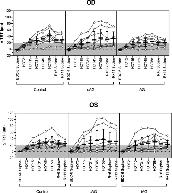
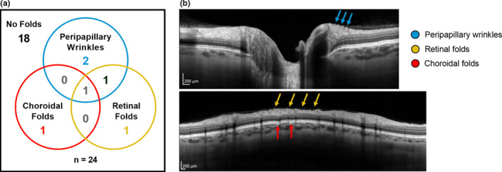
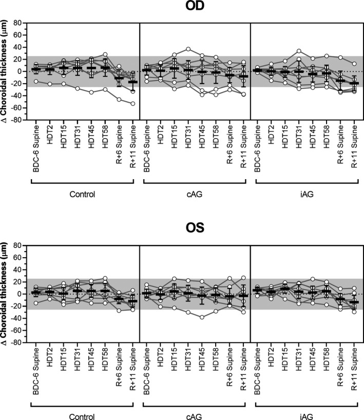
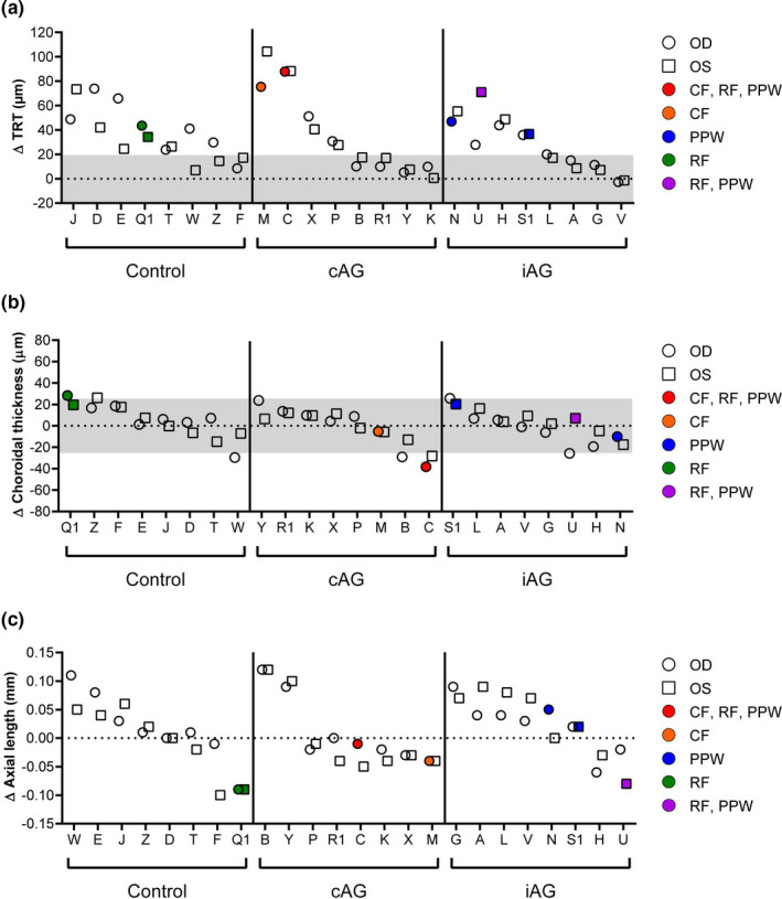
Spaceflight associated neuro-ocular syndrome (SANS) is hypothesized to develop as a consequence of the chronic headward fluid shift that occurs in sustained weightlessness. We exposed healthy subjects (n = 24) to strict 6° head-down tilt bed rest (HDTBR), an analog of weightlessness that generates a sustained headward fluid shift, and we monitored for ocular changes similar to findings that develop in SANS. Two-thirds of the subjects received a daily 30-min exposure to artificial gravity (AG, 1 g at center of mass, ~0.3 g at eye level) during HDTBR by either continuous (cAG, n = 8) or intermittent (iAG, n = 8) short-arm centrifugation to investigate whether this intervention would attenuate headward fluid shift-induced ocular changes. Optical coherence tomography images were acquired to quantify changes in peripapillary total retinal thickness (TRT), retinal nerve fiber layer thickness, and choroidal thickness, and to detect chorioretinal folds. Intraocular pressure (IOP), optical biometry, and standard automated perimetry data were collected. TRT increased by 35.9 µm (95% CI, 19.9-51.9 µm, p < 0.0001), 36.5 µm (95% CI, 4.7-68.2 µm, p = 0.01), and 27.6 µm (95% CI, 8.8-46.3 µm, p = 0.0005) at HDTBR day 58 in the control, cAG, and iAG groups, respectively. Chorioretinal folds developed in six subjects across the groups, despite small increases in IOP. Visual function outcomes did not change. These findings validate strict HDTBR without elevated ambient CO2 as a model for investigating SANS and suggest that a fluid shift reversal of longer duration and/or greater magnitude at the eye may be required to prevent or mitigate SANS.

Keywords: artificial gravity; bed rest; centrifugation; chorioretinal folds; retinal thickness; spaceflight analog; spaceflight associated neuro-ocular syndrome.


Conflict of interest statement

The authors declare that they have no competing interests.

Figures

FIGURE 1
FIGURE 1
Optic disc edema is induced by strict 6º HDTBR but not mitigated by artificial gravity. The change in total retinal thickness (TRT) from BDC‐6 seated baseline at each time point for the right (OD) and left (OS) eyes of control (n = 8), continuous artificial gravity (cAG, n = 8), and intermittent artificial gravity (iAG, n = 8) subjects. Data for BDC‐6, R+6, and R+11 are presented for the supine posture. TRT increased with HDTBR duration for all three groups and did not return to baseline values by R+11. No differences between either experimental group and the control group were evident, indicating that the 30‐min artificial gravity protocols had no effect on TRT at any phase of the study. Circles represent individual subject data, lines connect data for individual subjects, horizontal bars represent the mean value across subjects, error bars represent the 95% CI, and the shaded area represents the predefined range (±19.4 µm) of normal day‐to‐day variation. Statistical significance for each time point is indicated in Table 2. HDT, head‐down tilt; BDC, baseline data collection; R, recovery
FIGURE 2
FIGURE 2
Chorioretinal folds can develop during HDTBR. (a) Six of 24 subjects developed chorioretinal folds (peripapillary wrinkles, retinal folds, and/or choroidal folds) during HDTBR. Four subjects presented with a single subtype, 1 subject presented with both retinal and choroidal folds in the same eye, and 1 subject presented all three subtypes in the same eye. (b) OCT images show examples of peripapillary wrinkles (top, cyan arrows), retinal folds (bottom, yellow arrows), and choroidal folds (bottom, red arrows) in 1 HDTBR subject (Subject C). Scale bars represent 200 µm
FIGURE 3
FIGURE 3
Choroidal thickening is not induced by HDTBR. No significant change in mean choroidal thickness, as compared to seated baseline values (BDC‐6), was detected at any time point during HDTBR. During recovery after HDTBR (R+6 and R+11), the choroid thinned relative to baseline. No differences were detected between either experimental group and the control group for any phase of the study. Circles represent individual subject data, lines connect data for individual subjects, horizontal bars represent the mean value across subjects, error bars represent the 95% CI, and the shaded area represents the predefined range (±25.5 µm) of normal day‐to‐day variation. Statistical significance for each time point is indicated in Table 2. HDT, head‐down tilt; BDC, baseline data collection; R, recovery. OD, right eye; OS, left eye
FIGURE 4
FIGURE 4
Relationship between chorioretinal folds and changes in other ocular parameters on HDT58. (a) Chorioretinal folds only developed in eyes that showed subtle signs of optic disc edema based on increases in total retinal thickness (TRT) exceeding the predefined 19.4 µm threshold (shaded gray area), and the two instances of choroidal folds occurred in the 2 subjects who had the greatest increases in TRT. Only the subject with isolated retinal folds was affected bilaterally. (b) No consistent pattern was observed between chorioretinal fold development and changes in choroidal thickness. (c) The two subjects with retinal folds had decreases in axial length. OD, right eye (circles); OS, left eye (squares); CF, choroidal folds; RF, retinal folds; PPW, peripapillary wrinkles

Similar articles

Cited by

References

    1. Balasubramanian, S. , Tepelus, T. , Stenger, M. B. , Lee, S. M. C. , Laurie, S. S. , Liu, J. H. K. , Feiveson, A. H. , Sadda, S. R. , Huang, A. S. , & Macias, B. R. (2018). Thigh cuffs as a countermeasure for ocular changes in simulated weightlessness. Ophthalmology, 125, 459–460. 10.1016/j.ophtha.2017.10.023 - DOI - PubMed
    1. Costa, V. P. , & Arcieri, E. S. (2007). Hypotony maculopathy. Acta Ophthalmologica Scandinavica, 85, 586–597. 10.1111/j.1600-0420.2007.00910.x - DOI - PubMed
    1. Draeger, J. , Schwartz, R. , Groenhoff, S. , & Stern, C. (1995). Self‐tonometry under microgravity conditions. Aviation, Space and Environmental Medicine, 66, 568–570. - PubMed
    1. Fannin, L. A. , Schiffman, J. C. , & Budenz, D. L. (2003). Risk factors for hypotony maculopathy. Ophthalmology, 110, 1185–1191. - PubMed
    1. Feola, A. J. , Nelson, E. S. , Myers, J. , Ethier, C. R. , & Samuels, B. C. (2018). The impact of choroidal swelling on optic nerve head deformation. Investigative Ophthalmology & Visual Science, 59, 4172–4181. 10.1167/iovs.18-24463 - DOI - PubMed

Publication types