Review
. 2021 Jul 10;12(7):1059.
doi: 10.3390/genes12071059.

Role and Evolution of the Extracellular Matrix in the Acquisition of Complex Multicellularity in Eukaryotes: A Macroalgal Perspective

Affiliations
Review

Role and Evolution of the Extracellular Matrix in the Acquisition of Complex Multicellularity in Eukaryotes: A Macroalgal Perspective

Bernard Kloareg et al. Genes (Basel). .

Abstract

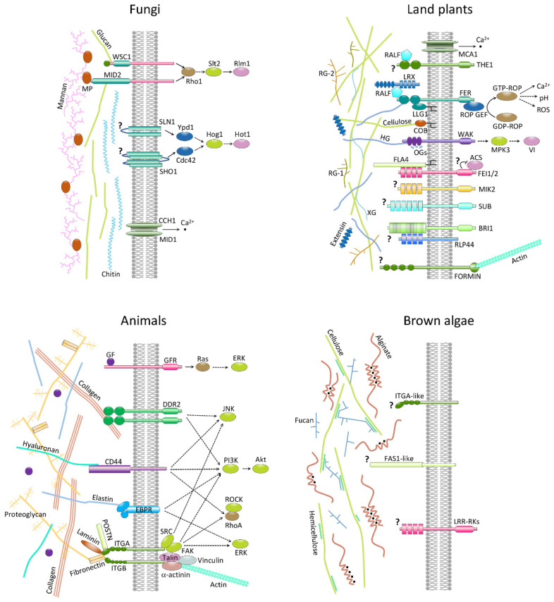
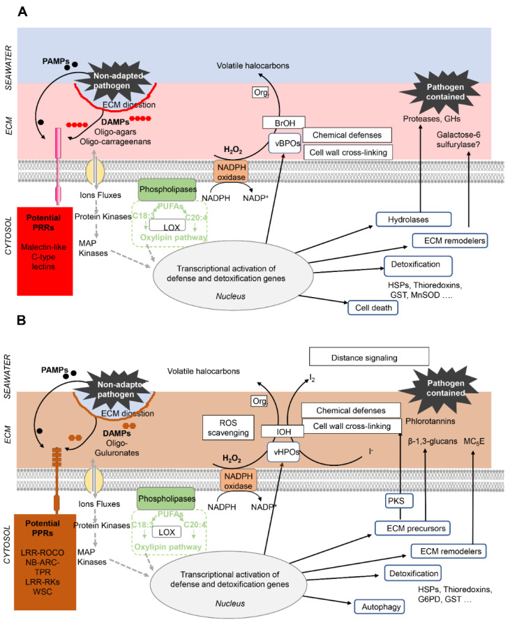
Multicellular eukaryotes are characterized by an expanded extracellular matrix (ECM) with a diversified composition. The ECM is involved in determining tissue texture, screening cells from the outside medium, development, and innate immunity, all of which are essential features in the biology of multicellular eukaryotes. This review addresses the origin and evolution of the ECM, with a focus on multicellular marine algae. We show that in these lineages the expansion of extracellular matrix played a major role in the acquisition of complex multicellularity through its capacity to connect, position, shield, and defend the cells. Multiple innovations were necessary during these evolutionary processes, leading to striking convergences in the structures and functions of the ECMs of algae, animals, and plants.

Keywords: biomechanical properties; cell wall; development; evolutionary convergences; extracellular matrix; hydric and salinity stress; innate immunity; marine macroalgae; multicellularity; sulfated polysaccharides.


Conflict of interest statement

The authors declare no conflict of interest.

Figures

Figure 1
Figure 1
Schematic tree of the eukaryotes showing the phylogenetic positions of the brown, green, and red macroalgal lineages. Gray sectors mark lineages that have given rise to complex multicellular species. Adapted from Coelho and Cock, 2020 [3].
Figure 2
Figure 2
Models of macroalgal extracellular matrices. Semicrystalline cellulose microfibrils are present in almost all types of macroalgae. They constitute a crucial structural component but are sparse in comparison to plant cell walls. Most cell wall structural proteins have yet to be characterized, but their identity is expected to differ in each macroalgal phylum. (A) Brown algae (example of a Fucales species). Alginates are the most abundant component of brown algal ECMs. Alginate chains auto-associate through their guluronate (G)-rich regions. This process is coordinated by Ca2+ ions. Fucose-containing sulfated polysaccharides (FCSPs) acts as cross-linkers between cellulose microfibrils. Hemicelluloses (β-1,4-1,3-glucans, β-1,3-glucans) have been recently identified in brown algae and might act as intermediates between the cellulose microfibrils. Phlorotannins (polyphenols) are likely to be associated with alginates and proteins. Iodine is generally abundant in brown algal ECM but its association mode with other components is unresolved. Arabinogalactan proteins and proteins containing WSC-domains have been identified among CW proteins. Model adapted from Deniaud and coworkers [26] and updated based on the following references [27,28,29]. (B) Red algae (example of a carrageenophyte). Sulfated galactans (carrageenans in this case) are the most abundant polymers of the red algal ECM. Chains containing 3,6-anhydro-D-galactose units associate through hydrogen-bonding and hydrophobic interactions. The resulting helices further cluster into microfibrils by ionic interactions between their sulfate groups and cations. Glucomannans have been shown to cross-link cellulose microfibrils. Sulfated glucans have been identified as a minor component, but their interactions with the other polysaccharides remain uncertain. Model adapted from the following references [32,33,34]. (C) Green algae (Ulva species). The major polysaccharide ulvan can form a weak gel in the presence of both boric acid and divalent cations (e.g., Ca2+). However, the exact mechanism of chain association is currently not understood. Two minor polysaccharides were also identified. Xyloglucans interact with cellulose, but their role as cross-linkers of cellulose microfibrils remains to be demonstrated. In contrast, glucuronans interact with ulvans and proteins. Model adapted from Lahaye and Robic [35].
Figure 3
Figure 3
Phylogenetic tree of the cellulose synthases (CESA) and cellulose synthase-like proteins CSL (family GT2). The phylogenetic tree was constructed using the Maximum Likelihood (ML) approach with the program MEGA-X [82]. Numbers indicate the bootstrap values in the ML analysis. Bootstrap values inferior to 50% are not shown. The tree was rooted by the clade of bacterial cellulose synthases. The full listing of the aligned proteins is reported in Supplementary Table S1. The uncompressed tree is shown in Supplementary Figure S1.
Figure 4
Figure 4
Unrooted phylogenetic tree of eukaryotic sulfotransferases. The phylogenetic tree was constructed using the Maximum Likelihood (ML) approach with the program MEGA-X [82]. Numbers indicate the bootstrap values in the ML analysis. Bootstrap values inferior to 50% are not shown. The sequence labels in bold correspond to biochemically characterized sulfotransferases. The full listing of the aligned proteins is reported in Supplementary Table S1. The uncompressed tree is shown in Supplementary Figure S2.
Figure 5
Figure 5
Correlation between the sulfated fucan content of isolated cell walls of brown algae and their position in the intertidal zone. (A). Fucose content (% w/w) vs. duration of emersion (% of time). (B) Sulfate substituent content (% w/w) vs. duration of emersion. The maximal duration of emersion in a daily tidal cycle was calculated for Roscoff (Brittany, France), based on species belt zonation in the intertidal (taken as the medium height relative to the lowest astronomical tide), using the spring tide mid-water mark for Pelvetia canaliculata, Fucus ceranoides, F. spiralis, F. vesiculosus, and Ascophyllum nodosum and the neap tide mid-water mark for F. serratus, Bifurcaria bifurcate, and Laminaria digitata. See Supplementary Table S2 for the corresponding data values.
Figure 6
Figure 6
Membrane-localized, ECM-sensing proteins in land plant, animals, and fungi. Related membrane proteins identified in brown algae are also shown. MCA1, MIDI1-complementing activity 1; THE1, THESEUS1; FER, FERONIA; WAK, wall-associated kinase; RALF, rapid alkalinization factor; LRX, leucine-rich repeat extensin; LLG1, LORELEI-like glycosylphosphatidylinositol-anchored protein 1; FLA4, fasciclin-like arabinogalactan protein 4; COB, extracellular glycosyl-phosphatidyl inositol-(GPI)-anchored protein COBRA; FEI1/2, leucine-rich repeat receptor kinases; MIK2, MALE DISCOVERER1-interacting receptor like kinase 2 I; SUB, STRUBBELIG; BRI1, brassinosteroid insensitive 1; RLP44, receptor-like protein 44; ROP, rho of plants; ROP GEF, rho of plants guanine-nucleotide exchange factor; MPK3, map kinase 3; VI, vacuolar invertase; ACS, aminocyclopropane 1-carboxylic acid synthase HG, homogalacturonan; OGs, oligogalacturonides; XG, xyloglucan; RG-1, rhamnogalacturonan I; RG-2, rhamnogalacturonan II; ROS, reactive oxygen species; GF, growth factor; GFR, growth factor receptor; DDR2, Discoidin domain-containing receptor 2; EBPR, Elastin-binding protein receptor, ITGA/B, Integrin A/B subunit; POSTN, fasciclin domain protein periostin; Ras, Ras GTPase; JNK, c-Jun N-terminal mitogen-activated protein kinase; PI3K, Phosphoinositide 3-kinase; Akt, RAC-α serine/threonine-protein kinase; RhoA, Ras homolog A GTPase; ROCK, Rho-associated protein kinase; ERK, Extracellular-signal-regulated mitogen-activated protein kinase; WSC1, Cell wall integrity and stress response component 1; MID2, Mating pheromone-induced death protein 2; SLN1, Synthetic lethal of N-end rule 1; SHO1, Synthetic high osmolarity-sensitive 1; CCH1/MID1, Stretch-activated voltage-gated high-affinity calcium channel composed of Calcium channel homolog 1 and Mating pheromone-induced death protein 1; Rho1, Ras homolog 1 GTPase; Slt2, Suppressor of the lytic phenotype mitogen-activated protein kinase 2; Rlm1, Resistance to lethality of MKK1P386 overexpression MADS-box transcription factor 1; Ypd1, Tyrosine phosphatase dependent 1; Cdc42, Cell division cycle 42 rho-like GTPase; Hog1, High-osmolarity glycerol mitogen-activated protein kinase 1; Hot1, High-osmolarity-induced transcription factor 1; MP, mannoprotein; FAS1-like, Fasciclin-like domain protein, LRR-RKs, Leucine-rich repeat domain receptor kinases; ?, ligand unknown or uncertain step of the signal transduction pathway; black dots, calcium atoms; branched black lines indicate GPI anchors. Adapted from [26,117,118].
Figure 7
Figure 7
Role of the extracellular matrix in the innate immunity of multicellular red (A) and brown (B) algae. Drawings adapted from in [136] and updated based on evidence provided by the authors of [7,139,140,149,152,161]. Note that the basic principles of innate immunity are shared by brown and red algae (as well as by plants and animals): recognition by Pattern Recognition Receptors (PPRs) of pathogen-derived PAMPs or cell-wall derived DAMPs triggers signaling cascades that activate defense and detoxification responses. Red (A) and brown algae (B) differ by the nature of their DAMPs and PPRs, highlighted in red and brown, respectively. In the ECM per se the main defense responses deal with ECM remodeling (thickening, strengthening) and the secretion of chemicals and enzymes that target the pathogen, eventually leading to the containment of non-adapted pathogens. Abbreviations: PAMPs, pathogen associated molecular patterns; DAMPs, Damage associated molecular patterns; PUFAs, poly unsaturated fatty acids; MAP Kinase, mitogen activated protein kinase; LOX, lipoxygenase; vHPO, vanadium-dependent haloperoxidase; IoH, hypoiodous acid; vBPO, vanadium-dependent bromoperoxidase; BrOH, hypobromous acid; ROS, reactive oxygen species. Org. organic compound. HSPs, heat shock proteins; G6PD, glucose-6-phospate-dehydrogenase; GST, Glutathion-S-transferase; SOD, superoxide dismutase; PKS, polyketate synthase; MC5E, mannuronan C5 epimerase, GHs, glycoside hydrolases.

Similar articles

Cited by

References

    1. Grosberg R.K., Strathmann R.R. The evolution of multicellularity: A minor major transition? Annu. Rev. Ecol. Evol. Syst. 2007;38:621–654. doi: 10.1146/annurev.ecolsys.36.102403.114735. - DOI
    1. Cock J.M., Collen J. Independent emergence of complex multicellularity in the brown and red algae. In: Ruiz-Trillo I., Nedelcu A.M., editors. Evolutionary Transitions to Multicellular Life. Springer; Dordrecht, The Netherlands: 2015. pp. 335–361.
    1. Coelho S.M., Cock J.M. Brown Algal Model Organisms. Annu. Rev. Genet. 2020;54:71–92. doi: 10.1146/annurev-genet-030620-093031. - DOI - PubMed
    1. Michel G., Tonon T., Scornet D., Cock J.M., Kloareg B. The cell wall polysaccharide metabolism of the brown alga Ectocarpus Siliculosus. Insights into the evolution of extracellular matrix polysaccharides in Eukaryotes. New Phytol. 2010;188:82–97. doi: 10.1111/j.1469-8137.2010.03374.x. - DOI - PubMed
    1. Cock J.M., Sterck L., Rouze P., Scornet D., Allen A.E., Amoutzias G., Anthouard V., Artiguenave F., Aury J.M., Badger J.H., et al. The Ectocarpus genome and the independent evolution of multicellularity in brown algae. Nature. 2010;465:617–621. doi: 10.1038/nature09016. - DOI - PubMed

LinkOut - more resources