Review
. 2021 Jul 10;11(7):1012.
doi: 10.3390/biom11071012.

Mitostasis, Calcium and Free Radicals in Health, Aging and Neurodegeneration

Affiliations
Review

Mitostasis, Calcium and Free Radicals in Health, Aging and Neurodegeneration

Juan A Godoy et al. Biomolecules. .

Abstract

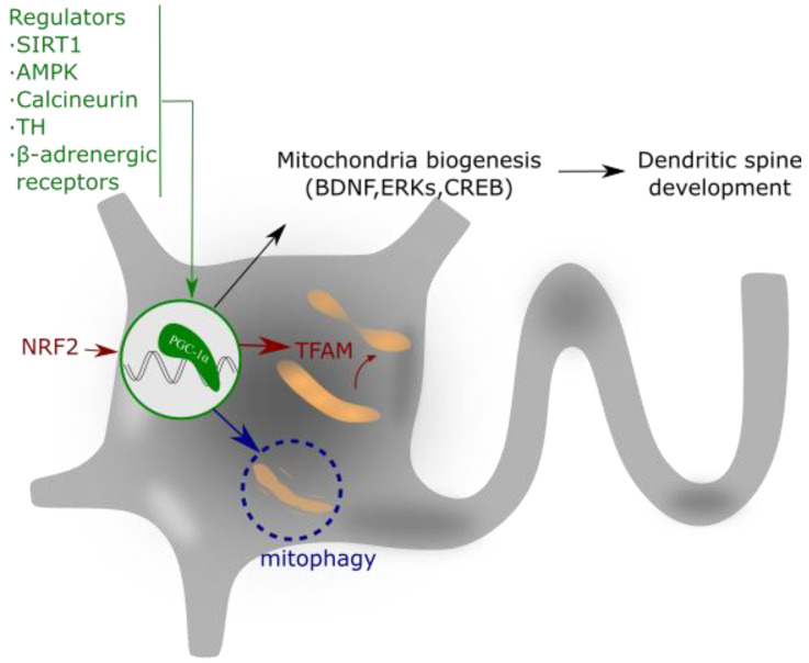
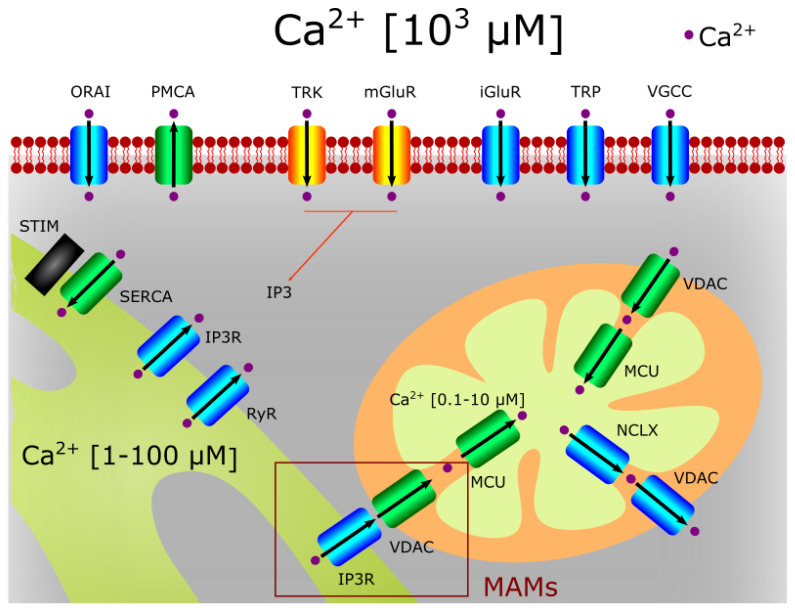
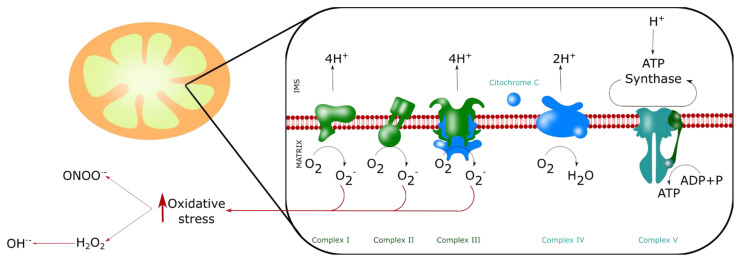
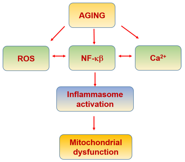
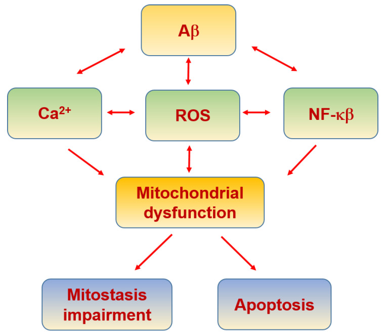
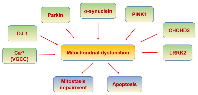
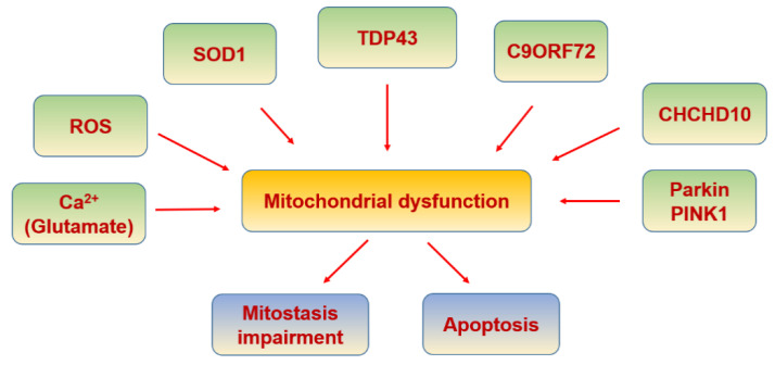
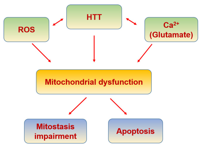
Mitochondria play key roles in ATP supply, calcium homeostasis, redox balance control and apoptosis, which in neurons are fundamental for neurotransmission and to allow synaptic plasticity. Their functional integrity is maintained by mitostasis, a process that involves mitochondrial transport, anchoring, fusion and fission processes regulated by different signaling pathways but mainly by the peroxisome proliferator-activated receptor-γ coactivator-1α (PGC-1α). PGC-1α also favors Ca2+ homeostasis, reduces oxidative stress, modulates inflammatory processes and mobilizes mitochondria to where they are needed. To achieve their functions, mitochondria are tightly connected to the endoplasmic reticulum (ER) through specialized structures of the ER termed mitochondria-associated membranes (MAMs), which facilitate the communication between these two organelles mainly to aim Ca2+ buffering. Alterations in mitochondrial activity enhance reactive oxygen species (ROS) production, disturbing the physiological metabolism and causing cell damage. Furthermore, cytosolic Ca2+ overload results in an increase in mitochondrial Ca2+, resulting in mitochondrial dysfunction and the induction of mitochondrial permeability transition pore (mPTP) opening, leading to mitochondrial swelling and cell death through apoptosis as demonstrated in several neuropathologies. In summary, mitochondrial homeostasis is critical to maintain neuronal function; in fact, their regulation aims to improve neuronal viability and to protect against aging and neurodegenerative diseases.

Keywords: aging; calcium; mitochondria; mitostasis; nitric oxide; oxidative stress.


Conflict of interest statement

The authors declare that they have no competing interests that might be perceived to influence the results and discussion reported in this paper.

Figures

Figure 1
Figure 1
Mitochondrial fission.This process is initiated by MFF forming a ring in the middle of the primary organelle to be divided. Drp1 GTPase translocates to the mitochondria and binds to MFF, inducing a conformational change by the hydrolysis of GTP. This conformational change generates the ring contraction, which starts mitochondrial fission. The reorganization of the cytoskeleton generates the driving forces that separate both new mitochondria.
Figure 2
Figure 2
Mitochondrial fusion. The fusion of two preexistent mitochondria is a protective mechanism based on the fusion of the OMM and IMM. The OMM fusion is mediated by MFN1 and MFN2, and the fusion of the IMM is mediated by OPA1. First, MFN1 and MFN2 GTPase activity induce the formation of homo- or hetero-oligomeric complexes in the fusion spot. Then, OPA1-mediated GTP hydrolysis promotes the membrane fusion.
Figure 3
Figure 3
PGC-1α regulates mitochondrial biogenesis and spine growth. Its activity is based on the interaction with NRF that activates to TFAM. This complex regulates mitochondrial gene expression and addresses the requirements of neuronal energy when needed for dendrite activity or growth. PGC-1α expression is regulated by different intracellular signaling pathways, as explained in the text.
Figure 4
Figure 4
Ca2+ homeostasis in neurons. The entrance of extracellular Ca2+ into neurons is due to the direct activation of neurotransmitter and neurotrophin receptors (such as NMDAR and TRK) and cationic channels (such as TRPs or voltage activated Ca2+ channels). The VDAC channel can import or export Ca2+ in the mitochondria when associated to MCU or NCLX, respectively. IP3R and VDAC channels found in MAMs can also transport Ca2+ from the ER to the mitochondria. The Ca2+ extrusion to the extracellular medium is mediated by ATPases (i.e., PMCA).
Figure 5
Figure 5
ROS production by mitochondria. Mitochondria produce ROS, mainly superoxide anion O2· by the MRC (at the complexes I, II and III), and indirectly hydroxyl radical (OH·), hydrogen peroxide (H2O2) andperoxynitrite (ONOO). These four molecules are the main reactive agents inducing nitro-oxidative stress.
Figure 6
Figure 6
Inflammaging and mitochondria.
Figure 7
Figure 7
Insulin resistance and mitochondria.
Figure 8
Figure 8
Alzheimer’s disease and mitochondria.
Figure 9
Figure 9
Parkinson’s disease and mitochondria.
Figure 10
Figure 10
Amyotrophic lateral sclerosis and mitochondria.
Figure 11
Figure 11
Huntington’s disease and mitochondria.

References

    1. Erbslöh F., Bernsmeier A., Hillesheim H.R. Der Glucoseverbrauch des Gehirns und seine Abhängigkeit von der Leber. Arch. für Psychiatr. und Nervenkrankheiten Ver. mit Zeitschrift für die Gesamte Neurol. und Psychiatr. 1958;196:611–626. doi: 10.1007/BF00344388. - DOI - PubMed
    1. Sokoloff L. Energetics of functional activation in neural tissues. Neurochem. Res. 1999;24:321–329. doi: 10.1023/A:1022534709672. - DOI - PubMed
    1. Lin M.Y., Sheng Z.H. Regulation of mitochondrial transport in neurons. Exp. Cell Res. 2015;334:35–44. doi: 10.1016/j.yexcr.2015.01.004. - DOI - PMC - PubMed
    1. Misgeld T., Schwarz T.L. Mitostasis in neurons: Maintaining mitochondria in an extended cellular architecture. Neuron. 2017;96:651–666. doi: 10.1016/j.neuron.2017.09.055. - DOI - PMC - PubMed
    1. Brini M., Calì T., Ottolini D., Carafoli E. Neuronal calcium signaling: Function and dysfunction. Cell. Mol. Life Sci. 2014;71:2787–2814. doi: 10.1007/s00018-013-1550-7. - DOI - PMC - PubMed

Publication types