. 2021 Jul 19;11(7):1057.
doi: 10.3390/biom11071057.

Interaction Mode of the Novel Monobactam AIC499 Targeting Penicillin Binding Protein 3 of Gram-Negative Bacteria

Affiliations

Interaction Mode of the Novel Monobactam AIC499 Targeting Penicillin Binding Protein 3 of Gram-Negative Bacteria

Stefan Freischem et al. Biomolecules. .

Erratum in

Abstract

Novel antimicrobial strategies are urgently required because of the rising threat of multi drug resistant bacterial strains and the infections caused by them. Among the available target structures, the so-called penicillin binding proteins are of particular interest, owing to their good accessibility in the periplasmic space, and the lack of homologous proteins in humans, reducing the risk of side effects of potential drugs. In this report, we focus on the interaction of the innovative β-lactam antibiotic AIC499 with penicillin binding protein 3 (PBP3) from Escherichia coli and Pseudomonas aeruginosa. This recently developed monobactam displays broad antimicrobial activity, against Gram-negative strains, and improved resistance to most classes of β-lactamases. By analyzing crystal structures of the respective complexes, we were able to explore the binding mode of AIC499 to its target proteins. In addition, the apo structures determined for PBP3, from P. aeruginosa and the catalytic transpeptidase domain of the E. coli orthologue, provide new insights into the dynamics of these proteins and the impact of drug binding.

Keywords: AIC499; Gram-negative bacteria; PBP3; antibiotic; monobactam; penicillin binding protein; structure; transpeptidase domain; β-lactam.


Conflict of interest statement

The authors declare no conflict of interest.

Figures

Figure 2
Figure 2
Design of an EcTPd* construct for structural studies. (A) X-ray structure of EcPBP3ΔTM published previously (PDB entry 4BJP, [13]). Secondary structure elements and intervening loops relevant to this paper are labeled. Note that the n-PBd of the elongated molecule has been traced only incompletely. (B) Sequence alignment of EcPBP3ΔTM (upper sequence) and the newly designed EcTPd* construct (bottom sequence) performed with Clustal Omega [41]. The structural elements labeled in (A) as well as the tri-glycine linkers (yellow background) are highlighted in the sequence. (C) Overlay of 2D 1H-15N TROSY-HSQC NMR spectra of uniformly [2H, 13C, 15N]-labeled EcPBP3ΔTM (blue) and EcTPd* (red) demonstrates the structural similarity of both proteins in solution.
Figure A1
Figure A1
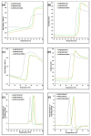
Raw data of the thermal shift experiments. In the upper row, the ratio of 350 nm/330 nm fluorescence (A), the turbidity (B) are plotted against temperature. Tm derived from turbidity measurements were determined to be 58.4 °C for apo-EcPBP3ΔTM, and 63.4 and 65.3 °C for EcPBP3ΔTM in complex with aztreonam and AIC499, respectively. In the middle row, the analogous data are shown for PaPBP3ΔTM (C,D). Tm were calculated to be 45.9, 53.8 and 54.0 °C for the DMSO control, PaPBP3ΔTM in complex with aztreonam and with AIC499, respectively. In the bottom row, the first derivatives of the turbidity are plotted for EcPBP3ΔTM and PaPBP3ΔTM, respectively (E,F). The reference experiments with addition of DMSO only are displayed in gray, whereas the graphs for complexes with aztreonam and AIC499 are colored green and gold, respectively.
Figure A2
Figure A2
Superposition of the X-ray structures of the EcTPd* protein introduced in this study (dark blue) and EcPBP3ΔTM published previously (PDB entry 4BJP, gold), demonstrating the similar coverage of ordered structure. The GGG linkers replacing the deleted segments in EcTPd* are colored black.
Figure 1
Figure 1
Structure of the monobactam AIC499, obtained by iterative optimization. The relevant functional groups are marked in color: benzamidine-based head group (R1), blue; β-lactam N-1 position (R2), red; β-lactam C-4 position (R3), orange; amino-thiazole (R4), gray; linker (R5), green.
Figure 3
Figure 3
X-ray structures of EcPBP3 determined in the absence and presence of AIC499 (red stick model). (A) The published apo-EcPBP3ΔTM structure (PDB entry 4BJP [13], gold) is used for superposition with the AIC499-complexed EcPBP3ΔTM (blue, this study). (B) The structure of apo-EcTPd* is shown in dark blue, while the complex with AIC499 is colored salmon. The GGG linkers replacing the removed segments are shown in black.
Figure 4
Figure 4
Structure of AIC499 within the active site of EcTPd*. (A) 3D representation of the covalently bound ligand together with the most relevant interacting side chains (gray) and their counterparts in the apo structure (green). The second conformation of the amidine-based head group, as well as the terminal sulfate moiety are shown in lighter color. Note that K499 has been truncated in the complex structure because of missing electron density. (B) LigPlot+ representation of individual contacts for one conformer. Hydrogen bonds (cyan) are plotted for donor-acceptor distances between 2.3 Å and 3.2 Å, while hydrophobic interactions (salmon) have distances between 3.0 Å and 4.0 Å. A complete list of distances between protein side chains and the AIC499 compound is provided in Table A1.
Figure 5
Figure 5
X-ray structures of PaPBP3 determined in the absence and presence of AIC499 (red stick model). (A) Crystal form 1 (blue), crystal form 2 (green) and crystal form 3 (bright orange) of PaPBP3ΔTM feature different unit cells, leading to slightly different orientations predominantly in the n-PBd. (B) The structure of apo-PaPBP3ΔTM crystal form 2 (cyan) is used for superposition with PaPBP3ΔTM:AIC499 chain B (dark red). Additionally, the head, anchor, and linker subdomains of the n-PBd are highlighted with black, dark blue, and light blue ellipses, respectively.
Figure 6
Figure 6
Structure of AIC499 within the active site of PaPBP3ΔTM. (A) 3D representation of the covalently bound ligand together with the most relevant interacting side chains (gray) and their counterparts in the apo structure (crystal form 2, green). Alternative conformations of the amidine-based head group and the terminal sulfate moiety are shown in lighter color. (B) LigPlot+ representation of individual contacts. Hydrogen bonds (cyan) are plotted for donor-acceptor distances between 2.3 Å and 3.2 Å, while hydrophobic interactions (salmon) have distances between 3.0 Å and 4.0 Å. A complete list of distances between protein side chains and the AIC499 compound is provided in Table A1.
Figure 7
Figure 7
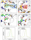
Comparison of aztreonam and AIC499 binding to PaPBP3 and EcPBP3. Panel (A) shows a close-up view of the linker (R5; green) and head group (R1; blue) of AIC499 in complex with PaPBP3ΔTM, while (B) focuses on the sulfate (R2; red) and dimethyl group (R3; orange). The analogous views of aztreonam in complex with PaPBP3ΔTM (PDB entry 3PBS [11]) are represented in (C) and (D), respectively. Refer to Table A2 for a complete list of distances between protein side chains and the aztreonam molecule. (E) DSF data obtained with EcPBP3ΔTM; the first derivative of the 350 nm/330 nm fluorescence ratio is plotted against the temperature. The control experiment with DMSO added to the protein (gray) yields a Tm of 58.6 °C, which increases to 63.4 and 65.4 °C after addition of 500 µM aztreonam (AZT, green) and AIC499 (AIC, gold), respectively. (F) Analogous experiments with PaPBP3ΔTM measured in complex with aztreonam (green) and AIC499 (gold) gave nearly identical Tm values of 54.0 and 54.1 °C, respectively. The Tm for the control experiment (gray) was determined to be 46.0 °C. Raw data of the thermal shift experiments are shown in Figure A1.

References

    1. O’Neill J. Antimicrobial Resistance: Tackling a Crisis for the Health and Wealth of Nations. London: Review on Antimicrobial Resistance. 2014. [(accessed on 15 May 2021)]. Available online: https://amr-review.org/Publications.html.
    1. Zervosen A., Sauvage E., Frère J.-M., Charlier P., Luxen A. Development of new drugs for an old target—The penicillin binding proteins. Molecules. 2012;17:12478–12505. doi: 10.3390/molecules171112478. - DOI - PMC - PubMed
    1. Sauvage E., Terrak M. Glycosyltransferases and transpeptidases/penicillin-binding proteins: Valuable targets for new antibacterials. Antibiotics. 2016;5:12. doi: 10.3390/antibiotics5010012. - DOI - PMC - PubMed
    1. Vollmer W., Bertsche U. Murein (peptidoglycan) structure, architecture and biosynthesis in Escherichia coli. Biochim. Biophys. Acta Biomembr. 2008;1778:1714–1734. doi: 10.1016/j.bbamem.2007.06.007. - DOI - PubMed
    1. Goffin C., Ghuysen J.-M. Biochemistry and comparative genomics of SxxK superfamily acyltransferases offer a clue to the mycobacterial paradox: Presence of penicillin-susceptible target proteins versus lack of efficiency of penicillin as therapeutic agent. Microbiol. Mol. Biol. Rev. 2002;66:702–738. doi: 10.1128/MMBR.66.4.702-738.2002. - DOI - PMC - PubMed

MeSH terms

LinkOut - more resources