. 2021 Jul 12;10(7):847.
doi: 10.3390/antibiotics10070847.

The Phagosome-Lysosome Fusion Is the Target of a Purified Quillaja saponin Extract (PQSE) in Reducing Infection of Fish Macrophages by the Bacterial Pathogen Piscirickettsia salmonis

Affiliations

The Phagosome-Lysosome Fusion Is the Target of a Purified Quillaja saponin Extract (PQSE) in Reducing Infection of Fish Macrophages by the Bacterial Pathogen Piscirickettsia salmonis

Hernán D Cortés et al. Antibiotics (Basel). .

Abstract

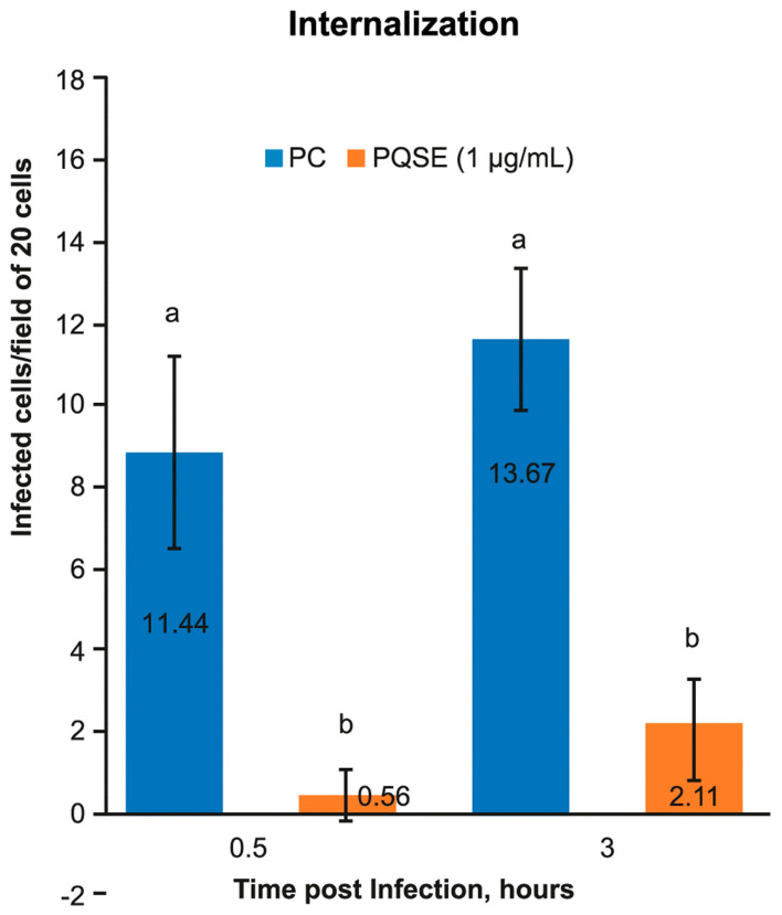
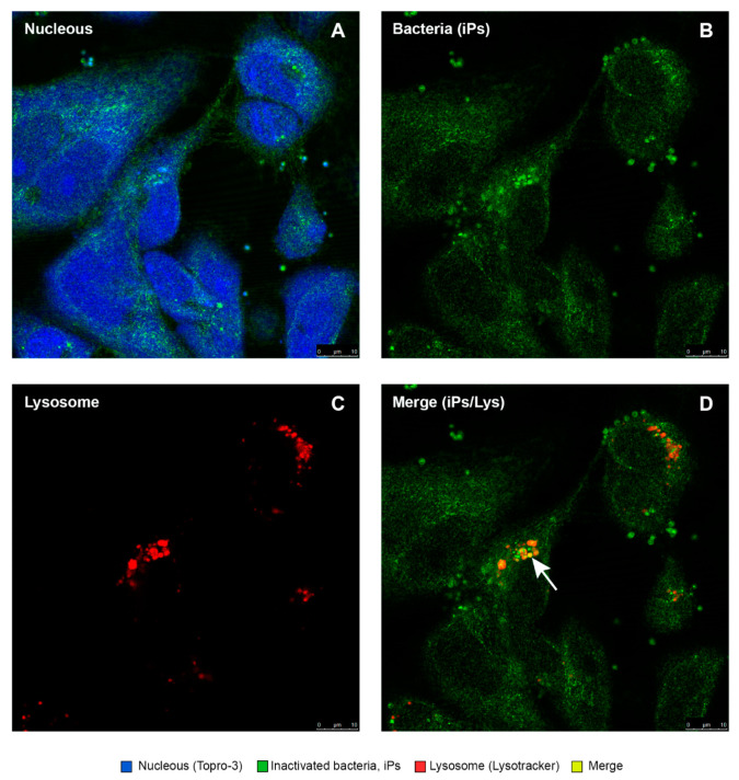
Piscirickettsia salmonis, the etiological agent of Piscirickettsiosis, is a Gram-negative and facultative intracellular pathogen that has affected the Chilean salmon industry since 1989. The bacterium is highly aggressive and can survive and replicate within fish macrophages using the Dot/Icm secretion system to evade the host's immune response and spread systemically. To date, no efficient control measures have been developed for this disease; therefore, the producers use large amounts of antibiotics to control this pathogen. In this frame, this work has focused on evaluating the use of saponins from Quillaja saponaria as a new alternative to control the Piscirickettsiosis. It has been previously reported that purified extract of Q. saponaria (PQSE) displays both antimicrobial activity against pathogenic bacteria and viruses and adjuvant properties. Our results show that PQSE does not present antimicrobial activity against P. salmonis, although it reduces P. salmonis infection in an in vitro model, promoting the phagosome-lysosome fusion. Additionally, we demonstrate that PQSE modulates the expression of IL-12 and IL-10 in infected cells, promoting the immune response against the pathogen and reducing the expression of pathogen virulence genes. These results together strongly argue for specific anti-invasion and anti-intracellular replication effects induced by the PQSE in macrophages.

Keywords: Piscirickettsia salmonis; Quillaja saponaria; bacterial; fish macrophage; phagosome–lysosome fusion; saponin.


Conflict of interest statement

The authors declare no conflict of interest. The funder had no role in the design of the study, decisions about protocols, developments of the trials, the collection of data, discussions, the analysis of results, the writing of the manuscript, or in the decision to publish the results.

Figures

Figure 1
Figure 1
UHPLC of purified Quillaja saponin extract (PQSE) (96% saponins, w/w). UHPLC of PQSE from which almost all non-saponin fraction components (phenolics, polysaccharides) have been separated.
Figure 2
Figure 2
Assay conditions to phagosome-lysosome fusion measure. Three conditions, Pre-treatment, Co-treatment, and Post-treatment of SHK-1 cells with Quillaja Extract (PQSE), and challenged with P. salmonis, were used to evaluate the phagosome-lysosome fusion.
Figure 3
Figure 3
Viability of SHK-1 cells exposed to a High Purified Quillaja saponin Extract (PQSE). The image corresponds to Table 1. cell viability was higher than 90% until doses of 12.5 µg/mL of the extract (equivalent to 12.0 µg/mL of saponin). But at levels more than 12.5 ug/mL, the cell viability was reduced to 60%. The cell viability of the Positive Control (Triton X-100) was 39%. The viability was estimated as (Optical density of treated cells, OD)/(OD Control cells) × 100.
Figure 4
Figure 4
Internalization of P. salmonis in SHK-1 cells pre-incubated 4 h with 0.5 µg/mL of Quillaja saponaria Extract (PQSE). The internalization test for P. salmonis in SHK-1 cells, showed a significant reduction in the number of cells infected with the bacterium in the groups pre-incubated with PQSE, either, at 0.5 hpi (early internalization) as a 3 hpi (late internalization), with respect to the Positive Control (PC) (cells infected with P. salmonis). The test considered, two (2) treatments (PQSE & PC), three (3) repetitions/treatment, sixty (60) cells/repetition. The statistical analysis was performed using a Manova, to evaluate, time and treatments, independent variables. Different letters, a and b represent statistical differences at p < 0.05.
Figure 5
Figure 5
Gene expression analysis of dotB (T4-BSS) in SHK1 cells pre-incubated with 0.5 µg/mL of Quillaja extract (PQSE) and challenged with P. salmonis. dotB gene expression at 1, 2 and 72 hpi in cells pre-incubated with PQSE. Results are expressed as fold change relative to Negative Control (NC), as average values of triplicate determinations. SHK-1 cells pre-incubated with PQSE for 4 h, prior to a challenge with P. salmonis, significantly reduce (p < 0.05) the gene expression of the virulence factor dotB, compared to the Positive Control (PC: P. salmonis), at 24 and 72 hpi. NC: Negative Control (without PQSE and without P. salmonis); PC: Positive Control (P. salmonis) PQSE+P.sal: SHK-1 cells pre-incubated with PQSE and then challenged with P. salmonis. Different letters, a, b and c depict statistically significant differences according to the Mann-Whitney test (p <0.05).
Figure 6
Figure 6
Gene expression analysis of chaPs at 24 and 72 hpi in SHK-1 cells pre-treated 4 h with 0.5 µg/mL of PQSE. Results are expressed as fold change relative to Negative Control (NC), as average values of triplicate determinations. SHK-1 cells pre-treated for 4 h with PQSE, prior to a challenge with P. salmonis, significantly reduced (p <0.05) the gene expression of ChaPs, compared to the PC, at 24 and 72 hpi. NC: Negative Control (without PQSE and without P. salmonis); PC: Positive Control (P. salmonis); PQSE/P.sal: SHK-1 cells pre-treated with PQSE and then challenged with P. salmonis. Different letters a and b depict statistically significant differences according to the Mann-Whitney test (p <0.05).
Figure 7
Figure 7
Gene expression analysis of IL-10 at 1, 24 and 72 hpi, in SHK-1 cells pre-treated with 0.5 µg/mL of PQSE. SHK-1 cells pre-incubated 4 h with PQSE, prior to a challenge with P. salmonis, significantly reduced (p < 0.05) the gene expression of Interleukin 10 (IL-10), with respect to NC and PC, at 24 and 72 hpi. The results are expressed as fold change relative to the NC, as average values of triplicate determinations. NC: Negative Control (without PQSE and without P. salmonis); PC: Positive Control (P. salmonis); PQSE/P.sal: SHK-1 cells pre-treated with PQSE and challenged with P. salmonis. Different letters, a, b and c depict statistically significant differences according to the Mann-Whitney test (p < 0.05).
Figure 8
Figure 8
Gene expression analysis of IL-12 at 1, 24 and 72 hpi in SHK-1 cells pre-treated with 0.5 µg/mL of PQSE. SHK-1 cells pre-treated for 4 h with PQSE, prior to a challenge with P. salmonis, significantly increased (p < 0.05) the gene expression of IL-12, with respect to NC and PC, at 24 and 72 hpi. The results are expressed as fold change relative to the NC, as average values of triplicate determinations. NC: Negative Control (without PQSE and without P. salmonis); PC: Positive Control (P. salmonis); PQSE/P.sal: SHK-1 cells pre-treated with PQSE and challenged with P. salmonis. Different letters, a, b and c depict statistically significant differences according to the Mann-Whitney test (p < 0.05).
Figure 9
Figure 9
Colocalization of P. salmonis (virulent and inactivated) with lysosomes in SHk-1 cells. Intracellular localization of virulent P. salmonis LF89 (vPs) and formaldehyde-inactivated (iPs) strains within SHK-1 macrophages was evaluated by confocal microscopy. The confocal micrograph (panel (A)) shows inactivated P. salmonis (iPs) (in green) colocalized within lysosomes (red). The colocalization is visualized with a color between yellow and orange. This indicates the phagosome maturation of infected cells. (Panel (B)) shows a virulent strain of P. salmonis (vPs) (in green) that does not colocalize with lysosomes. VPs strains remain in their phagosome and do not mature to the P-L fusion, surviving and proliferating in macrophages.
Figure 10
Figure 10
Colocalization of P. salmonis with lysosomes, in SHk-1 under Quillaja (PQSE). Intracellular localization of P. salmonis LF89 strains within SHK-1 macrophages was evaluated by confocal microscopy under three PQSE addition strategies. For the cell count, the design included 5 Treatments, 2 Rep/Treatment, 3 fields/Rep and 20 cells/field, for a total of 60 cells/Rep and 120 cells/treatment. It is observed that the treatments with the addition of 0.5 µg/mL of PQSE to the cells, independent of the time of its addition (Pre, Co, or post challenge with vPs), colocalized significantly more than the inactivated (iPs) and virulent (vPs) P. salmonis. No statistical differences (p > 0.05) were observed between the three PQSE addition strategies. The lowest colocalization was observed in cells challenged with the virulent strain (vPs), without PQSE. Different letters a, b and c depict statistically significant differences (p < 0.05).
Figure 11
Figure 11
Intracellular colocalization of inactivated P. salmonis in SHK-1 cells. (A) The blue color shows the nuclei of the cells marked with TOPRO-3. (B) The green color shows inactivated bacteria (iPs). (C) Lysosomes were observed and are marked with Lysotracker Red DND-99. (D) The colocalization of the iPs within lysosomes is displayed in a yellow-orange color. This colocalization is a sign of phagosome–lysosome fusion.
Figure 12
Figure 12
Location of P. salmonis in SHK-1 cells pre-treated with 0.5 µg/mL of PQSE (24 hpi). Photomicrographs show the location of the virulent P. salmonis (vPs) strain LF89 in SHK-1 cells pre-incubated with PQSE for 4 h, prior to challenge with the bacterium. (A) Nuclei of cells marked with TOPRO-3 (blue) can be observed. (B) Virulent bacteria (vPs) (green) can be observed. (C) Lysotracker Red DND-99-labeled lysosomes distributed throughout the cell (red) can be observed, and (D) vPs bacteria co-located within lysosomes were observed to be yellow-orange as an indicator that there was maturation of the phagosome that contained the bacterium and phagosome–lysosome fusion.
Figure 13
Figure 13
Location of P. salmonis in SHK-1 cells in a co-treatment with 0.5 µg/mL of PQSE (24 hpi). Photomicrographs show the location of the virulent P. salmonis (vPs) strain LF89 in SHK-1 cells in a co-treatment with PQSE and simultaneous challenge with the bacterium (vPs) for 4 h. (A) The nuclei of the cells marked with TOPRO-3 (blue) can be observed. (B) Virulent bacteria (vPs) (green) can be observed. (C) Lysosomes marked with Lysotracker Red DND-99 (red) can be observed, and vPs bacteria located inside lysosomes were observed to be orange-yellow as an indicator that there was maturation of the phagosome that contained the bacterium and phagosome–lysosome fusion.
Figure 14
Figure 14
Location of P. salmonis in SHK-1 cells in post-treatment with 0.5 µg/mL of PQSE (24 hpi). The microphotographs show the location of the virulent P. salmonis (vPs) strain LF89 in SHK-1 cells treated with PQSE, 3 h after challenge with vPs. (A) The nuclei of the cells marked with TOPRO-3 (blue) can be observed. (B) Virulent bacteria (vPs) (green) can be observed. (C) The Lysotracker Red-labeled Lysosomes can be observed (red), and vPs bacteria located inside lysosomes were observed to be orange-yellow as an indicator that there was maturation of the phagosome that contained the bacterium and phagosome–lysosome fusion.

Similar articles

Cited by

References

    1. Figueroa J., Cárcamo J., Yañez A., Olavarria V., Ruiz P., Manríquez R., Muñoz C., Romero A., Avendaño-Herrera R. Addressing viral and bacterial threats to salmon farming in Chile: Historical contexts and perspectives for management and control. Rev. Aquac. 2019;11:299–324. doi: 10.1111/raq.12333. - DOI
    1. Servicio Nacional de Pesca y Acuicultura (Sernapesca). Informe Sanitario de Salmonicultura en Centros Marinos. Primer Semestre; Valparaíso, Chile: 2018.
    1. Tandberg J.I., Lagos L.X., Langlete P., Berger E., Rishovd A.-L., Roos N., Varkey D., Paulsen I., Winther-Larsen H.C. Comparative analysis of membrane vesicles from three Piscirickettsia salmonis isolates reveals differences in vesicle characteristics. PloS ONE. 2016;11:e0165099. doi: 10.1371/journal.pone.0165099. - DOI - PMC - PubMed
    1. Miranda C.D., Godoy F.A., Lee M.R. Current status of the use of antibiotics and the antimicrobial resistance in the chilean salmon farms. Front. Microbiol. 2018;9:1284. doi: 10.3389/fmicb.2018.01284. - DOI - PMC - PubMed
    1. Cabello F.C., Godfrey H., Buschmann A.H., Dölz H.J. Aquaculture as yet another environmental gateway to the development and globalisation of antimicrobial resistance. Lancet Infect. Dis. 2016;16:e127–e133. doi: 10.1016/S1473-3099(16)00100-6. - DOI - PubMed

LinkOut - more resources