Review
. 2021 Jul 2;9(7):770.
doi: 10.3390/biomedicines9070770.

Glycobiology of the Epithelial to Mesenchymal Transition

Affiliations
Review

Glycobiology of the Epithelial to Mesenchymal Transition

Michela Pucci et al. Biomedicines. .

Abstract

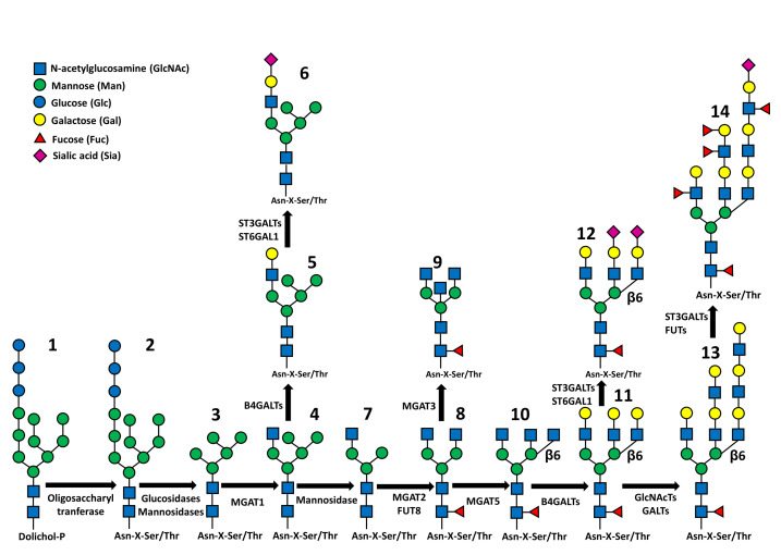
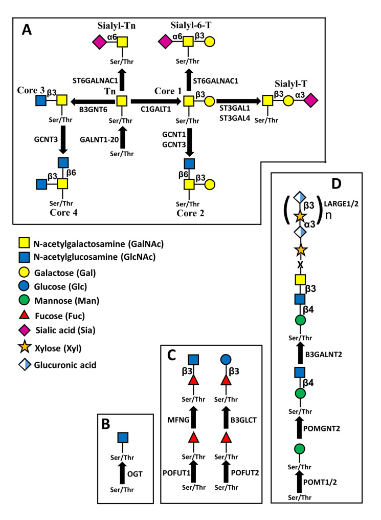
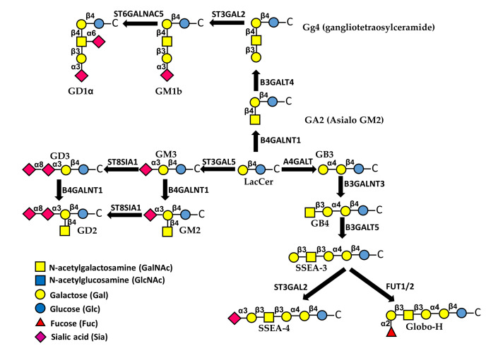
Glycosylation consists in the covalent, enzyme mediated, attachment of sugar chains to proteins and lipids. A large proportion of membrane and secreted proteins are indeed glycoproteins, while glycolipids are fundamental component of cell membranes. The biosynthesis of sugar chains is mediated by glycosyltransferases, whose level of expression represents a major factor of regulation of the glycosylation process. In cancer, glycosylation undergoes profound changes, which often contribute to invasion and metastasis. Epithelial to mesenchymal transition (EMT) is a key step in metastasis formation and is intimately associated with glycosylation changes. Numerous carbohydrate structures undergo up- or down-regulation during EMT and often regulate the process. In this review, we will discuss the relationship with EMT of the N-glycans, of the different types of O-glycans, including the classical mucin-type, O-GlcNAc, O-linked fucose, O-linked mannose and of glycolipids. Finally, we will discuss the role in EMT of galectins, a major class of mammalian galactoside-binding lectins. While the expression of specific carbohydrate structures can be used as a marker of EMT and of the propensity to migrate, the manipulation of the glycosylation machinery offers new perspectives for cancer treatment through inhibition of EMT.

Keywords: carbohydrate antigens; galectins; glycolipids; glycosylation; glycosyltransferases.


Conflict of interest statement

The authors declare no conflict of interest.

Figures

Figure 1
Figure 1
Synthetic representation of N-glycan biosynthesis. Structures 1 and 2: the Glc3Man9GlcNAc2 structure attached to dolichol phosphate (1) or to polypeptide (2). Structure 3: Man5GlcNAc2 structure product of the trimming of 4 Man and 3 Glc residues. Structure 4: addition of the first outer β1,2-linked GlcNAc. Structures 5 and 6: hybrid type structures in which 5 Man residues coexist with a fully processed branch. Structure 7: trimming of the last 2 Man residues. Structure 8: addition of the second outer β1,2-linked GlcNAc and of “core-linked” fucose. Structure 9: addition of the “bisecting” β1,4-linked GlcNAc. Structure 10: addition of the β1,6-linked GlcNAc. Structure 11: addition of β1,4-linked Gals. Structure 12: addition of some Sia residues either α2,3 or α2,6-linked. Structure 13: Elongation of the branches by sequential action of GlcNAcTs and GalTs. Structure 14: addition of sialic acids and external fucose residues. The fucosylated structure present on the left branch could be either the Lea or the Lex structure, depending on the Fuc linkage to GlcNAc (α1,4 for Lea; α1,3 for Lex). The fucosylated structure present on the internal branch could be either the Leb or the Ley structure, depending on the Fuc linkage to GlcNAc as above. The β1,6-linked branch on the right is terminated by a sialylated Lewisa or Lewisx antigen.
Figure 2
Figure 2
Structures of different types of O-linked chains. (A) structure and biosynthesis of mucin-type O-linked chains. Four “core” structures are shown, which can be elongated by other sugars. (B) Addition of O-linked GlcNAc, mediated by O-GlcNAc transferase. Removal of the sugar is catalyzed by O-GlcNAc ase (not shown). (C) O-fucosylation. The O-linked fucose mounted on EGF repeats by POFUT1 can be elongated by β3GlcNAc through manic fringe (MFNG) and also by β4Gal and α6Sia (not shown). The O-linked fucose mounted on TSRs repeats by POFUT2 can be elongated by β3Glc (D) O-mannosylation. The structure shown is referred to as M3 and can be elongated by the repeated units of xylose and glucuronic acid.
Figure 3
Figure 3
Biosynthesis and structure of major glycolipids. The first sugar attached to ceramide (C) is always Glc, followed by a β1,4-linked Gal. This basic structure can be elongated through different pathways. Sialylated glycolipids are referred to as gangliosides.

References

    1. Gomes Ferreira I., Pucci M., Venturi G., Malagolini N., Chiricolo M., Dall’Olio F. Glycosylation as a Main Regulator of Growth and Death Factor Receptors Signaling. Int. J. Mol. Sci. 2018;19:580. doi: 10.3390/ijms19020580. - DOI - PMC - PubMed
    1. Bellis S.L. Variant glycosylation: An underappreciated regulatory mechanism for b1 integrins. Biochim. Biophys. Acta. 2004;1663:52–60. doi: 10.1016/j.bbamem.2004.03.012. - DOI - PubMed
    1. Dall’Olio F., Vanhooren V., Chen C.C., Slagboom P.E., Wuhrer M., Franceschi C. N-glycomic biomarkers of biological aging and longevity: A link with inflammaging. Ageing Res. Rev. 2013;12:685–698. doi: 10.1016/j.arr.2012.02.002. - DOI - PubMed
    1. Takahashi M., Kizuka Y., Ohtsubo K., Gu J., Taniguchi N. Disease-associated glycans on cell surface proteins. Mol. Asp. Med. 2016;51:56–70. doi: 10.1016/j.mam.2016.04.008. - DOI - PubMed
    1. Varki A. Biological roles of glycans. Glycobiology. 2017;27:3–49. doi: 10.1093/glycob/cww086. - DOI - PMC - PubMed

LinkOut - more resources