. 2021 Jul 8;9(7):792.
doi: 10.3390/biomedicines9070792.

Occluded Superior Mesenteric Artery and Vein. Therapy with the Stable Gastric Pentadecapeptide BPC 157

Affiliations

Occluded Superior Mesenteric Artery and Vein. Therapy with the Stable Gastric Pentadecapeptide BPC 157

Mario Knezevic et al. Biomedicines. .

Abstract

Background: We investigated the occluded essential vessel tributaries, both arterial and venous, occluded superior mesenteric vein and artery in rats, consequent noxious syndrome, peripherally and centrally. As therapy, we hypothesized the rapidly activated alternative bypassing pathways, arterial and venous, and the stable gastric pentadecapeptide BPC 157 since it rapidly alleviated venous occlusion syndromes.

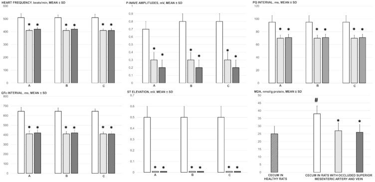
Methods: Assessments were performed for 30 min (gross recording, venography, ECG, pressure, microscopy, biochemistry, and oxidative stress), including portal hypertension, caval hypertension, aortal hypotension, and centrally, the superior sagittal sinus hypertension; systemic arterial and venous thrombosis, ECG disturbances, MDA-tissue increase, the multiple organs lesions, heart, lung, liver, kidney and gastrointestinal tract, including brain (swelling, and cortex (cerebral, cerebellar), hypothalamus/thalamus, hippocampus lesions). Rats received BPC 157 medication (10 µg/kg, 10 ng/kg) intraperitoneally at 1 min ligation-time.

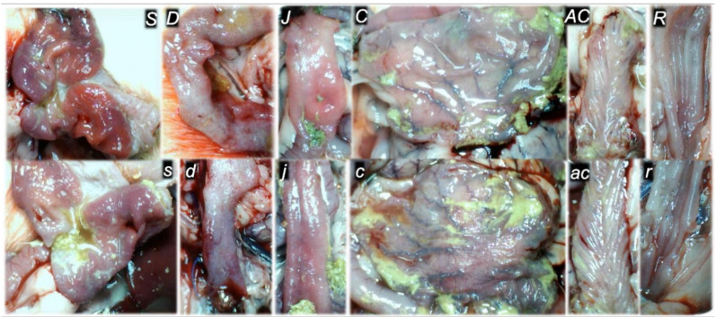
Results: BPC 157 rapidly activated collateral pathways. These collateral loops were the superior mesenteric vein-inferior anterior pancreaticoduodenal vein-superior anterior pancreaticoduodenal vein-pyloric vein-portal vein pathway, an alternative pathway toward inferior caval vein via the united middle colic vein and inferior mesenteric vein through the left colic vein, and the inferior anterior pancreaticoduodenal artery and inferior mesenteric artery. Consequently, BPC 157 counteracted the superior sagittal sinus, portal and caval hypertension, aortal hypotension, progressing venous and arterial thrombosis peripherally and centrally, ECG disturbances attenuated. Markedly, the multiple organs lesions, heart, lung, liver, kidney, and gastrointestinal tract, in particular, as well as brain lesions, and oxidative stress in tissues were attenuated.

Conclusions: BPC 157 therapy rapidly recovered rats, which have complete occlusion of the superior mesenteric vein and artery.

Keywords: BPC 157; rats; superior mesenteric vein and artery occlusion; vascular recruitment.


Conflict of interest statement

The authors declare that there are no conflict of interest.

Figures

Figure 1
Figure 1
Blood pressure, mm Hg (in the superior sagittal sinus (SSS), portal vein (PV), abdominal aorta (AA), inferior caval vein (ICV)), thrombus mass, g (in the superior sagittal sinus (SSS), portal vein (PV), inferior caval vein (ICV), superior mesenteric vein (SMV), external jugular vein (EJV), hepatic veins (HV), abdominal aorta (AA) and superior mesenteric artery (SMA)), relative volume of arteries (superior mesenteric artery (SMA), proximal to occlusion and distal to occlusion, inferior mesenteric artery (IMA), abdominal aorta (AA) and brain) and relative volume of veins (portal vein (PV), pyloric vein (PyV), inferior caval vein (ICV), inferior anterior pancreaticoduodenal vein (IAPDV), superior mesenteric vein (SMV), inferior mesenteric vein (IMV), middle colic vein (MDV) and left colic vein (LCV)) presentation in the rats with ligated superior mesenteric vein and artery at the 30 min ligation-time, following medication (BPC 157 10 µg/kg (light gray bars), 10 ng/kg (dark gray bars); saline 5 mL/kg (white bars) given intraperitoneally. Especially, as described previously [5,6,7], pressure recordings were made in deeply anesthetised rats with a cannula (BD Neoflon™ Cannula, Franklin Lakes, NJ, USA) connected to a pressure transducer (78534C MONITOR/TERMINAL; Hewlett Packard, Houston, TX, USA) inserted into the portal vein, inferior caval vein and superior sagittal sinus, and abdominal aorta at the level of the bifurcation at 30 min post-ligation after 5 min of recording (see, Section 2.5). Six rats/group/interval. Mean ± SD, * p ˂ 0.05, at least vs. control.
Figure 2
Figure 2
ECG changes (heart frequency (beats/min), P-wave amplitude (mV), PQ interval (ms), QTc interval (ms), ST-elevation (mV)) at 5 min (A), 15 min (B) and 30 min (C) ligation-time, oxidative stress (MDA, nmol/g protein), in the rats with ligated superior mesenteric vein and artery at 30 min ligation-time following medication (BPC 157 10 µg/kg (light gray bars), 10 ng/kg (dark gray bars); saline 5 mL/kg (white bars) given intraperitoneally. Six rats/group/interval. Mean ± SD, * p ˂ 0.05, at least vs. control, # p ˂ 0.05, at least vs. healthy (dashed bar).
Figure 3
Figure 3
Gross gastrointestinal lesions presentation (sum of the longest lesions diameters, mm, mean ± SD), gastrointestinal lesions microscopy (scoring (0–15, Min/Med/Max), organs (lung, liver, kidney, and heart) lesions microscopy (scoring (0–3, Min/Med/Max), neuropathologic scoring (cerebral cortex, cerebellar cortex, hypothalamus, hippocampus, 0–8), enzymes serum values (IU/L, mean ± SD), liver and spleen relative weight (% of total body weight, mean ± SD) at 30 min ligation-time following medication (BPC 157 10 µg/kg (light gray bars), 10 ng/kg (dark gray bars); saline 5 mL/kg (white bars) given intraperitoneally. Six rats/group/interval. * p ˂ 0.05, at least vs. control.
Figure 4
Figure 4
Serosal disturbances (hemorrhage, congestion, arterial filling, ramification), scored 0–4, Min/Med/Max, at 5 min, 15 min and 30 min ligation-time in rats with occluded superior mesenteric vein and artery following medication (BPC 157 10 µg/kg (light gray bars), 10 ng/kg (dark gray bars); saline 5 mL/kg (white bars) given intraperitoneally. Six rats/group/interval. * p ˂ 0.05, at least vs. control.
Figure 5
Figure 5
Superior mesenteric vein (below ligation) venography, and veins presentation (jejunal branches of superior mesenteric vein (1), ileal branches of superior mesenteric vein (2), superior mesenteric vein (3), ileocolic/right colic vein truncus (4), ileocolic vein (5), right colic vein (6), inferior anterior pancreaticoduodenal vein (7), superior anterior pancreaticoduodenal vein (8), pyloric vein (9), portal vein (10), middle colic vein (11)) in rats with the occlusion of the superior mesenteric vein and artery immediately following medication (BPC 157 10 µg/kg (upper), or 10 ng/kg (low) (right) or saline 5 mL/kg (left)) given intraperitoneally at 15 min ligation-time.
Figure 6
Figure 6
Inferior caval vein venography, and vessels presentation (abdominal aorta (1), common iliac artery (2), renal artery (3), superior mesenteric artery distal to occlusion (4), jejunal branches of superior mesenteric artery (5), ileal branches of superior mesenteric artery (6), ileocolic-right colic truncus (7), middle colic artery (8), ileocolic artery (9), caudal epigastric artery (10), pancreaticoduodenal artery (11), coeliac truncus (12), superior mesenteric artery proximal to occlusion (13), internal iliac artery (14)) in rats with the occlusion of the superior mesenteric vein and artery immediately following medication (BPC 157 10 µg/kg (upper), or 10 ng/kg (low) (right) or saline 5 mL/kg (left)) given intraperitoneally at 15 min ligation-time.
Figure 7
Figure 7
Illustrative gross vessel presentation in the rats with the occluded superior mesenteric vein and artery, without therapy (low, control) and BPC 157 therapy (upper, 10 ng/kg, providing the same presentation with the 10 µg/kg). Left. Pyloric vein (dashed arrow (black (BPC 157), white (control)) inflow into the portal vein (full arrow (black (BPC 157), white (control)), proximal to occlusion of the end of the superior mesenteric vein (red arrow). Reestablished blood flow (upper, BPC 157), congestion, and vascular failure (low, control). Middle. Superior mesenteric artery (full arrow (black (BPC 157), white (control)) inflow into the portal vein (full arrow (black (BPC 157), white (control)), proximal to occlusion and distal from occlusion (red arrow). Congested and tortuous vein of the ascending colon (white dashed arrow). Dashed black arrows indicate collaterals (big dashed arrow (medial colic artery), small dashed arrow (inferior anterior pancreaticoduodenal artery). Reestablished blood flow to part of superior mesenteric artery distal from occlusion filled with blood (upper, BPC 157), vascular failure, and empty superior mesenteric artery (low, control). Right. Inferior caval vein and abdominal aorta with presentation close to normal (black arrows (full (inferior caval vein), dashed (abdominal aorta), and congested inferior caval vein (full white arrow), and tinny empty abdominal aorta (dashed white arrow). Camera attached to a VMS-004 Discovery Deluxe USB microscope (Veho, Dayton, OH, USA).
Figure 8
Figure 8
Illustrative gross vessel presentation in the rats with the occluded superior mesenteric vein and artery, without therapy (low, control) and BPC 157 therapy (upper, 10 ng/kg, providing the same presentation with the 10 µg/kg). Left. Inferior anterior pancreaticoduodenal vein (dashed arrow (black (BPC 157), white (control)) with prominent duodenal congestion (low, control). Reestablished blood flow (upper, BPC 157), congestion, and vascular failure (low, control). Middle. Superior mesenteric vein (red dashed arrows), inferior mesenteric vein (full red arrows), middle colic vein (yellow arrows), left colic vein (black (BPC 157), white (control) distal from occlusion. Tinny veins of the transverse colon (low, control), and exaggerated veins indicating activated collaterals (BPC 157) as a part of the reestablished blood flow to compensate for occlusion (upper, BPC 157). Right. Inferior mesenteric artery and abdominal aorta with presentation close to normal (black arrows (BPC 157), or tinny empty inferior mesenteric artery and abdominal aorta (low, control). Tortuous veins presentation of the rectal veins (control, low). Camera attached to a VMS-004 Discovery Deluxe USB microscope (Veho, Dayton, OH, USA).
Figure 9
Figure 9
Illustrative brain presentation in the rats with the occluded superior mesenteric vein and artery, in the calvarial window immediately after vessels occlusion, and subsequent medication saline (5 mL/kg ip) (low, control) or BPC 157 (10 ng/kg ip) (upper)) (left) and after sacrifice (right), at the 30 min ligation-time. Saline (5 mL/kg ip) (low, control) or BPC 157 (10 ng/kg ip) (upper). Prominent brain swelling in control rats (low), completely reversed in BPC 157 rats (upper). Camera attached to a VMS-004 Discovery Deluxe USB microscope (Veho, Dayton, OH, USA).
Figure 10
Figure 10
At 30 min ligation-time, gross presentation of the mucosa of the stomach (s,S), duodenum (d,D), jejunum (j,J), cecum (c,C), ascending colon (ac,AC), and rectum (r,R), in the rats with the occluded superior mesenteric vein and artery, in the rats that received saline medication (5 mL/kg ip) (control, low, small letters) or BPC 157 medication (10 ng/kg ip) (upper, capitals). Camera attached to a VMS-004 Discovery Deluxe USB microscope (Veho, Dayton, OH, USA).
Figure 11
Figure 11
At 30 min ligation-time, microscopy presentation of the congestion of the stomach (a,A) and small intestine mucosa (b,B) and rectum (c,C) and perirectal vascular plexus (d,D) (HE, ×100) in the rats with occluded superior mesenteric vein and artery, controls (small letters) and BPC 157 treated (capitals). Control rats with occluded superior mesenteric vein and artery exhibited the congestion and dilatation of the small vessels in the stomach (a) and small intestine mucosa (b), congestion and dilatation of the small vessels of the rectum wall (c) along with congestion of perirectal blood vessels (d). Contrarily, BPC 157 treated rats showed no changes in the stomach (A) and small intestine mucosa (B) and no changes in the rectum (C) and perirectal vascular plexus (D).
Figure 12
Figure 12
At 30 min ligation-time, microscopy presentation of the congestion of the heart (a,A) (HE, ×200), lung (b,B) (HE, ×100), liver (c,C) (HE, ×100), and the renal parenchyma (d,D) (HE, ×100) in the rats with occluded superior mesenteric vein and artery, controls (small letters) and BPC 157 treated (capitals). Control rats with occluded superior mesenteric vein and artery exhibited the subendocardial infarct as well as congestion within the myocardium (a) and lung septa along with intra-alveolar hemorrhage (b), liver congestion and dilatation of sinusoids, portal vein, and central veins (c) along with congestion of renal parenchyma and glomeruli (d). BPC 157 treated rats showed no heart congestion and heart infarct (A) along with minimal lung congestion and intra-alveolar hemorrhage (B) and no changes in the liver (C) and renal parenchyma (D).
Figure 13
Figure 13
At 30 min ligation-time, microscopy presentation of the neuropathologic changes in the cerebral cortex (a,A (HE, ×200), b,B (HE, ×400)) and cerebellar cortex (c,C (HE, ×100), d,D (HE, ×400)) areas of the rats with occluded superior mesenteric vein and artery, controls (small letters) and BPC 157 treated (capitals). Cerebral cortex (a,A,b,B). While control rats with occluded superior mesenteric vein and artery exhibited marked edema and congestion (a), BPC 157 treated rats presented only mild edema (A). A scant karyopyknosis presented control rats with occluded superior mesenteric vein and artery in the cerebral cortex (arrows) (b) while no changes appeared in BPC 157 treated rats (B). Cerebellar cortex (c,C,d,D). Control rats with occluded superior mesenteric vein and artery exhibited subarachnoid hemorrhage at the base of the brain in the cerebellar area (c) and multiple karyopyknosis of Purkinje cells of the cerebellar cortex (arrows) (d), unlike BPC 157 treated rats which remained without change (C,D).
Figure 14
Figure 14
At 30 min ligation-time, microscopy presentation of the neuropathologic changes in the hypothalamus (a,A (HE, ×100), b,B (HE, ×200)) and hippocampus (c,C (HE, ×200)) in the rats with occluded superior mesenteric vein and artery, controls (small letters) and BPC 157 treated (capitals). Control rats with occluded superior mesenteric vein and artery exhibited in the hypothalamus and hippocampus edema (a,c) and karyopyknosis of the hypothalamic (arrows) (b) and hippocampal (arrows) (c) cells; only minimal hypothalamic edema in BPC 157 (A) and no karyopyknosis of the hypothalamic cells (B), and no changes in the hippocampus (C).

References

    1. Amaravathi U., Balamurugan N., Muthu Pillai V., Ayyan S.M. Superior mesenteric arterial and venous thrombosis in COVID-19. J. Emerg. Med. 2021;60:e103–e107. doi: 10.1016/j.jemermed.2020.12.016. - DOI - PMC - PubMed
    1. Oldenburg W.A., Lau L.L., Rodenberg T.J., Edmonds H.J., Burger C.D. Acute mesenteric ischemia: A clinical review. Arch. Intern. Med. 2004;164:1054–1062. doi: 10.1001/archinte.164.10.1054. - DOI - PubMed
    1. Sikiric P., Rucman R., Turkovic B., Sever M., Klicek R., Radic B., Drmic D., Stupnisek M., Misic M., Vuletic L.B., et al. Novel cytoprotective mediator, stable gastric pentadecapeptide BPC 157. Vascular recruitment and gastrointestinal tract healing. Curr. Pharm. Des. 2018;24:1990–2001. doi: 10.2174/1381612824666180608101119. - DOI - PubMed
    1. Sikiric P., Hahm K.B., Blagaic A.B., Tvrdeic A., Pavlov K.H., Petrovic A., Kokot A., Gojkovic S., Krezic I., Drmic D., et al. Stable Gastric Pentadecapeptide BPC 157, Robert’s stomach cytoprotection/adaptive cytoprotection/organoprotection, and Selye’s stress coping response: Progress, achievements, and the future. Gut Liver. 2020;14:153–167. doi: 10.5009/gnl18490. - DOI - PMC - PubMed
    1. Vukojevic J., Siroglavic M., Kasnik K., Kralj T., Stancic D., Kokot A., Kolaric D., Drmic D., Sever A.Z., Barisic I., et al. Rat inferior caval vein (ICV) ligature and particular new insights with the stable gastric pentadecapeptide BPC 157. Vascul. Pharmacol. 2018;106:54–66. doi: 10.1016/j.vph.2018.02.010. - DOI - PubMed