Review
. 2021 Jul 6;57(7):692.
doi: 10.3390/medicina57070692.

High-Resolution CT Findings of Myositis-Related Interstitial Lung Disease

Affiliations
Review

High-Resolution CT Findings of Myositis-Related Interstitial Lung Disease

Ryoko Egashira. Medicina (Kaunas). .

Abstract

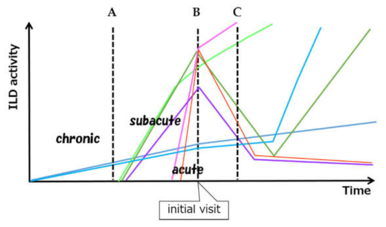
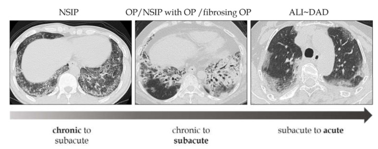
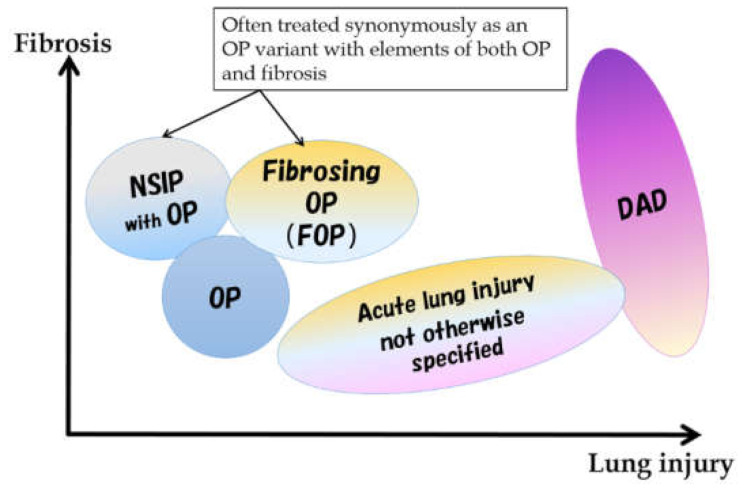
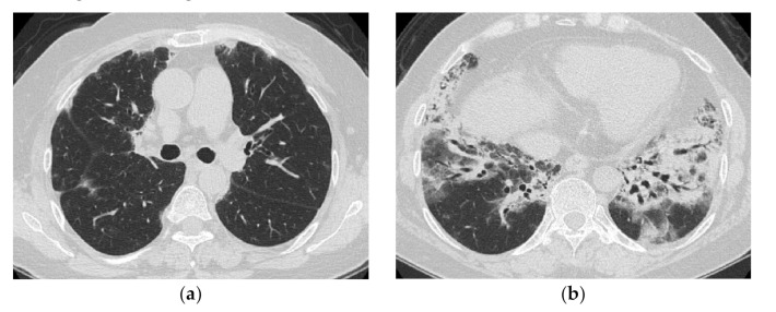
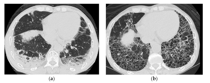
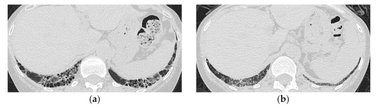
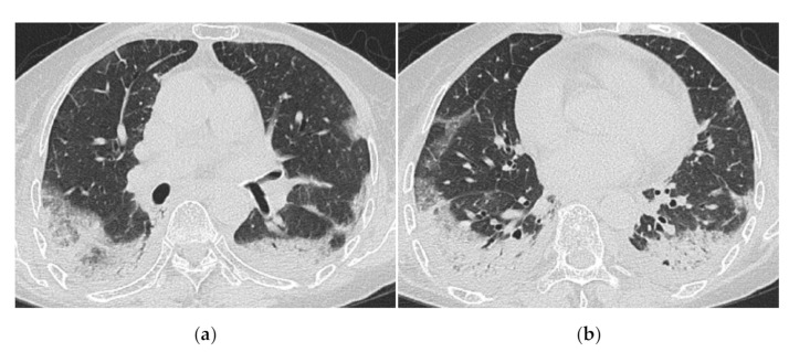
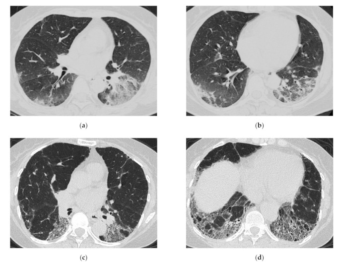
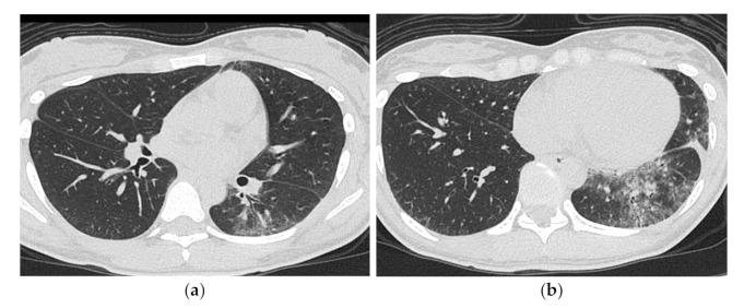
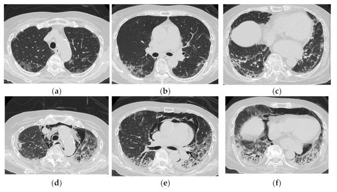
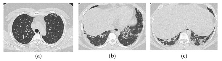
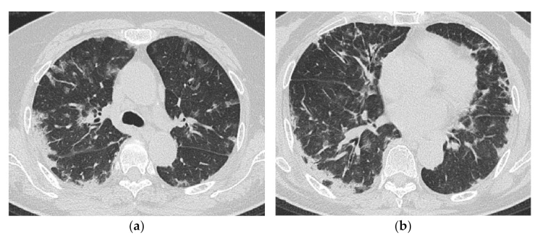
Myositis-related interstitial lung disease presents with a wide variety of lesions, ranging from chronic to acute. It can be divided into two main forms by the types of onsets, namely, chronic to subacute type showing nonspecific interstitial pneumonia (NSIP) or NSIP with an organizing pneumonia (OP)/fibrosing OP (FOP) pattern and acute type showing acute lung injury (ALI) to diffuse alveolar damage (DAD) pattern. Anti-aminoacyl tRNA Synthetase antibody-positive cases mainly show an NSIP or FOP pattern, whereas anti-melanoma differentiation-associated gene 5 antibody-positive cases show ALI to DAD pattern. Bilateral consolidation with or without ground-glass opacification with lower lobe predominance is common as a major pattern in all types, but the distribution or extent is sometimes different. The early detection of findings that indicate a rapid progressive course is vital. Diffuse cranio-caudal distribution and multiple ground-glass opacifications with random distribution might indicate a poorer prognosis.

Keywords: anti- synthetase syndrome; high-resolution CT; interstitial lung disease; interstitial pneumonia; myositis-specific antibody; polymyositis/dermatomyositis.


Conflict of interest statement

The author declares no conflict of interest.

Figures

Figure 1
Figure 1
Schematic view of considerable variation of ILD activity and clinical course. A, B and C mean the time of the diagnosis of myositis. A: Subclinical onset of chronic type of ILD found at the time of the diagnosis of myositis. B: Simultaneous onset and diagnosis of acute/subacute type of ILD C: Preceding ILD and delayed onset of myositis. ILD can develop subclinically, can relapse after remission. (ILD: interstitial lung disease).
Figure 2
Figure 2
Relationship between morphologic patterns and the types of onset. The phenotypes of these lesions are difficult to distinguish clearly, given that varying degrees of inflammation and lung injury exist on the same spectrum. NSIP: nonspecific interstitial pneumonia, OP: organizing pneumonia, ALI: acute lung injury, DAD: diffuse alveolar damage.
Figure 3
Figure 3
Spectrum of morphological patterns in subacute to acute pathologies. NSIP: nonspecific interstitial pneumonia, OP: organizing pneumonia, DAD: diffuse alveolar damage.
Figure 4
Figure 4
A 50-year-old woman with subacute onset interstitial lung disease associated with dermatomyositis. There is a bilateral basal consolidation extending along the bronchovascular bundles with mild ground-glass opacification and mild traction bronchiectasis within (b). Patchy subpleural consolidation is also seen at the level of the pulmonary artery trunk (a).
Figure 5
Figure 5
A 51-year-old man with subacute onset interstitial lung disease associated with polymyositis. At the time of initial diagnosis (a), there was a subpleural dense consolidation to ground-glass opacification, accompanied by a decreased lower lobe volume. Then, 13 years later (b), the lesion became more extensive, and the areas of consolidation had changed as an area with reticular and small cystic lesion, some of which resembled a honeycomb lung.
Figure 6
Figure 6
A 59-year-old woman with dermatomyositis under long-term follow-up. The most recent high-resolution computed tomography (HRCT) (a) shows bilateral honeycomb-like cystic lesion just below the pleura, and a review of the HRCT scan at the time of initial presentation 17 years ago (b) shows that the same area was previously ground-glass opacification with mild reticulation showing NSIP pattern.
Figure 7
Figure 7
A 70-year-old woman with rapidly progressive interstitial lung disease associated with clinically amyopathic dermatomyositis (antibody unknown). HRCT images (a,b) show diffuse ground-glass opacification and consolidation just below the pleura. Crazy-paving appearance, interlobular septal thickening and bilateral pleural effusions are also seen. The patient died 2 weeks after admission.
Figure 8
Figure 8
A 64-year-old woman with subacute onset interstitial lung disease associated with polymyositis. At the time of initial diagnosis (a,b), there was bilateral consolidation to ground-glass opacification with lower and peribronchovascular predominance. Then, 13 years later (c,d), the lesion became less extensive, and the lesion had changed into reticulation with traction bronchiectasis. Decreased lung volume of the lower lobes was visible.
Figure 9
Figure 9
A 30-year-old man with nonspecific interstitial pneumonia pattern associated with dermatomyositis (anti-ARS Ab positive). HRCT images (a,b) shows bilateral ground-glass opacification and reticulation with peribronchovascular predominance. Mild traction bronchiectasis and decreased volume in the left lower lobe was also seen.
Figure 10
Figure 10
An 83-year-old woman with dermatomyositis related interstitial lung disease (anti-MDA5 Ab positive). At the time of initial diagnosis (ac), there was diffuse ground-glass opacification and consolidation with basal and subpleural predominance. Subpleural parenchymal band-like opacity was also seen. One month later (df), the lesion became more extensive, and increase of reticulation with traction bronchiectasis, lower volume loss was visible. Note the severe mediastinal emphysema.
Figure 11
Figure 11
A 19-year-old man with interstitial lung disease associated with dermatomyositis (anti-MDA5 Ab positive). On HRCT (ac), bilateral subpleural patchy consolidation are seen with basal predominance. Peribronchovascular component is not predominant. The onset was subacute, but the clinical course was stable.
Figure 12
Figure 12
A 69-year-old woman with rapidly progressive interstitial lung disease associated with dermatomyositis (antibody unknown). On HRCT (a,b), bilateral diffuse patchy ground-glass opacification and consolidation are seen in both lungs. The distribution of the lesions is unbiased both in the vertical and horizontal directions. A transbronchial lung biopsy specimen showed diffuse edematous thickening of the alveolar septa and fibrin deposition, which was considered to correspond to acute lung injury (not shown). The patient died three weeks after admission.

References

    1. Yousem S., Gibson K., Kaminski N., Oddis C.V., Ascherman D.P. The pulmonary histopathologic manifestations of the anti-Jo-1 tRNA synthetase syndrome. Mod. Pathol. 2010;23:874–880. doi: 10.1038/modpathol.2010.65. - DOI - PubMed
    1. Tazelaar H.D., Viggiano R.W., Pickersgill J., Colby T.V. Interstitial lung disease in polymyositis and dermatomyositis. Clinical features and prognosis as correlated with histologic findings. Am. Rev. Respir. Dis. 1990;141:727–733. doi: 10.1164/ajrccm/141.3.727. - DOI - PubMed
    1. Travis W.D., Costabel U., Hansell D.M., King T.E., Jr., Lynch D.A., Nicholson A.G., Ryerson C.J., Ryu J.H., Selman M., Wells A.U., et al. An official American Thoracic Society/European Respiratory Society statement: Update of the international multidisciplinary classification of the idiopathic interstitial pneumonias. Am. J. Respir. Crit. Care Med. 2013;188:733–748. doi: 10.1164/rccm.201308-1483ST. - DOI - PMC - PubMed
    1. Beardsley B., Rassl D. Fibrosing organising pneumonia. J. Clin. Pathol. 2013;66:875–881. doi: 10.1136/jclinpath-2012-201342. - DOI - PubMed
    1. Fujisawa T., Suda T., Nakamura Y., Enomoto N., Ide K., Toyoshima M., Uchiyama H., Tamura R., Ida M., Yagi T., et al. Differences in clinical features and prognosis of interstitial lung diseases between polymyositis and dermatomyositis. J. Rheumatol. 2005;32:58–64. - PubMed

MeSH terms

Substances