. 2021 Jul 4;10(7):843.
doi: 10.3390/pathogens10070843.

Melinacidin-Producing Acrostalagmus luteoalbus, a Major Constituent of Mixed Mycobiota Contaminating Insulation Material in an Outdoor Wall

Affiliations

Melinacidin-Producing Acrostalagmus luteoalbus, a Major Constituent of Mixed Mycobiota Contaminating Insulation Material in an Outdoor Wall

Aino Maria A Andersson et al. Pathogens. .

Abstract

Occupants may complain about indoor air quality in closed spaces where the officially approved standard methods for indoor air quality risk assessment fail to reveal the cause of the problem. This study describes a rare genus not previously detected in Finnish buildings, Acrostalagmus, and its species A. luteoalbus as the major constituents of the mixed microbiota in the wet cork liner from an outdoor wall. Representatives of the genus were also present in the settled dust in offices where occupants suffered from symptoms related to the indoor air. One strain, POB8, was identified as A. luteoalbus by ITS sequencing. The strain produced the immunosuppressive and cytotoxic melinacidins II, III, and IV, as evidenced by mass spectrometry analysis. In addition, the classical toxigenic species indicating water damage, mycoparasitic Trichoderma, Aspergillus section Versicolores, Aspergillus section Circumdati, Aspergillus section Nigri, and Chaetomium spp., were detected in the wet outdoor wall and settled dust from the problematic rooms. The offices exhibited no visible signs of microbial growth, and the airborne load of microbial conidia was too low to explain the reported symptoms. In conclusion, we suggest the possible migration of microbial bioactive metabolites from the wet outdoor wall into indoor spaces as a plausible explanation for the reported complaints.

Keywords: Acrostalagmus luteoalbus; cork liner; guttation droplets; indoor dust; melinacidin; mixed mycobiota; outdoor wall.


Conflict of interest statement

The authors declare no conflict of interest. The funders had no role in the design of the study; in the collection, analyses, or interpretation of data; in the writing of the manuscript; or in the decision to publish the results.

Figures

Figure 1
Figure 1
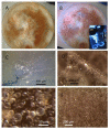
Stereomicrographs of major fungi colonizing moist cork liner. (A,a) Aspergillus-like conidiophores; (B,b) ascomata characteristic for Chaetomium-like fungi; (C,c) mycoparasitic Trichoderma-like colonies; and (D,d) a dominant colonizer, a fungus with straight erect repeatedly branched orange conidiophores, 100 × 4–4.5 μm.
Figure 2
Figure 2
Pieces of moist cork liner and mineral wool cultivated on malt extract agar for 14 d at 22 °C. (AC,G,H); dominant colonies from cork liner (samples 1P61,1POB,1K). (DF) Major colonies from mineral wool (sample 3MW). Origin of isolates characterized/identified are marked with red arrows and representative strain codes. (K) Colonies of white, spore-forming actinobacteria; (L) unidentified toxigenic and antagonistic fungicidic colony type appearing on this plate only.
Figure 3
Figure 3
Settled dust collected from six rooms cultivated on MEA for 14 d. Origin of characterized/identified isolates are marked with red arrows and representative strain codes. (AG) Major fungal colonies from dust collected from problematic rooms. (H,I) Dust cultivated from nonproblematic rooms in ordinary use.
Figure 4
Figure 4
Acrostalagmus luteoalbus strain POB8 cultivated on malt extract agar for 10 d. (A) Pure-cultured strain in visible light. (B) Same plate exhibiting blue fluorescing exudates when excited with UV light (insert). (CF) Stereomicroscopic views of plate in (A): (C,D) Exudates emitted from fungal hyphae. (E,F) Liberated vesicles.
Figure 5
Figure 5
MS spectra of compounds of Acrostalagmus luteoalbus POB8 ethanol extract. (A) TIC of three compounds at retention times of 8.4 (1), 12.2 (2), and 17.8 min (3). (BD) MS spectra of compounds 1, 2, and 3 had protonated mass ions at m/z 729.2, 713.2, and 697.2; sodiated mass ions at m/z 751.2, 735.2, and 719.2; and sodiated mass ions of dimers at m/z 1479.0, 1447.0, and 1415.0, respectively.
Figure 6
Figure 6
MS/MS spectra of compounds of A. luteoalbus POB8 ethanol extract: (A) compound 1 precursor ion m/z at 729.7; (B) compound 2 precursor ion m/z at 713.8; (C) compound 3 precursor ion m/z at 697.8.
Figure 7
Figure 7
Floor plan of the public building investigated for mold growth. Samples of settled indoor dust and pieces of cork liner used as insulation inside plinth and mineral wool inside the outer brick wall were collected from selected rooms. The first number (1 to 3) in the sample code indicates the floor (first, second, third floor). Rooms 131a, 131b, and 335 were abandoned in 2010. Occupants in room 145b complained about indoor air and reported symptoms. Occupants in room 146 did not report symptoms until 2014 but had an air cleaner installed in the room. Rooms 134 and 223 were nonproblematic and in ordinary use.
Figure 8
Figure 8
Sampling of building materials from water-damaged outdoor wall (upper panels), and micrographs of obtained samples (lower panels). Upper panels show (A) drilling of holes in plinth of outer wall; (B) used tools; and (C) obtained samples of wet cork liners stored in plastic bags. Lower panels show pieces of sampled wet cork liner with Hoechst 33342 nucleic acid stain + propidium iodide staining microbial structures containing DNA or RNA blue and red. (D) Degraded surface contaminated with microbes. (E,F) Growing actinobacterium and growing fungal hyphae.
Figure 9
Figure 9
Scheme illustrating experimental design for tracking diversity of major microbial constituents in building materials and dust. After three weeks of incubation, colonies on primary isolation plates (not yet single-spored) were numbered and screened for toxicity. Toxic colonies were streaked pure, characterized, and separated into morphotypes. Representatives of morphotypes were identified by ITS sequencing or by comparison with reference strains according to [106].

References

    1. Andersen B., Frisvad J.C., Dunn R.R., Thrane U. A pilot study on baseline fungi and moisture indicator fungi in Danish homes. J. Fungi. 2021;7:71. doi: 10.3390/jof7020071. - DOI - PMC - PubMed
    1. Shukla S., Budden K., Neal R., Hansbro P. Microbiome effects on immunity, health and disease in the lung. Clin. Translat. Immunol. 2017;6:e133. doi: 10.1038/cti.2017.6. - DOI - PMC - PubMed
    1. Horve P., Lloyd S., Mhuireach G., Dietz L., Fretz M., MacCrone G., Van Den Wymelenberg K., Ishaq S.L. Building upon current knowledge and techniques of indoor microbiology to construct the next era of theory into microorganisms, health, and the built environment. J. Expo. Sci. Environ. Epidemiol. 2020;30:219–235. doi: 10.1038/s41370-019-0157-y. - DOI - PMC - PubMed
    1. Leung M.H.Y., Lee P.K.H. The roles of the outdoors and occupants in contributing to a potential pan-microbiome of the built environment: A review. Microbiome. 2016;4:21. doi: 10.1186/s40168-016-0165-2. - DOI - PMC - PubMed
    1. Haahtela T., Alenius H., Lehtimäki J., Sinkkonen A., Fyhrquist N., Hyöty H., Ruokolainen L., Mäkelä M. Immunological resilience and biodiversity for prevention. Allergy. 2021 doi: 10.1111/all.14895. - DOI - PubMed

LinkOut - more resources