Review
. 2021 Jul 7;10(7):859.
doi: 10.3390/pathogens10070859.

From Jekyll to Hyde: The Yeast-Hyphal Transition of Candida albicans

Affiliations
Review

From Jekyll to Hyde: The Yeast-Hyphal Transition of Candida albicans

Eve Wai Ling Chow et al. Pathogens. .

Abstract

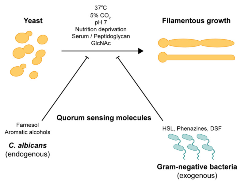
Candida albicans is a major fungal pathogen of humans, accounting for 15% of nosocomial infections with an estimated attributable mortality of 47%. C. albicans is usually a benign member of the human microbiome in healthy people. Under constant exposure to highly dynamic environmental cues in diverse host niches, C. albicans has successfully evolved to adapt to both commensal and pathogenic lifestyles. The ability of C. albicans to undergo a reversible morphological transition from yeast to filamentous forms is a well-established virulent trait. Over the past few decades, a significant amount of research has been carried out to understand the underlying regulatory mechanisms, signaling pathways, and transcription factors that govern the C. albicans yeast-to-hyphal transition. This review will summarize our current understanding of well-elucidated signal transduction pathways that activate C. albicans hyphal morphogenesis in response to various environmental cues and the cell cycle machinery involved in the subsequent regulation and maintenance of hyphal morphogenesis.

Keywords: cell cycle regulation; hyphal activation; hyphal morphogenesis; polymorphism; signal transduction pathways.


Conflict of interest statement

The authors declare no conflict of interest.

Figures

Figure 1
Figure 1
External hyphal-inducing signals. The yeast-to-hyphae transition in C. albicans can be triggered by various environmental cues such as high temperature (37 °C), high CO2 concentration (~5%), pH 7, nutrition deprivation, serum, peptidoglycan, N-acetylglucosamine, and inhibited by quorum-sensing molecules from endogenous and exogenous sources.
Figure 2
Figure 2
N-acetylglucosamine (GlcNAc) and amino acid-induced signal transduction pathways in C. albicans. Ngt1, localized in the plasma membrane, transports GlcNAc into the cell. However, when GlcNAc is present in high concentrations, it can enter the cell through diffusion. The main signal transduction pathway for GlcNAc-induced hyphal growth was initially thought to be the cAMP-PKA pathway. Recently, the transcription factors Ngs1 and Rep1, which are involved in GlcNAc catabolism, were found to stimulate hyphal growth via a cAMP-independent pathway. GlcNAc catabolism also increases the extracellular pH, which favors the hyphal growth via the alternate Rim101 pathway. Extracellular amino acids are detected via the plasma membrane-localized SPS (Ssy1-Ptr3-Ssy5) complex. The SPS-sensor activation leads to endoproteolytic processing at the nuclear exclusion domain of transcription factors Stp1 and Stp2. Processed Stp1 regulates the expression of secreted aspartyl proteinase (e.g., SAP2) and oligopeptide transporters (e.g., OPT1 and OPT3), while processed Stp2 regulates the expression of amino acid permeases (APPs).
Figure 3
Figure 3
Signal transduction pathways that govern hyphal growth in C. albicans. Activation of filamentous growth in C. albicans by various environmental cues and signal transduction pathways; the cAMP-PKA pathway, the Cek1-mediated pathway, the PKC pathway, and the embedded matrix.
Figure 4
Figure 4
Regulation of hyphal elongation requires mechanisms for initiation and long-term maintenance. Initiation of hypha growth requires transcription factors such as Efg1, Cph1, Czf1, and Flo8. Subsequent elongation process and maintenance require the involvement of Hgc1, Eed1, and Ume6. Both Eed1 and Ume6 are negatively regulated by Tup1 and Nrg1.
Figure 5
Figure 5
Schematic representation of polarized growth in C. albicans hyphal cells. Polarized growth is driven by Spitzenkörper, a vesicle supply center maintained at a fixed distance from the hyphal tip. Post-Golgi membrane-bound secretory vesicles are continuously delivered to the site of polarized growth. Secretory vesicles, tethered by the Rab-type GTPase Sec4 and the GEF Sec2, are transported to the hyphal tip via actin cables with the class V myosin Myo2 complexed to the regulatory light chain Mlc1, providing the motive force. The vesicles accumulate in Spitzenkörper before docking with the exocyst, which consists of Sec3, Sec5, Sec6, Sec8, Sec10, Sec15, Exo70, and Exo84 subunits, before fusing with the plasma membrane. Spa2, Bni1, and Bud6 coordinate the functions of the Spitzenkörper and the polarisome complex at the apical site of the hyphal tip. Endocytosis, endocytic recycling of polarity proteins, involves the cortical actin patches at the apical site of the hyphal tip. Actin patch organization and dynamics involve the actin cytoskeletal proteins Sla1 and Sla2, the actin skeleton-regulatory protein Pan1, and the Vpr1-Wal1-Myo5 complex, which activates the Arp2/3 complex. The landmark GTPase Rsr1, upon activation by its GEF Bud2, localizes Cdc24 to the site of tip growth, in addition to Ca2+ binding of the EF-hand motif in Cdc24.
Figure 6
Figure 6
C. albicans morphogenesis is tightly regulated by the cell cycle-associated cyclins and cyclin-dependent kinase (CDK). The essential CDK Cdc28 serves as the master regulator that controls the cell cycle progression at G1/S and G2/M phases by forming CDK complexes with specific cyclins. Levels of Cdc28 are relatively stable throughout the cell cycle and deplete during hyphal growth. In contrast, levels of the G1 and B-type mitotic cyclins oscillate during the cell cycle. G1 cyclins Cln3 and Ccn1 peak in the G1 phase and decline in the early G2 phase, while B-type mitotic cyclin Clb2 peaks in the early G2/M phase and declines in the M phase. Upon hyphal induction, Fkh2 is phosphorylated by Cln3-Cdc28 and Ccn1-Cdc28 complexes in a cell cycle-dependent manner to enhance the expression of hyphal-specific genes. The Hgc1-Cdc28 complex is essential for the maintenance of hyphal growth. The exocyst subunit Exo84 is phosphorylated by the Hgc1-Cdc28 complex for the regulation of polarized secretion. Phosphorylation of the septin subunit Cdc11 (by Ccn1-Cdc28 and Hgc1-Cdc28 complexes), GAP Rga2 (by the Hgc1-Cdc28 complex), and the polarisome protein Spa2 (by Hgc1-Cdc28 and Clb2-Cdc28 complexes) promote polarized growth. Rga2 is phosphorylated and inactivated by Hgc1-Cdc28, which relives the repression of the GTPase Cdc42. Phosphorylation of the transcription factor Efg1 and protein kinase Gin4 inhibit cell separation. Phosphorylated Efg1 binds to promoters of Ace2 target genes, inhibiting their transcription. Phosphorylated Gin4 modifies the dynamics of the septin ring by subsequent phosphorylation of Sep7 and deactivating the cell separation program via inhibition of the protein phosphatase Cdc14.

References

    1. Scully C., EI-Kabir M., Samaranayake L.P. Candida and Oral Candidosis: A Review. Crit. Rev. Oral Biol. Med. 1994;5:125–157. doi: 10.1177/10454411940050020101. - DOI - PubMed
    1. Achkar J.M., Fries B.C. Candida Infections of the Genitourinary Tract. Clin. Microbiol. Rev. 2010;23:253–273. doi: 10.1128/CMR.00076-09. - DOI - PMC - PubMed
    1. Rosenbach A., Dignard D., Pierce J.V., Whiteway M., Kumamoto C.A. Adaptations of Candida albicans for Growth in the Mammalian Intestinal Tract. Eukaryot. Cell. 2010;9:1075–1086. doi: 10.1128/EC.00034-10. - DOI - PMC - PubMed
    1. Kühbacher A., Burger-Kentischer A., Rupp S. Interaction of Candida Species with the Skin. Microorganisms. 2017;5:32. doi: 10.3390/microorganisms5020032. - DOI - PMC - PubMed
    1. Pappas P.G., Lionakis M.S., Arendrup M.C., Ostrosky-Zeichner L., Kullberg B.J. Invasive candidiasis. Nat. Rev. Dis. Primers. 2018;4:18026. doi: 10.1038/nrdp.2018.26. - DOI - PubMed

LinkOut - more resources