. 2021 Sep 2;81(17):3481-3495.e7.
doi: 10.1016/j.molcel.2021.07.019. Epub 2021 Aug 5.

Molecular basis for substrate recruitment to the PRMT5 methylosome

Affiliations

Molecular basis for substrate recruitment to the PRMT5 methylosome

Kathleen M Mulvaney et al. Mol Cell. .

Abstract

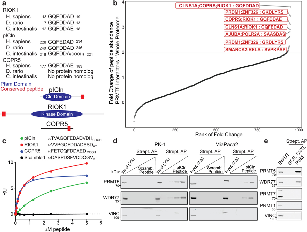
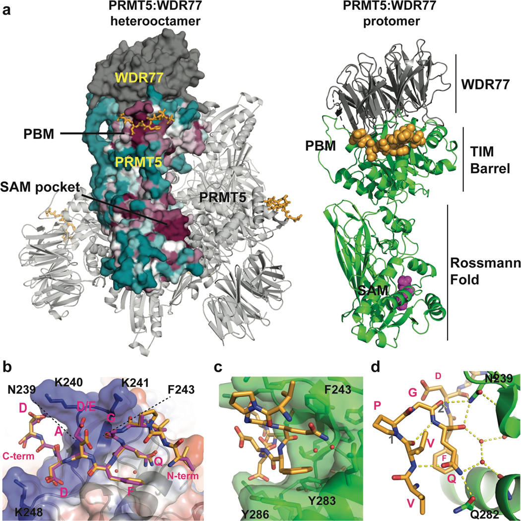
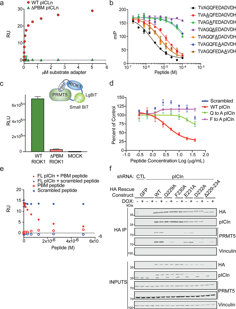
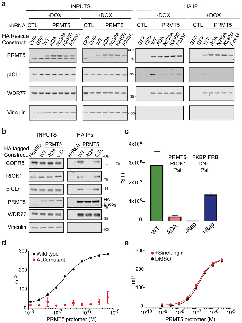
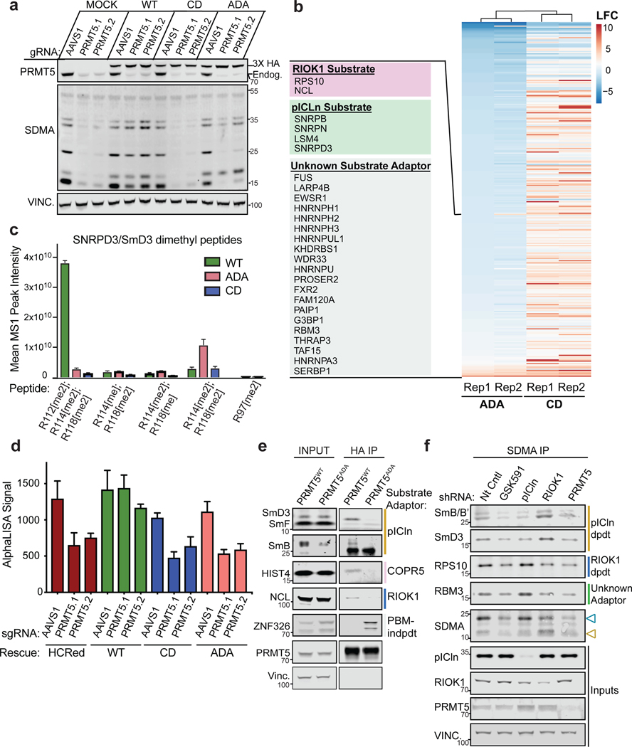
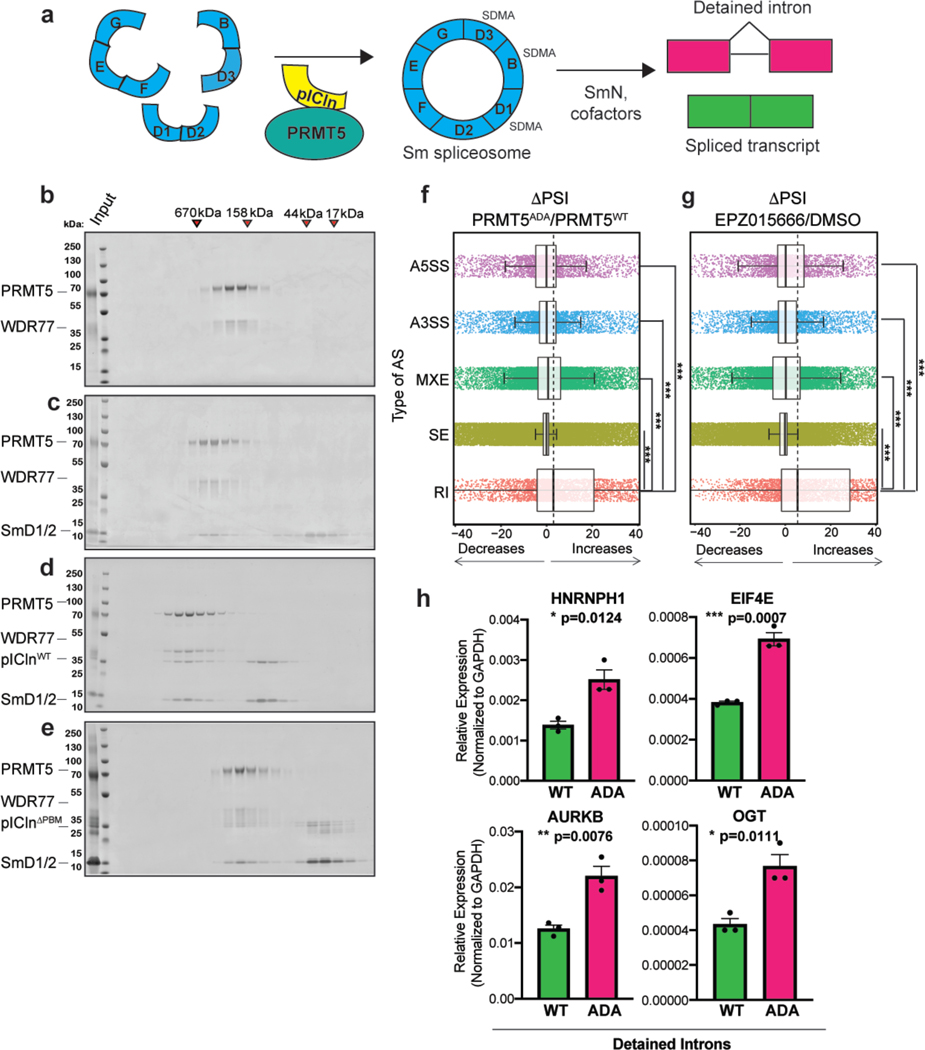
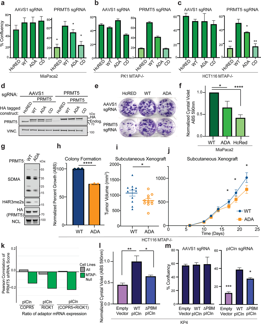
PRMT5 is an essential arginine methyltransferase and a therapeutic target in MTAP-null cancers. PRMT5 uses adaptor proteins for substrate recruitment through a previously undefined mechanism. Here, we identify an evolutionarily conserved peptide sequence shared among the three known substrate adaptors (CLNS1A, RIOK1, and COPR5) and show that it is necessary and sufficient for interaction with PRMT5. We demonstrate that PRMT5 uses modular adaptor proteins containing a common binding motif for substrate recruitment, comparable with other enzyme classes such as kinases and E3 ligases. We structurally resolve the interface with PRMT5 and show via genetic perturbation that it is required for methylation of adaptor-recruited substrates including the spliceosome, histones, and ribosomal complexes. Furthermore, disruption of this site affects Sm spliceosome activity, leading to intron retention. Genetic disruption of the PRMT5-substrate adaptor interface impairs growth of MTAP-null tumor cells and is thus a site for development of therapeutic inhibitors of PRMT5.

Keywords: CDKN2A; COPR5; MTAP; PRMT5; RIOK1; Sm protein; arginine methylation; histone; pICln; splicing.


Conflict of interest statement

Declaration of interests Materials related to this article are in a provisional patent application by the Broad Institute. W.R.S. is a board or scientific advisory board (SAB) member and holds equity in Peloton Therapeutics, Ideaya Biosciences, Civetta Therapeutics, and Bluebird bio; has consulted for Array, Astex, Dynamo Therapeutics, Ipsen, Merck Pharmaceuticals, PearlRiver Therapeutics, Sanofi, Scorpion Therapeutics, and Servier; and receives research funding from Pfizer Pharmaceuticals, Merck Pharmaceuticals, Ideaya Biosciences, and Deerfield Management. B.J.M. is an employee and equity holder of Tango Therapeutics. D. Porter is an employee and equity holder of Cedilla Therapeutics. A.J.A. has consulted for Oncorus Inc., Arrakis Therapeutics, and Merck & Company and has research funding from Mirati Therapeutics and Deerfield Management.

Figures

Figure 1.
Figure 1.. Identification of a conserved PRMT5 substrate adaptor binding motif: GQF[D/E]DA[D/E].
a. Amino acid sequence alignment of known substrate adaptor proteins of PRMT5 (COPR5, pICln, and RIOK1) identified an evolutionarily conserved 7-mer peptide that lies at the N-terminus of RIOK1 and C-termini of pICln and COPR5. b. Peptide distribution of 7 amino acid sequence motifs across all PRMT5 binding proteins (BioGrid) is shown. GQF[D/E]DA[D/E] was enriched among PRMT5 binding proteins compared to other 7-mer peptide sequences across the human proteome. c. Interactions between the PRMT5 complex and the conserved 7-mer peptides described in (a) were measured by SPR. The PRMT5 complex was immobilized by biotin-neutravidin and the indicated peptides titrated as analyte. The RU response at equilibrium is shown. d. Streptavidin affinity purification (AP) from cell lysates derived from MiaPaca2 and PK1 cells that were incubated with increasing concentrations of either the pICLn 17-mer peptide containing the 7-mer sequence in (A) or a scrambled control peptide. Immunoblots show the amount of PRMT5 and WDR77 that were co-purified with the peptides. Vinc. is a loading control. e. Streptavidin AP from MiaPaca2 cell lysates that were incubated with 1μg of either the pICLn 17-mer peptide (“PBM”) containing the 7-mer sequence in (A) or a scrambled control peptide. Immunoblots show the amount of PRMT5, WDR77, PRMT1 and PRMT7 that were co-purified with the peptides. Vinc. is a loading control.
Figure 2.
Figure 2.. Structural basis for recognition of the PRMT5-binding motif (PBM)
a. The PRMT5:WDR77 complex bound to the RIOK1 PBM peptide. Left, the PRMT5 surface for the “front” protomer is colored by amino acid conservation on a sliding scale from cyan (least conserved) to maroon (most conserved). The RIOK1 PBM peptide is colored orange and shown as a stick representation. Right, a single protomer unit of the complex is represented in cartoon form with PRMT5 in green and WDR77 in grey. The RIOK1 peptide and SAM are shown in orange and purple, respectively. b. PRMT5 (from the RIOK1 bound structure) is represented in cartoon form with a transparent surface colored by electrostatic potential from negative (red) to positive (blue). PBM peptides from RIOK1 (orange) and pICln (purple) are represented as sticks. Structures were aligned by minimization of global RMSD using Pymol. c. The PBM binding groove of PRMT5 (green) is shown as a cartoon with transparent surface. Two ordered water molecules are represented as red spheres. The RIOK1 peptide is shown in orange. d. Key hydrogen bonds of the RIOK1:PRMT5 interaction are shown as yellow dotted lines. Hydrogen bonds representing i+3 bonding pattern for β-turns labeled as “1” or “2” and described in the main text.
Figure 3.
Figure 3.. The PBM is required for PRMT5 binding to substrate adaptor proteins.
a. Interactions between the PRMT5 complex and pICln protein were measured by SPR. The PRMT5 complex was immobilized by biotin-neutravidin and then full-length or mutant pICln protein was titrated as analyte. The RU response at equilibrium is shown. b. Competitive fluorescence polarization (FP) assay using a fluorescent RIOK1 peptide as probe. The indicated peptides were titrated as competitor molecules. c. The relative interaction between PRMT5 and RIOK1 or RIOK1ΔPBM was measured in cells via NanoBiT assay in HEK293T cells following transient transfection. The N-termini of RIOK1 and PRMT5 were tagged with complementary “LargeBiT” and “SmallBiT” sequences, respectively. Relative luciferase signal was assayed as a measure of functional complementation. d. The NanoBiT PRMT5:RIOK1 system was used to test the ability of PBM peptides to compete for PRMT5 binding. Prior to addition of peptides, cells were permeabilized with 0.05% NP-40. Peptides of WT or point mutant PBM sequence were titrated into the permeabilized cells as indicated. Relative luciferase signal was assayed as a measure of functional complementation. e. Competition between full-length pICln and PBM peptide as measured by SPR. The PRMT5 complex was immobilized by biotin-neutravidin interaction. 125 nM pICln was co-injected with increasing concentrations of the indicated peptides. For comparison, the RU response with peptide alone is also shown. The RU response at equilibrium is shown. f. Co-immunoprecipitation (co-IP) of HA-tagged WT or PBM mutant pICln in the presence or absence of endogenous pICln depletion (dox inducible shRNA). Immunoblot detects the amounts of immunoprecipitated PRMT5 and pICln. Two exposures of PRMT5 are shown to visualize the dynamics of the binding between pIClnWT and pICln mutants. Vinc. is a loading control.
Figure 4.
Figure 4.. The PBM groove is required for PRMT5 binding to substrate adaptor proteins.
a. Co-IP of HA tagged WT or PBM groove mutant PRMT5 in the presence or absence of endogenous PRMT5 depletion (dox. inducible shRNA) was performed. Immunoblot detects amounts of immunoprecipitated PRMT5, WDR77 and pICln. Vinc. is a loading control. Blots were cropped as indicated and one mutant which did not express (Y286A) was eliminated. b. Co-IP of HA tagged WT, PBM groove mutant (ADA), or catalytically dead (CD) PRMT5 in the presence or absence of endogenous PRMT5 depletion (CRISPr Cas9 KO). Immunoblot detects the amount of immunoprecipitated PRMT5, WDR77, pICln, RIOK1, and COPR5. Vinc. is a loading control. c. Mutations of the PBM interface on PRMT5 were tested via cell-based NanoBiT as previously described. PRMT5WT or PRMT5ADA mutant (N239A/K240D/F243A) and RIOK1 were tagged with complementary NanoBiT sequences and co-expressed. For comparison, NanoBiT complementation between the manufacturers’ rapamycin inducible control pair (FKBP-FRB; +/− Rap.) is also shown. Relative luciferase signal was assayed as a measure of functional complementation. d. Polarization of a fluorescent PBM peptide derived from RIOK1 was measured across the indicated concentrations of either WT or ADA PRMT5. e. Polarization of a fluorescent PBM peptide as in (d), co-incubated with or without SAM analog sinefungin.
Figure 5.
Figure 5.. The PBM-PBM groove interaction is required for methylation of a subset of PRMT5 substrates.
a. Symmetric dimethyl arginine (SDMA) detected by immunoblot in a PRMT5 KO-rescue system utilizing PRMT5WT, PRMT5ADA or PRMT5CD in HCT116 MTAP KO cells. b. SDMA-modified peptides immunoprecipitated from CD, ADA, and WT PRMT5 rescued MiaPaca2 cells following KO of endogenous PRMT5, and quantified by label-free MS1 quant of LC-MS/MS data. Each row represents a distinct SDMA peptide detected. Peptides are clustered by ADA Replicate 1. Peptides with a 2-fold decrease in ADA versus WT are labeled; list is in ordered by FC except RIOK1 and pICln substrates which were isolated for visual clarity. c. The mean MS1 peak intensity for all dimethylated peptides of SmD3 that were detected by LC-MS/MS in (b) are shown for each the WT, ADA and CD PRMT5 cell lines. Peptides are ordered 1–5 based on their log fold-change WT/CD ~strength as a PRMT5 substrate. d. The relative amount of SDMA-modified SmB protein was measured by AlphaLISA proximity assay, as described in methods, for each HCT116 MTAP−/−: HcRed, WT, ADA and CD-PRMT5 expressing stable cell lines in the presence or absence of 2 PRMT5 targeting sgRNAs. e. Co-IP of HA-PRMTWT or PRMT5ADA from HCT116 MTAP−/− cells depleted for endogenous PRMT5 was performed and inputs and purified protein complexes resolved by SDS-PAGE and detected by immunoblotting are shown. Anti-SmD3, anti-SmB measure pICln substrate binding; anti-NCL measures RIOK1 substrate binding; anti-Hist4 measures COPR5 substrate binding; anti-ZNF326 measures PBM groove-independent substrate binding; Vinc is a loading control. f. Denaturing SDMA immunoprecipitations from lysates of MiaPaca2 cells 6 days after shRNA induction with doxycycline for the indicated shRNA or PRMT5 inhibitor control (GSK591, 20nM). Immunoblot detection with the relevant antibodies is as indicated.
Figure 6.
Figure 6.. The pIClnPBM-PRMT5PBM groove interaction is required for Sm substrate recruitment and subsequent Sm complex formation and splicing activity.
a. Illustration of the pICln-PRMT5 dependent process of Sm methylation, spliceosome complex formation, and subsequent splicing activity. Sm proteins require PRMT5 methylation and pICln-PRMT5 scaffolding to assemble. Sm spliceosome assembly leads to proficient splicing of detained introns. b. In vitro reconstitution of human PRMT5-WDR77 complexes. PRMT5-WDR77 were incubated for 30 minutes at 4°C and protein complexes were resolved by gel filtration chromatography. Protein fractions were separated on SDS-PAGE gels and visualized by Coomassie stain. Estimated molecular mass elution volumes are shown for standards run on this column. Co-elution indicates binding (or shared mass), increased mass indicates larger protein complexes being formed. Expected sizes: PRMT5-WDR77 octamer ~440kDa; PRMT5 ~72kDa; WDR77 ~37kDa; SmD1,2 ~15kDa; pICln ~29kDa. c. In vitro reconstitution of human PRMT5-WDR77 complexes in the presence of SmD1/2, resolved as in (b). d. In vitro reconstitution of human PRMT5-WDR77 complexes in the presence of pIClnWT and SmD1/2, resolved as in (b). e. In vitro reconstitution of human PRMT5-WDR77 complexes in the presence of pIClnΔPBM and SmD1/2, resolved as in (b). f. Change in percent spliced in (PSI) comparing PRMT5WT and PRMT5ADA expressing HCT116 MTAP−/− cells is shown. A5SS and A3SS=alternative 5’ and 3’ splice sites, respectively; MXE=mutually exclusive exons; SE=Skipped Exon; RI= retained intron. Vertical lines within boxes= median, edges of boxes- 1st and 4th quartiles, whiskers = min/max values, outliers (>/<1.5x interquartile range) excluded. Dashed line=PSI shift in median RI. ***=p<0.001 for Wilcoxon Rank test between groups. g. Change in PSI between 72h 10μM EPZ015666 (PRMT5 inhibitor) or DMSO control treated U-87 MG cells in shown for published data by Braun, et al 2017. Plotted as in (f). ***=p<0.001 for Wilcoxon Rank test between groups. h. qRT-PCR of previously reported detained introns (DI) using RNA purified from MTAP−/− HCT116 cells expressing PRMT5WT or PRMT5ADA and depleted for endogenous PRMT5 as a template for cDNA generation and subsequent real-time PCR. Amplicons flank the intron-exon junction of PRMT5-responsive DI. Increased signal represents intron retention.
Figure 7.
Figure 7.. Mutation of the PBM:PRMT5 interaction site impairs growth in MTAP null cells
a. Incucyte cell growth plots of MiaPaca2 Cas9 expressing cells infected with PRMT5-targeting guide RNA to KO endogenous PRMT5 or AAVS1 guide RNA control are shown. Cell lines are stably expressing HA-tagged HcRed, PRMT5WT, PRMT5ADA or PRMT5CD. 5 days after KO and 2 days after selection with puromycin, cells are seeded into growth assays. Growth was measured in the presence of 10μM MTA. Day 6 (T=156h) is plotted. Mean +/− SEM are shown for n=5 replicates; *=p<0.05 paired student’s t-test. b. Incucyte plots of PK1 MTAP−/−Cas9 expressing cells infected with PRMT5-targeting guide RNA to KO endogenous PRMT5. Cell lines are stably expressing HA-tagged HcRed, PRMT5WT, PRMT5ADA or PRMT5CD. Growth was measured in the presence of 10μM MTA. Day 6 (T=156h) is plotted; n=1. c. Incucyte plots of HCT116 MTAP−/− Cas9 expressing cells infected with PRMT5-targeting guide RNA to KO endogenous PRMT5 or AAVS1 guide RNA control. Cell lines are stably expressing HA-tagged HcRed, PRMT5WT, PRMT5ADA or PRMT5CD. Growth was measured in the presence of 10μM MTA. Day 6 (T=152h) is plotted; Mean +/− SEM are shown for n=3 replicates.*=p<0.05; **=p<0.01; student’s t-test. d. Immunoblots of the genotypes from (A) MiaPaca2 cells. Immunoblotting was used to detect endogenous and HA-tagged PRMT5 in each cell line: HcRed, PRMT5WT, PRMT5ADA or PRMT5CD in the presence of AAVS1 control or PRMT5 targeting sgRNA. e. Crystal violet-stained colony formation assay (CFA) of MiaPaca2 Cas9 expressing cells infected with AAVS1 control or PRMT5-targeting guide RNA to KO endogenous PRMT5 and rescue with either HcRed, PRMT5WT, or PRMT5ADA in the presence of 10μM MTA; 14 days. One representative experiment is shown, n=5. f. Quantification of CFA in (e) using acid-solubilization and ABS reading. Normalized mean ABS is shown +/− SEM of n=5 experiments. *=p<0.05 student’s t-test; ****=p<0.0001 student’s t-test. g. Immunoblot of total PRMT5, SDMA and H4R3 SDMA levels in HCT116 MTAP−/− PRMT5WT or PRMT5ADA cells. NCL serves as a loading control. h. Quantification of CFA in HCT116 MTAP−/− PRMT5WT or PRMT5ADA cells using acid-solubilization and ABS reading on normalized mean ABS is shown +/− SEM of n=3 experiments. CFA image shown in Figure S6A. ****=p<0.0001 student’s t-test. i. HCT116 MTAP−/− PRMT5WT or PRMT5ADA cells were injected subcutaneously in nude mice and formed tumors. 11 tumors per group are shown at Day 22. 2 mice (1 from each group) were eliminated from statistical analysis due to poor engraftment. *=p<0.05 1-sided student’s t-test. j. HCT116 MTAP−/− PRMT5WT or PRMT5ADA cells were injected subcutaneously in nude mice and formed tumors. 12 tumors per group are shown over time (Day 6–22). *=p<0.05 1-sided student’s t-test. k. Pearson correlations of PRMT5 shRNA Demeter2 score and the mRNA ratios of the 3 substrate adaptors are shown for all MTAP WT and MTAP null cells in the Cancer Dependency Map. l. Quantification and statistical analysis of the biological replicate CFA in Figure S6B (EV vs pIClnWT p=0.0077; pIClnWT vs pIClnΔPBM p=0.043; n=4, student’s t-test). m. Incucyte plots of KP4 Cas9 expressing cells stably expressing V5-tagged pICln and EV constructs infected with pICln-targeting guide RNA to KO endogenous pICln or AAVS1 control guide RNA are shown T=5 days after KO. Mean +/− SEM are shown for n=3 replicates. (*=p<0.05; ***=p<0.001, student’s t-test.

References

    1. Adams PD, Afonine PV, Bunkóczi G, Chen VB, Davis IW, Echols N, Headd JJ, Hung LW, Kapral GJ, Grosse-Kunstleve RW, et al. (2010). PHENIX: A comprehensive Python-based system for macromolecular structure solution. Acta Crystallogr. Sect. D Biol. Crystallogr. - PMC - PubMed
    1. Alinari L, Mahasenan KV, Yan F, Karkhanis V, Chung JH, Smith EM, Quinion C, Smith PL, Kim L, Patton JT, et al. (2015). Selective inhibition of protein arginine methyltransferase 5 blocks initiation and maintenance of B-cell transformation. Blood. - PMC - PubMed
    1. Antonysamy S (2017). The structure and function of the PRMT5:MEP50 complex. Subcell. Biochem. - PubMed
    1. Arribas-Layton M, Dennis J, Bennett EJ, Damgaard CK, and Lykke-Andersen J (2016). The C-Terminal RGG Domain of Human Lsm4 Promotes Processing Body Formation Stimulated by Arginine Dimethylation. Mol. Cell. Biol. - PMC - PubMed
    1. Ashkenazy H, Abadi S, Martz E, Chay O, Mayrose I, Pupko T, and Ben-Tal N (2016). ConSurf 2016: an improved methodology to estimate and visualize evolutionary conservation in macromolecules. Nucleic Acids Res. - PMC - PubMed

Publication types

MeSH terms