. 2021 Jul 14;11(7):1256.
doi: 10.3390/diagnostics11071256.

Post-Traumatic Intraparenchymal Renal Hemorrhages: Correlation between CT and DSA Vascular Findings for Superselective Embolization Procedures

Affiliations

Post-Traumatic Intraparenchymal Renal Hemorrhages: Correlation between CT and DSA Vascular Findings for Superselective Embolization Procedures

Francesco Giurazza et al. Diagnostics (Basel). .

Abstract

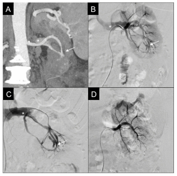
Background: This study aims to investigate the correlation between computed tomography (CT) and digital subtraction angiography (DSA) findings in patients affected by acute post-traumatic intraparenchymal renal hemorrhages and evaluate their conservative management with superselective embolization.

Methods: This retrospective multicenter analysis focuses on patients affected by renal bleedings detected by contrast-enhanced CT and treated with superselective endovascular embolization. CT findings were compared to DSA. Embolization procedural data were analyzed and renal function was evaluated before and after the intervention.

Results: Twenty-seven patients were retrospectively evaluated in one year. Compared to DSA, CT showed 96.3% diagnostic accuracy in terms of hemorrhage recognition; concerning the type of vascular lesion, there was discrepancy between CT and DSA in five cases. The technical success rate of embolization was 100%, while primary clinical success was 88.9%. The inferior parenchymal third was the most frequent site of renal injury. Microcoils were the most adopted embolics. Renal function did not change significantly before and after embolization.

Conclusions: CT has elevated diagnostic accuracy in detecting post-traumatic intraparenchymal renal hemorrhages; in a small percentage, the type of vascular lesion may differ from the findings observed at DSA. In this scenario, superselective embolization presents high clinical success with a low complication rate.

Keywords: computed tomography; intraparenchymal; renal hemorrhage; superselective embolization; trauma.


Conflict of interest statement

The authors declare no conflict of interest. The funders had no role in the design of the study; in the collection, analyses, or interpretation of data; in the writing of the manuscript, or in the decision to publish the results.

Figures

Figure 1
Figure 1
46-year-old male, affected by iatrogenic PA. (A) CT reconstruction with MIP and coronal oblique MPR, in venous phase, shows the PA (white arrow) in correspondence with the point of entrance of the nephrostomy into the renal cortex; amount of intracaliceal contrast agent residual from the urological procedure. (B) Selective right renal DSA confirms PA (white arrow). (C) Microcatheter positioning into the feeder of the PA (white arrow). (D) Final DSA showing PA resolution after embolization with a microvascular plug (MVP-5Q), renal parenchyma normally vascularized.
Figure 2
Figure 2
57-year-old female, affected by iatrogenic arteriovenous fistula (AVF). (A) CT reconstruction with MIP and coronal MPR, in arterial phase: intraparenchymal AVF (white arrow) at the inferior third of the left kidney, with early opacification of the left renal vein (white asterisk). (B) Selective right renal DSA confirms AVF (white arrow), left renal vein opacified in early phase of injection (white asterisk). (C) Selective catheterization of the inferior third, up to the AVF (white arrow) with evidence of the renal vein (white asterisk). (D) Final DSA showing AVF resolution after embolization with two pushable microcoils (MicroNester 4 mm), renal parenchyma being vascularized, renal vein no longer opacified.
Figure 3
Figure 3
50-year-old male, affected by iatrogenic active bleeding. (A) CT reconstruction with MIP and coronal MPR, in arterial phase: intraparenchymal active bleeding (white arrow) at the superior third of the left kidney; homolateral urological stent is evident. (B) Selective left renal DSA confirms active bleeding (white arrow). (C) Superselective microcatheterization and embolization with two detachable coils (Concerto 4 mm × 8 cm). (D) Final DSA showing active bleeding resolution, renal parenchyma vascularized.

References

    1. Martin J.G., Shah J., Robinson C., Dariushnia S. Evaluation and Management of Blunt Solid Organ Trauma. Tech. Vasc. Interv. Radiol. 2017;20:230–236. doi: 10.1053/j.tvir.2017.10.001. - DOI - PubMed
    1. Mucelli F.P., Mucelli R.P., Marrocchio C., Tollot S., Cova M. Endovascular Interventional Radiology of the Urogenital Tract. Medicina. 2021;57:278. doi: 10.3390/medicina57030278. - DOI - PMC - PubMed
    1. Coccolini F., Panel W.-A.E., Moore E.E., Kluger Y., Biffl W., Leppaniemi A., Matsumura Y., Kim F., Peitzman A.B., Fraga G.P., et al. Kidney and uro-trauma: WSES-AAST guidelines. World J. Emerg. Surg. 2019;14:1–25. doi: 10.1186/s13017-019-0274-x. - DOI - PMC - PubMed
    1. Chong S.T., Cherry-Bukowiec J.R., Willatt J.M., Kielar A.Z. Renal trauma: Imaging evaluation and implications for clinical management. Abdom. Radiol. 2016;41:1565–1579. doi: 10.1007/s00261-016-0731-x. - DOI - PubMed
    1. Iacobellis F., Ierardi A.M., Mazzei M.A., Magenta Biasina A., Carrafiello G., Nicola R., Scaglione M. Dual-phase CT for the assessment of acute vascular injuries in high-energy blunt trauma: The imaging findings and management implications. Br. J. Radiol. 2016;89:20150952. doi: 10.1259/bjr.20150952. - DOI - PMC - PubMed

LinkOut - more resources