Review
. 2021 Jul 22;13(15):3695.
doi: 10.3390/cancers13153695.

Lobular Breast Cancer: Histomorphology and Different Concepts of a Special Spectrum of Tumors

Affiliations
Review

Lobular Breast Cancer: Histomorphology and Different Concepts of a Special Spectrum of Tumors

Matthias Christgen et al. Cancers (Basel). .

Abstract

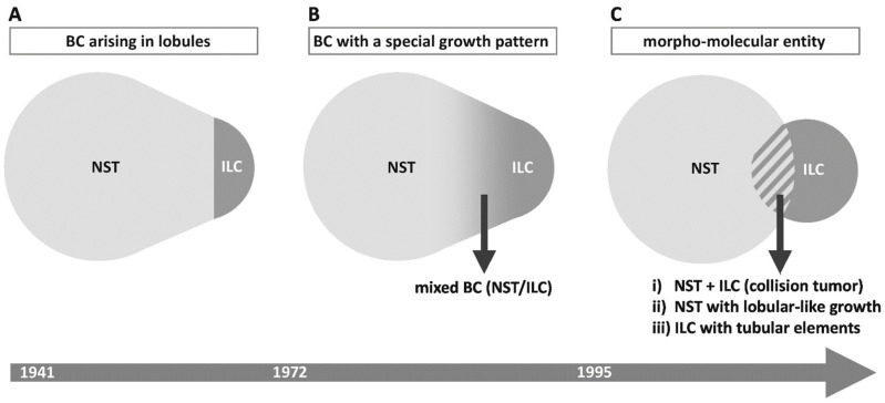
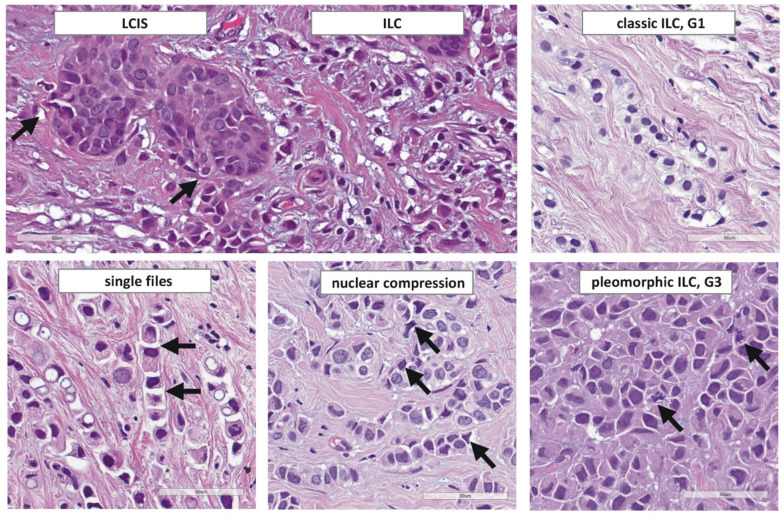
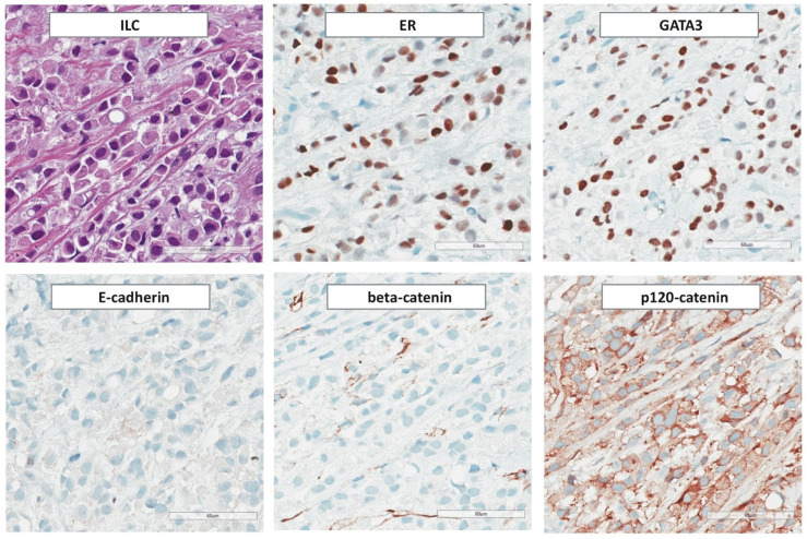
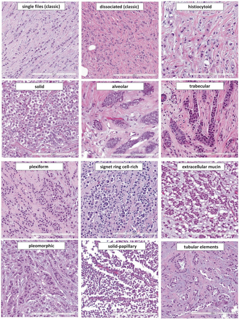
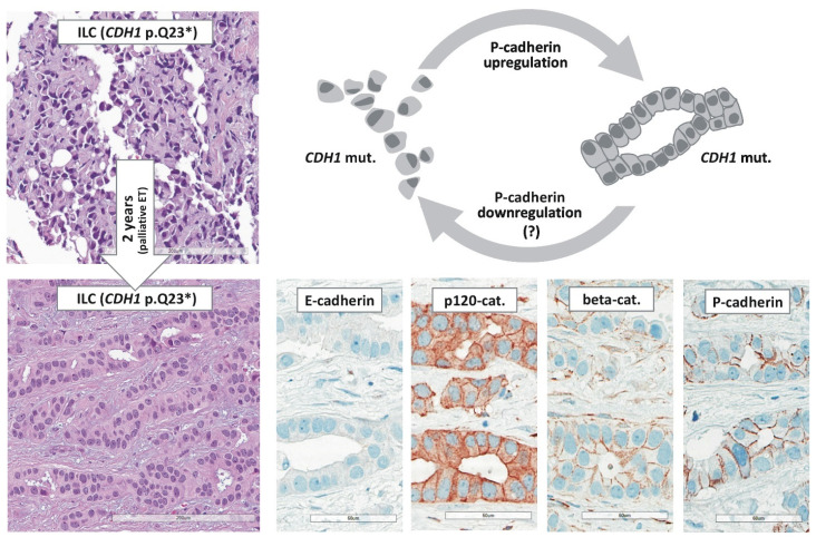
Invasive lobular breast cancer (ILC) is the most common special histological type of breast cancer (BC). This review recapitulates developments in the histomorphologic assessment of ILC from its beginnings with the seminal work of Foote and Stewart, which was published in 1941, until today. We discuss different concepts of ILC and their implications. These concepts include (i) BC arising from mammary lobules, (ii) BC growing in dissociated cells and single files, and (iii) BC defined as a morpho-molecular spectrum of tumors with distinct histological and molecular characteristics related to impaired cell adhesion. This review also provides a comprehensive overview of ILC variants, their histomorphology, and differential diagnosis. Furthermore, this review highlights recent advances which have contributed to a better understanding of the histomorphology of ILC, such as the role of the basal lamina component laminin, the molecular specificities of triple-negative ILC, and E-cadherin to P-cadherin expression switching as the molecular determinant of tubular elements in CDH1-deficient ILC. Last but not least, we provide a detailed account of the tumor microenvironment in ILC, including tumor infiltrating lymphocyte (TIL) levels, which are comparatively low in ILC compared to other BCs, but correlate with clinical outcome. The distinct histomorphology of ILC clearly reflects a special tumor biology. In the clinic, special treatment strategies have been established for triple-negative, HER2-positive, and ER-positive BC. Treatment specialization for patients diagnosed with ILC is just in its beginnings. Accordingly, ILC deserves greater attention as a special tumor entity in BC diagnostics, patient care, and cancer research.

Keywords: HER2; LCIS; LIN; beta-catenin; p120-catenin; pleomorphic; solid; tubulolobular.


Conflict of interest statement

The authors declare no conflict of interest. The funders had no role in the writing of the manuscript.

Figures

Figure 1
Figure 1
Different concepts of ILC as a special tumor entity and their implications. This is a Venn diagram-like schematic presentation of the relation between BC of NST and ILC, according to different concepts. (A) ILC as BC arising in lobules and terminal ducts (Foote and Stewart in 1941) [7]. BC of NST and ILC are clearly separated (based on the presence or absence of LCIS). For details, see text Section 2.1. (B) ILC as BC with a special growth pattern (1972 until today, consistent with the 5th edition of the WHO classification of tumors) [19,56]. The boundary between BC of NST and ILC is less clearly defined compared to concept “A”. BCs with indefinite histomorphology may be classified as mixed BC NST/ILC. For further details, please see text Section 2.2 and Appendix A Table A1. (C) ILC as a morpho-molecular entity (a concept unofficially prevalent in clinical research since the discovery of CDH1 mutations in 1995) [11]. BC of NST and ILC share a morphologic overlap. Cases in the morphologic overlap may be interpreted as (i) collision tumors, (ii) NST with lobular-like growth pattern but no loss of cell adhesion, and (iii) ILC with tubular elements [58,59]. For details, see text Section 2.3. An approximate time scale showing the origin of these concepts is given at the bottom.
Figure 2
Figure 2
Histomorphology of ILC. From top left to lower right, photomicrographs (×400 magnification) illustrate: transition from LCIS to ILC (arrows indicate LCIS cells that breach through the basal lamina), classical ILC (G1), ILC arranged in single files with gaps between individual cells (arrows), ILC with prominent nuclear compression (arrows), pleomorphic ILC (G3) with brisk mitotic activity (arrows).
Figure 3
Figure 3
Immunohistochemical features of ILC. From Photomicrographs (×200 magnification) illustrate: expression of ER and GATA3, loss of E-cadherin and β-catenin, and aberrant cytoplasmic localization of p120-catenin.
Figure 4
Figure 4
Histomorphology of ILC variants reported in the scientific literature. From top left to lower right: single file growth pattern (classical ILC), dissociated growth pattern (classical ILC), histiocytoid ILC, solid ILC, alveolar growth pattern, trabecular growth pattern, plexiform growth pattern, signet ring cell-rich ILC, ILC with extracellular mucin, pleomorphic ILC, solid-papillary ILC, ILC with tubular elements (×200 magnification).
Figure 5
Figure 5
E-cadherin to P-cadherin switching in ILC with tubular elements [59]. Photomicrographs (×200 magnification) illustrate an ILC diagnosed by CNB (upper left) and the resection specimen after primary endocrine therapy (ET). Both specimens were E-cadherin-negative and harbored the same deleterious CDH1 mutation (p.Q23*). Note that the resection specimens displayed tubular elements which were associated with expression of P-cadherin and β-catenin, while p120-catenin showed a partially aberrant cytoplasmic localization and a partially regular membranous localization. The cartoon illustrates that ILC cells form focal tubular elements by dynamic upregulation of P-cadherin (id est, E-cadherin to P-cadherin switching) [59].

Similar articles

Cited by

References

    1. Hoadley K.A., Yau C., Hinoue T., Wolf D.M., Lazar A.J., Drill E., Shen R., Taylor A.M., Cherniack A.D., Thorsson V., et al. Cell-of-Origin Patterns Dominate the Molecular Classification of 10,000 Tumors from 33 Types of Cancer. Cell. 2018;173:291–304.e296. doi: 10.1016/j.cell.2018.03.022. - DOI - PMC - PubMed
    1. Hoberger M., von Laffert M., Heim D., Klauschen F. Histomorphological and molecular profiling: Friends not foes! Morpho-molecular analysis reveals agreement between histological and molecular profiling. Histopathology. 2019;75:694–703. doi: 10.1111/his.13930. - DOI - PubMed
    1. Hyman D.M., Piha-Paul S.A., Won H., Rodon J., Saura C., Shapiro G.I., Juric D., Quinn D.I., Moreno V., Doger B., et al. HER kinase inhibition in patients with HER2- and HER3-mutant cancers. Nature. 2018;554:189–194. doi: 10.1038/nature25475. - DOI - PMC - PubMed
    1. Cheatle G.L., Cutler M. Tumors of the Breast. Their Pathology, Symptoms, Diagnosis and Treatment. Lippincott Company; Philadelphia, PA, USA: Montreal, QC, Canada: 1930.
    1. Cornil V. Les Tumeurs du Sein. Librairie Germer Bailliere and Co.; Paris, France: 1908.