Review
. 2021 Jul 8;10(7):1722.
doi: 10.3390/cells10071722.

How Influenza Virus Uses Host Cell Pathways during Uncoating

Affiliations
Review

How Influenza Virus Uses Host Cell Pathways during Uncoating

Etori Aguiar Moreira et al. Cells. .

Abstract

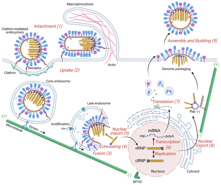
Influenza is a zoonotic respiratory disease of major public health interest due to its pandemic potential, and a threat to animals and the human population. The influenza A virus genome consists of eight single-stranded RNA segments sequestered within a protein capsid and a lipid bilayer envelope. During host cell entry, cellular cues contribute to viral conformational changes that promote critical events such as fusion with late endosomes, capsid uncoating and viral genome release into the cytosol. In this focused review, we concisely describe the virus infection cycle and highlight the recent findings of host cell pathways and cytosolic proteins that assist influenza uncoating during host cell entry.

Keywords: EPS8; HDAC6; M1; TNPO1; capsid uncoating; influenza; pandemic; ubiquitin; virus–host interaction.


Conflict of interest statement

The authors declare the absence of any commercial or financial relationships that could be construed as a potential conflict of interest.

Figures

Figure 1
Figure 1
IAV structure and genome. Influenza is an enveloped virus in which structural proteins can be found associated with the virus envelope, a lipid bilayer derived from the plasma membrane of the host cell. The viral envelope contains three of the viral transmembrane proteins: hemagglutinin (HA), neuraminidase (NA), and the matrix ion channel M2. HA and NA proteins are the main proteins at the virus surface and HA is four times more abundant than NA. M2 also penetrates the envelope but represents a minor component of the envelope, with about 20 molecules per virus particle. The matrix protein M1 is found beneath the lipid membrane, and forms a rigid single-helical layer shell, the virus capsid. The nuclear export protein (NEP) is found in the interior of the virus. The IAV genome consists of eight negative-sense RNA segments that form distinct viral ribonucleoproteins (vRNPs). vRNPs are assembled as virus RNA segments where the termini of viral RNAs associate with the viral RNA-dependent RNA polymerase complex, PB1, PB2 and PA, while the rest of the viral RNAs are bound by oligomers of the nucleoprotein, NP. The virus has an asymmetric internal structure, maintained by vRNPs-vRNPs and M1-vRNPs interactions. Not shown in the figure, the interior of IAV bears a substantial number of host proteins (ubiquitin, tubulin, actin, annexin, among others). IAV is known to display a number of shapes. The spherical form of IAVs is typically about 100 nm in diameter. Filamentous forms of IAVs can be over a few μm in length.
Figure 2
Figure 2
Overview of the IAV replication cycle. The influenza virus life cycle can be divided into several stages: (1) Virus binding to the target cell. HA binds to sialic acid found on the surface of the host cell’s membrane. (2) Entry into the host cell: a clathrin-mediated endocytosis or macropinocytosis takes place. Early endosome containing viruses is transported by dynein along microtubules to the perinuclear region close to the microtubule-organizing center (MTOC). (3) Fusion of the virus envelope with the endosomal membrane. Acidification increases progressively from endocytic vesicles to late endosomes and induces a HA conformational change to a fusion-competent state. M2, an acid-activated viral ion channel, is required for efficient viral envelope fusion with the endosomal membrane and nucleocapsid release. (4) Uncoating of the virus capsid by disassembly of the M1 proteins and release of the viral ribonucleoproteins (vRNPs) to the cytosol. (5) Entry of vRNPs into the nucleus by an active nuclear import pathway. (6) Transcription and replication of the viral genome. The IAV genome is composed of negative-sense strand RNAs. The genome is first converted into positive-sense RNAs, forming complementary ribonucleoprotein (cRNP) complexes, that serve as templates to produce viral RNAs. The transcription of the vRNA generates mature viral messenger RNAs (mRNAs) that have a 5′ methylated cap and a poly(A) tail. (7) Viral protein translation occurs by free ribosomes or ribosomes on the rough endoplasmic reticulum. Some of these proteins enter the nucleus where they assemble with viral RNAs. (8) Export of the vRNPs from the nucleus. vRNPs are exported out of the nucleus via the CRM1 dependent pathway through the nuclear pores. (9) Transport of viral components, assembly and budding at the host cell plasma membrane. Viral glycoproteins, HA and NA, associate with lipid rafts, membrane microdomains comprised of densely packed cholesterol and sphingolipids. vRNP complexes are transported as sub-bundles on Rab11 to recycling endosomes close to ER exit sites toward the plasma membrane and are incorporated as a complex of eight different vRNPs into budding viruses. Finally, the plasma membrane containing the viral structural proteins at the assembly site bends releasing infectious virus into the extracellular environment.
Figure 3
Figure 3
Protein ubiquitination, ubiquitin chains, proteasomal and aggresome-autophagy degradation. Ubiquitin (Ub) is a small, 76 amino acid protein present in all eukaryotic cells that plays a key role in the cellular defense mechanism by functioning as a proteolytic signal for the proteasome. The process of covalent Ub attachment to target proteins is called ubiquitination (also known as ubiquitylation). This post-translational modification forms by an isopeptide bond between the carboxyl terminus of Ub and a lysine residue on the target protein. First, Ub is covalently conjugated to the E1 (Ub-activating enzyme) in an active ATP-dependent reaction and transferred to the E2 (Ub-conjugating enzyme). The E3 (Ub–protein ligase) transfers the Ub from E2 to the target protein and determines the specificity. A monoubiquitinated protein can have the Ub chain elongated by E3 that creates Ub–Ub isopeptide bonds. Chain extension can happen through seven lysine (K) residues on Ub: K6, K11, K27, K29, K33, K48 and K63. Proteasomes recognize K48 chains leading to target protein degradation (upper part). Other lysine chains are involved in different biological functions. K63 chains do not specify degradation but usually facilitate the recruitment of other proteins in the formation of functional complexes involved in cellular signaling such as aggresome formation (lower part). HDAC6 can bind misfolded proteins entangled with Ub K63 chains and bridges to dynein motors, mediating transport to and formation of the aggresome compartment. Free poly-Ub chains, referred to as unanchored Ub chains, have been found in virus particles. Unanchored poly-Ub chains arise when a deubiquitinase (DUB) removes an intact chain from a protein, or they can be generated through E1/E2/E3 cycles. They can be recognized by HDAC6 and activate the aggresome pathway as well IAV capsid uncoating. Ub Protein Data Bank (PDB): 1UBI; NH2 and COOH termini are labeled N and C, respectively. HDAC6 zinc finger (ZnF) and catalytic domain (CD) PDB: 3C5K and 5G0I, respectively.
Figure 4
Figure 4
IAV capsid uncoating, genome release and nuclear import. Endosome acidification occurs progressively from the cell periphery toward the microtubule-organizing center (MTOC). Late endosomal acidification (pH~6) triggers change of the homotrimeric glycoprotein HA mediating fusion between the viral envelope and the endosome membrane. Influx of protons and efflux of potassium from the virus core happen through the acid-activated viral ion channel M2. The pH drop triggers the activation of a histidine cluster in the virus capsid, contributed by three sequential M1 monomers, and promotes the capsid disassembly. Further the vRNPs dissociate from the M1 proteins. Free ubiquitin (Ub) chains derived from virus particles activate the aggresome processing pathway (APP) and recruit HDAC6 through its Ub-binding zinc finger domain (HDAC6 ZnF). Deubiquitinases (DUBs) could be involved in unanchored Ub formation. HDAC6 binds to M1 and to NP from vRNPs. HDAC6 by a region between its catalytic domains also binds motor proteins in microtubules and myosin II in actin microfilaments generating physical forces that help dissociate the M1 proteins, disassembling the virus capsid. The epidermal growth factor receptor pathway substrate 8 (EPS8) and transportin-1 (TNPO1) interact with M1 from the capsid and vRNPs, contributing to the disaggregation of the vRNP-associated M1 and vRNP debundling in the cytosol. In this way, vRNPs are transported by importin α/β to the nucleus as individual rod-shaped structures. PDB: TNPO1 (2Z5J), EPS8 (2E8M), importin α (4B18), DUB (6K9P).

References

    1. Carrasco-Hernandez R., Jácome R., López Vidal Y., Ponce de León S. Are RNA Viruses Candidate Agents for the Next Global Pandemic? A Review. ILAR J. 2017;58:343–358. doi: 10.1093/ilar/ilx026. - DOI - PMC - PubMed
    1. Burrell C.J., Howard C.R., Murphy F.A. Emerging Virus Diseases. Fenner White’s Med. Virol. 2017 doi: 10.1016/B978-0-12-375156-0.00015-1. - DOI
    1. Watkins K. Emerging Infectious Diseases: A Review. Curr. Emerg. Hosp. Med. Rep. 2018;6:86–93. doi: 10.1007/s40138-018-0162-9. - DOI - PMC - PubMed
    1. Ryu W.-S. Chapter 21—New Emerging Viruses. In: Ryu W.-S., editor. Molecular Virology of Human Pathogenic Viruses. Academic Press; Boston, MA, USA: 2017. pp. 289–302. - DOI
    1. Doherty P.C. What have we learnt so far from COVID-19? Nat. Rev. Immunol. 2021;21:67–68. doi: 10.1038/s41577-021-00498-y. - DOI - PMC - PubMed

Publication types

LinkOut - more resources