Review
. 2021 Jul 11;10(7):1750.
doi: 10.3390/cells10071750.

TMEM135 is a Novel Regulator of Mitochondrial Dynamics and Physiology with Implications for Human Health Conditions

Affiliations
Review

TMEM135 is a Novel Regulator of Mitochondrial Dynamics and Physiology with Implications for Human Health Conditions

Heather K Beasley et al. Cells. .

Abstract

Transmembrane proteins (TMEMs) are integral proteins that span biological membranes. TMEMs function as cellular membrane gates by modifying their conformation to control the influx and efflux of signals and molecules. TMEMs also reside in and interact with the membranes of various intracellular organelles. Despite much knowledge about the biological importance of TMEMs, their role in metabolic regulation is poorly understood. This review highlights the role of a single TMEM, transmembrane protein 135 (TMEM135). TMEM135 is thought to regulate the balance between mitochondrial fusion and fission and plays a role in regulating lipid droplet formation/tethering, fatty acid metabolism, and peroxisomal function. This review highlights our current understanding of the various roles of TMEM135 in cellular processes, organelle function, calcium dynamics, and metabolism.

Keywords: TMEM135; aging; fission; mitochondrial dynamics.


Conflict of interest statement

The authors declare no conflict of interest.

Figures

Figure 1
Figure 1
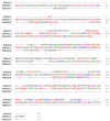
Comparison of the sequence homology of TMEM135 (A,B) across 11 different species using Clustal Omega Multiple Sequence alignment software (v.1.2.4). Clustal Omega designates the following colors for amino acid groups: AVFPMILW-Red: Small (small + hydrophobic [includes aromatic –Y])DE-Blue: Acidic, RHK-Magenta: Basic -H,STYHCNGQ -Green: Hydroxly + sulfhydryl + amine + G Others-Gray: Unusual amino/imino acids etc. (Accession numbers for TMEM135 species: OUZ99344.1, JAD00441.1, NP_508800.2, NP_001082887.1, NP_001085541.1, XP_040514949.1, NP_001013918.1, NP_082619.3, AAI03394.1, NP_075069.3, and XP_002708692.1 ) [36]. Figure created with BioRender.com (accessed on 15 June 2021).
Figure 1
Figure 1
Comparison of the sequence homology of TMEM135 (A,B) across 11 different species using Clustal Omega Multiple Sequence alignment software (v.1.2.4). Clustal Omega designates the following colors for amino acid groups: AVFPMILW-Red: Small (small + hydrophobic [includes aromatic –Y])DE-Blue: Acidic, RHK-Magenta: Basic -H,STYHCNGQ -Green: Hydroxly + sulfhydryl + amine + G Others-Gray: Unusual amino/imino acids etc. (Accession numbers for TMEM135 species: OUZ99344.1, JAD00441.1, NP_508800.2, NP_001082887.1, NP_001085541.1, XP_040514949.1, NP_001013918.1, NP_082619.3, AAI03394.1, NP_075069.3, and XP_002708692.1 ) [36]. Figure created with BioRender.com (accessed on 15 June 2021).
Figure 2
Figure 2
Comparison of the conserved sequence homology of TMEM135 (homo sapien) [NP_075069.3] with TMEM70 (homo sapien) [AAH02748.2] and TMEM242 (homo sapien) [NP_060922.2] using Clustal Omega Multiple Sequence alignment (v.1.2.4.) [36]. Clustal Omega designates the following colors for amino acid groups: AVFPMILW-Red: Small (small + hydrophobic [includes aromatic –Y])DE-Blue: Acidic, RHK-Magenta: Basic -H,STYHCNGQ -Green: Hydroxly + sulfhydryl + amine + G Others-Gray: Unusual amino/imino acids, etc. Figure created with BioRender.com (accessed on 15 June 2021).
Figure 3
Figure 3
This plausible model of TMEM135 demonstrates the many interactions for TMEM135 in several organelles, including the mitochondria [11] and the mitochondrial endoplasmic reticulum contact sites (MERCs). Additionally, TMEM135 is also a peroxisomal protein [66]. TMEM135 has been shown to co-localize with DRP1 as referenced in Wei Lee et al., 2016 [12]. Here, we show the plausible interaction between TMEM135 and DRP1 in the mitochondria and TMEM135 in the peroxisome [53], lysosome [66], lipid droplets [11], and the endoplasmic reticulum/ sarcoplasmic reticulum (ER/SR) [11]. Figure created with BioRender.com (accessed on 15 June 2021).
Figure 4
Figure 4
We hypothesize a role of TMEM135 in fission due to Ca2+ influx into the mitochondria and stimulation of DRP1-dependent mitochondrial fission. Mitochondrial dynamics are split into two processes: Fusion and Fission. Mitochondrial fusion is coordinated by Mitofusin 1 (MFN-1) and Mitofusin 2 (MFN-2) [light blue color], Optic Atrophy 1 (OPA-1) [light pink], Mitochondrial contact site and cristae organizing system (MICOS) [light yellow], and Cardiolipin [light green]. Mitochondrial fission proteins are coordinated by Mitochondrial Fission 1 protein (FIS1) [green], Mitochondrial dynamic protein of 51 kDa homolog (MiD51) [magenta] and Mitochondrial Fission Factor of 49kDA homology (MiD49) [purple], Mitochondria fission factor (Mff) [dark yellow], Transmembrane Protein 135 (TMEM135) [red] on the outer membrane of the mitochondria; whereas, Dynamin-1-like protein 1 (DRP1) [blue] is located in the inner membrane of the mitochondria. The endoplasmic reticulum (ER) [black] is represented in the fission process. It cannot be ruled out that TMEM135 plays a role in regulating the balance between mitochondrial fusion and fission since it has been proposed that TMEM135 activates DRP1 [12]. Figure created with BioRender.com (accessed on 15 June 2021).
Figure 5
Figure 5
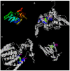
Predicted ligands for TMEM135 using I-TASSER software [71,72,107]. (A) The different colors represent the different alpha helixes. (BD) I-TASSER (version 5.1) software provides biological annotations of the target ligand by COACH based on the I-TASSER structure prediction, represented by the magenta-colored amino acid residues. Figure created with BioRender.com (accessed on 15 June 2021).

References

    1. Marx S., Maso T.D., Chen J.-W., Bury M., Wouters J., Michiels C., Le Calvé B. Transmembrane (TMEM) protein family members: Poorly characterized even if essential for the metastatic process. Semin. Cancer Biol. 2020;60:96–106. doi: 10.1016/j.semcancer.2019.08.018. - DOI - PubMed
    1. Fuller C.M. Time for TMEM? J. Physiol. 2012;590:5931–5932. doi: 10.1113/jphysiol.2012.245563. - DOI - PMC - PubMed
    1. Vinothkumar K.R., Henderson R. Structures of membrane proteins. Q. Rev. Biophys. 2010;43:65–158. doi: 10.1017/S0033583510000041. - DOI - PMC - PubMed
    1. Von Heijne G. Membrane-protein topology. Nat. Rev. Mol. Cell Biol. 2006;7:909–918. doi: 10.1038/nrm2063. - DOI - PubMed
    1. Alberts B., editor; Alberts B., editor. Molecular Biology of the Cell. 5th ed. Garland Science; New York, NY, USA: 2008.

Publication types

LinkOut - more resources