Review
. 2021 Jul 15;10(7):1794.
doi: 10.3390/cells10071794.

Implant Fibrosis and the Underappreciated Role of Myofibroblasts in the Foreign Body Reaction

Affiliations
Review

Implant Fibrosis and the Underappreciated Role of Myofibroblasts in the Foreign Body Reaction

Nina Noskovicova et al. Cells. .

Abstract

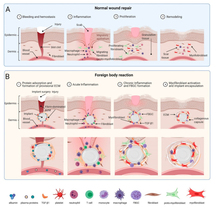
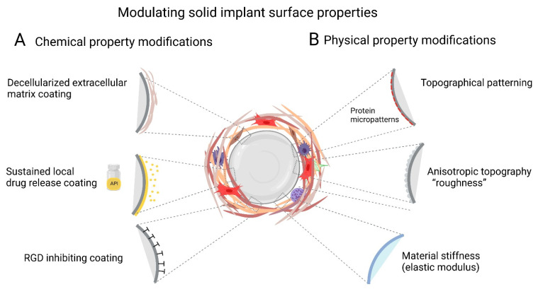
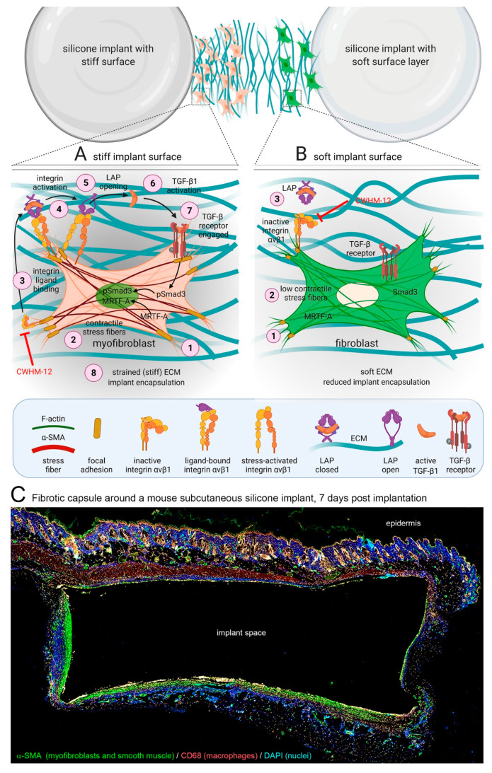
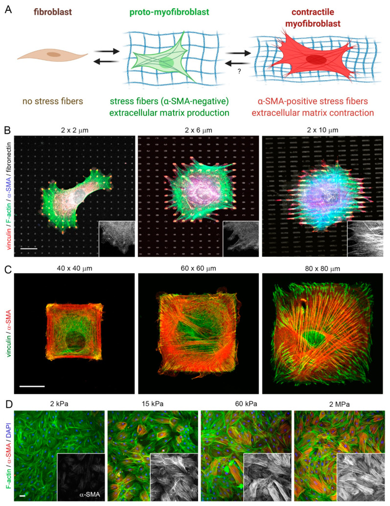
Body implants and implantable medical devices have dramatically improved and prolonged the life of countless patients. However, our body repair mechanisms have evolved to isolate, reject, or destroy any object that is recognized as foreign to the organism and inevitably mounts a foreign body reaction (FBR). Depending on its severity and chronicity, the FBR can impair implant performance or create severe clinical complications that will require surgical removal and/or replacement of the faulty device. The number of review articles discussing the FBR seems to be proportional to the number of different implant materials and clinical applications and one wonders, what else is there to tell? We will here take the position of a fibrosis researcher (which, coincidentally, we are) to elaborate similarities and differences between the FBR, normal wound healing, and chronic healing conditions that result in the development of peri-implant fibrosis. After giving credit to macrophages in the inflammatory phase of the FBR, we will mainly focus on the activation of fibroblastic cells into matrix-producing and highly contractile myofibroblasts. While fibrosis has been discussed to be a consequence of the disturbed and chronic inflammatory milieu in the FBR, direct activation of myofibroblasts at the implant surface is less commonly considered. Thus, we will provide a perspective how physical properties of the implant surface control myofibroblast actions and accumulation of stiff scar tissue. Because formation of scar tissue at the surface and around implant materials is a major reason for device failure and extraction surgeries, providing implant surfaces with myofibroblast-suppressing features is a first step to enhance implant acceptance and functional lifetime. Alternative therapeutic targets are elements of the myofibroblast mechanotransduction and contractile machinery and we will end with a brief overview on such targets that are considered for the treatment of other organ fibroses.

Keywords: TGF-β1; collagen; contracture; elastic modulus; fibroblast; macrophage; mechanosensing; micro-pattern; tissue repair; topography; wound healing.


Conflict of interest statement

The authors declare no conflict of interest.

Figures

Figure 1
Figure 1
Comparison between normal wound healing stages and phases of the FBR. (A) Normal wound healing is compromised of the same molecular players as the foreign body reaction (FBR) to implants (B) In fact, the FBR begins as a normal wound healing response, but the persistent presence of the biomaterial results in sustained fibrosis and scar tissue formation. Following the initial blood-biomaterial interaction and provisional ECM formation, acute inflammation, followed by chronic inflammation and fusion of macrophages into foreign body giant cells (FBGCs) occur in a subsequent fashion. Scheme was prepared using Biorender with kind support from Ronen Schuster.
Figure 2
Figure 2
Modulating implant surface properties is a common strategy to enhance tissue integration while simultaneously reducing the incidence and severity of implant FBR and fibrosis. Protein coverage and cell interactions with the surface both determine the ability of cells to attach and spread on implants. Various physical and chemical modifications to the Implants surface are used to inhibit FBR. (A) Chemical modifications of surface properties include coating with local and slow releasing anti-fibrotic drugs to prevent implant fibrosis. In addition, subcutaneously implanted tissue expanders coated with decellularized ECM into non-human primates display minimal fibrotic. Inhibiting αv-integrin binding, e.g., with the RGD-peptidomimetic inhibitor CWHM-12 attenuate implant encapsulation by preventing mechanical activation of latent TGF-β1 and myofibroblasts. (B) In addition to or combined with altering implant surface chemistry, other treatments include modulation of physical parameters such as surface hydrophilicity or wettability, porosity, stiffness (elastic modulus), anisotropic ‘roughness’, and regular topographies. Perceived stiffness and ‘true’ modulus of the implant surface can be used to control cell responses. Restriction of focal adhesion sizes by micropatterning stiff surfaces with adhesion sized fibronectin islets limits myofibroblast spreading, intracellular stress and α-SMA stress fiber incorporation. Modulating implant geometry (size, thickness, shape, and pore size) or coating with low fouling material such as zwitterions reduces protein adsorption to implant surfaces and reduces implant fibrosis. Scheme was prepared using Biorender.
Figure 3
Figure 3
Myofibroblast mechanisms of fibrotic encapsulation of implanted stiff materials. (A) During the FBR, inflammatory cells secrete profibrotic cytokines which recruit fibroblasts to the implantation site. The stiff implant surface and surrounding extracellular matrix (ECM) mechanically activate fibroblasts into myofibroblasts at different levels. (1) Formation of focal adhesions with the ECM allows (2) formation of α-SMA-positive stress fibers and development of high contractile forces, evidenced by translocation of myocardin-related transcription factor (MRTF)-A from the cytosol into the nucleus. (3) Following integrin binding to ligands such as the latency associate peptide (LAP), (4) cell contraction and mechanically resisting stiff ECM enhance activation of integrins in αv integrin-containing focal adhesions, including integrin β1. (5) Mechanically activated αvβ1 integrin binds with high affinity to the LAP portion of the ECM-bound large latent TGF-β1 complex (latent TGF-β1 binding protein LTBP not shown). (6) Transmission of cell forces to the stable connection results in unfolding of LAP and release of active TGF-β1. (7) Active TGF-β1 binds to the TGF-β receptor complex, promoting phosphorylation of Smad and translocation to the nucleus. (8) Both, TGF-β1/pSmad3 and MRTF-A signaling drive pro-fibrotic programs that further enhance myofibroblast activation and encapsulation of stiff implants with a stiff ECM capsule. (B) Reducing implant surface stiffness and/or inhibiting αv-integrin binding with the RGD-peptidomimetic inhibitor CWHM-12 at the beginning of this cascade both attenuate implant encapsulation by preventing mechanical activation of β1 integrin, latent TGF-β1, and myofibroblasts. Scheme was prepared using Biorender. Reproduced with permission from [69]. (C) Immunostaining of a cross-section through the peri-implant tissue forming after 7 days around 2 MPa-stiff silicone disks, implanted under the dorsal skin of a mouse. Myofibroblasts (α-SMA, green) accumulate at the implant surface together with CD68-positive macrophages (red) and in deeper layers of fibrotic tissue. Diameter of the implant: 6 mm.
Figure 4
Figure 4
Control of fibroblast-to-myofibroblast activation by adhesion patterns and substrate stiffness. (A) Activation of non-contractile (‘quiescent’) tissue fibroblasts into highly contractile, α-SMA stress fiber forming myofibroblasts passes over consecutive activation stages, one of which is characterized by α-SMA-negative stress fibers. This so-called pro-myofibroblast produces extracellular matrix and is the prevalent fibroblast phenotype in conventional cell culture. Myofibroblast activation stages can be controlled in culture by altering substrate adhesion patterns or stiffness. (B–D) Myofibroblasts from various origins were seeded onto different 2D culture substrates, followed by immunostaining for various proteins, color-coded as indicated. Acute incorporation of the myofibroblast marker α-SMA into stress fibers is increasing as a function of (B) the size of small fibronectin attachment islets that accommodate single focal adhesions, (C) area of large fibronectin adhesive islands that house single cells, and (D) stiffness of fibronectin-coated silicone elastomer substrates (elastic modulus in kPa). All these factors directly affect the ability of myofibroblasts to develop intracellular stress (actomyosin contractility). Scale bars: 20 µm. Scheme produced with Biorender.com.

References

    1. Ratner B.D. Biomaterials: Been There, Done That, and Evolving into the Future. Annu. Rev. Biomed. Eng. 2019;21:171–191. doi: 10.1146/annurev-bioeng-062117-120940. - DOI - PubMed
    1. Anderson J.M., Rodriguez A., Chang D.T. Foreign body reaction to biomaterials. Semin. Immunol. 2008;20:86–100. doi: 10.1016/j.smim.2007.11.004. - DOI - PMC - PubMed
    1. Anderson J.M. Exploiting the inflammatory response on biomaterials research and development. J. Mater. Sci. Mater. Electron. 2015;26:121. doi: 10.1007/s10856-015-5423-5. - DOI - PubMed
    1. Witherel C.E., Abebayehu D., Barker T.H., Spiller K.L. Macrophage and Fibroblast Interactions in Biomaterial-Mediated Fibrosis. Adv. Health Mater. 2019;8:e1801451. doi: 10.1002/adhm.201801451. - DOI - PMC - PubMed
    1. Veiseh O., Vegas A.J. Domesticating the foreign body response: Recent advances and applications. Adv. Drug Deliv. Rev. 2019;144:148–161. doi: 10.1016/j.addr.2019.08.010. - DOI - PMC - PubMed

Publication types

MeSH terms

LinkOut - more resources