. 2021 Jul 16;10(7):1800.
doi: 10.3390/cells10071800.

Trafficking to the Cell Surface of Amino Acid Transporter SLC6A14 Upregulated in Cancer Is Controlled by Phosphorylation of SEC24C Protein by AKT Kinase

Affiliations

Trafficking to the Cell Surface of Amino Acid Transporter SLC6A14 Upregulated in Cancer Is Controlled by Phosphorylation of SEC24C Protein by AKT Kinase

Vasylyna Kovalchuk et al. Cells. .

Abstract

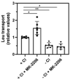
Cancer cells need a constant supply of nutrients. SLC6A14, an amino acid transporter B0,+ (ATB0,+) that is upregulated in many cancers, transports all but acidic amino acids. In its exit from the endoplasmic reticulum (ER), it is recognized by the SEC24C subunit of coatomer II (COPII) for further vesicular trafficking to the plasma membrane. SEC24C has previously been shown to be phosphorylated by protein kinase B/AKT, which is hyper-activated in cancer; therefore, we analyzed the influence of AKT on SLC6A14 trafficking to the cell surface. Studies on overexpressed and endogenous transporters in the breast cancer cell line MCF-7 showed that AKT inhibition with MK-2206 correlated with a transient increase of the transporter in the plasma membrane, not resulting from the inhibition of ER-associated protein degradation. Two-dimensional electrophoresis demonstrated the decreased phosphorylation of SLC6A14 and SEC24C upon AKT inhibition. A proximity ligation assay confirmed this conclusion: AKT inhibition is correlated with decreased SLC6A14 phosphothreonine and SEC24C phosphoserine. Augmented levels of SLC6A14 in plasma membrane led to increased leucine transport. These results show that the inactivation of AKT can rescue amino acid delivery through SLC6A14 trafficking to the cell surface, supporting cancer cell survival. The regulation of the ER export of the amino acid transporter seems to be a novel function of AKT.

Keywords: AKT kinase; SEC24C; SLC6A14; amino acid transporter; breast cancer.


Conflict of interest statement

The authors declare no conflict of interest.

Figures
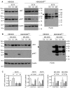
Figure 1
Figure 1
Effect of AKT on overexpressed SLC6A14. HEK293 cells or HEK293 cells transfected for 24 h with p3xFLAG-CMV14/B0,+ vector were treated for the indicated time with either 5 µg/mL SC79 (AKT activator) (A) or 5 µM MK-2206 (AKT inhibitor) (B). The blots were probed with the corresponding antibodies for the presence of AKT, AKT phosphorylated at Ser473 (pAKT), β-actin, and SLC6A14 (anti-FLAG). The representative blots out of 4 (A) or 3 (B) independent cell cultures are shown. Arrows indicate the fully glycosylated SLC6A14. (C) The integration of the indicated blots from (A,B) after transfection with p3xFLAG-CMV14/B0,+ vector. Significance of the respective controls of the lower and upper bands: * p < 0.01, ** p < 0.05.
Figure 2
Figure 2
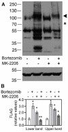
Effect of proteasomal degradation on SLC6A14. HEK293 cells were transfected for 24 h with p3xFLAG-CMV14/B0,+. Where indicated with “+” sign, 50 nM bortezomib was given for 24 h, while 5μM MK-2206 was added for the last 30 min, “−” means lack of the specified compound. (A) The upper panel shows the detection of SLC6A14 with anti-FLAG antibodies, while lower panel shows the detection of β-actin. The arrow indicates the fully glycosylated protein, the asterisk shows the core glycosylated species. (B) Integration of blots with FLAG detection. The analysis was performed with cells from 2 independent passages, and repeated for 2 different lysates from each passage. Significance of the respective control of the lower and upper bands: * p < 0.01, ** p < 0.05.
Figure 3
Figure 3
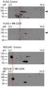
Two-dimensional analysis of SLC6A14 and SEC24C phosphorylation. HEK293 cells transfected for 24 h with p3xFLAG-CMV14/B0,+ were treated, where indicated, for 30 min with 5 μM MK-2206. The cells were lysed, as described in Section 2.2, and the lysates were analyzed via 2D electrophoresis. The change in pI is marked by a red line. The arrows show the migration of a fully glycosylated SLC6A14. Representative blots out of 4 independent experiments are shown.
Figure 4
Figure 4
PLA analysis detecting phosphorylation with serine and threonine moieties on SEC24C and overexpressed SLC6A14. HEK293 cells transfected for 24 h with p3xFLAG-CMV14/B0,+ were treated, where indicated, for 30 min with 5 μM MK-2206, then fixed and subjected to PLA analysis. The following primary antibodies were used: rabbit anti-FLAG (1:1000) with either mouse anti-phosphoserine (1:250) or mouse anti-phosphothreonine (1:200), and rabbit anti-SEC24C (1:250) with either mouse anti-phosphoserine (1:250) or mouse anti-phosphothreonine (1:200), as indicated on the left. The presence of phosphorylated amino acid in the studied protein is visualized by the red dots, while the nuclei are shown in blue. NC, negative control; PLA, without the primary antibodies. The experiment was performed with 2 independent cell passages subcultured in 3 different coverslips each. The selected images come from the same passage. Bar: 20 μm.
Figure 5
Figure 5
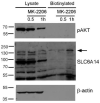
Effect of AKT inhibition on endogenous SLC6A14. MCF-7 cells were treated, where indicated, with 5 µM MK-2206. The cells were subjected to the biotinylation of surface proteins, as described in Section 2.5. The biotinylated fraction (from 800 µg protein) and the lysates (30 µg protein) were subjected to Western blot analysis and probed with anti-phosphoAKT antibodies (pAKT) and antibodies directed against SLC6A14 and β-actin. The representative blot out of 3 independent experiments is shown. The arrow indicates the presence of SLC6A14 in the plasma membrane.
Figure 6
Figure 6
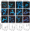
PLA analysis detecting phosphorylation on the serine and threonine moieties of endogenous SLC6A14 and SEC24C in breast cancer cells. MCF-7 cells were treated, where indicated, for 1 h with 5 μM MK-2206, then fixed and subjected to PLA analysis. The following primary antibodies were used: biotin rabbit anti-SLC6A14 antibody (1:250) with either mouse anti-phosphoserine (1:250) or mouse anti-phosphothreonine (1:200), and rabbit anti-SEC24C (1:250) with either mouse anti-phosphoserine (1:250) or mouse anti-phosphothreonine (1:200), as indicated. The presence of phosphorylated amino acid in the studied protein is visualized via the red dots, while nuclei are shown in blue. NC, negative control; PLA, without the primary antibodies. The representative images out of 2 independent cell cultures subcultured in 3 different coverslips each are shown. The selected images come from the same passage. Bar: 40 μm. The summarized results of the PLA assays are shown below. Statistical significance compared to the control at * p < 0.05.
Figure 7
Figure 7
Immunocytochemistry analysis of SEC24C phosphorylation in MCF-7 cells. Untreated cells or cells incubated for the indicated time with 5 μM MK-2206 were fixed and subjected to the immunocytochemistry procedure, as described in Section 2.4. The following primary antibodies were used: rabbit anti-SEC24C (1:250, red) with either mouse anti-phosphoserine (1:250, green) or mouse anti-phosphothreonine (1:200, green), as indicated on the left. The selected areas are shown as magnified images either above or below the corresponding panels. The detected co-localizations of SEC24C and phosphorylated amino acid are indicated with yellow arrows. Representative images from 2 independent cell passages subcultured in 3 different coverslips each are shown. The selected images come from the same passage. Bar: 20 μm.
Figure 8
Figure 8
Immunocytochemistry analysis of SLC6A14 phosphorylation in MCF-7 cells. Untreated cells or cells incubated for the indicated time with 5 μM MK-2206 were fixed and subjected to the immunocytochemistry procedure, as described in Section 2.4. The following primary antibodies were used: rabbit anti-SLC6A14 (1:250, red) with either mouse anti-phosphoserine (1:250, green) or mouse anti-phosphothreonine (1:200, green), as indicated on the left. The selected areas are shown as magnified images either above or below the corresponding panels. The detected co-localizations of transporter and phosphorylated amino acid are indicated with yellow arrows, and the surface presence of SLC6A14 with red arrows. Representative images from 2 independent cell passages subcultured in 3 different cover slips each are shown. The selected images come from the same passage. Bar: 20 μm.
Figure 9
Figure 9
Leucine transport in MCF7 cells. Transport of [3H]leucine was measured for 10 min, as described in Section 3.5, either in PBS (+Cl) or in gluconate solution (−Cl), with or without 30 min treatment with MK-2206, where indicated. The results of 6 independent measurements with different cell cultures are shown. * p < 0.01, ** p < 0.05.

Similar articles

Cited by

References

    1. Hanahan D., Weinberg R.A. Hallmarks of Cancer: The Next Generation. Cell. 2011;144:646–674. doi: 10.1016/j.cell.2011.02.013. - DOI - PubMed
    1. Nałęcz K.A. Amino Acid Transporter SLC6A14 (ATB0,+)–A Target in Combined Anti-cancer Therapy. Front. Cell Dev. Biol. 2020;8:594464. doi: 10.3389/fcell.2020.594464. - DOI - PMC - PubMed
    1. Scalise M., Pochini L., Galluccio M., Indiveri C. Glutamine transport. From energy supply to sensing and beyond. Biochim. Biophys. Acta (BBA) Bioenerg. 2016;1857:1147–1157. doi: 10.1016/j.bbabio.2016.03.006. - DOI - PubMed
    1. Scalise M., Pochini L., Galluccio M., Console L., Indiveri C. Glutamine transporters as pharmacological targets: From function to drug design. Asian J. Pharm. Sci. 2020;15:207–219. doi: 10.1016/j.ajps.2020.02.005. - DOI - PMC - PubMed
    1. Kandasamy P., Gyimesi G., Kanai Y., Hediger M.A. Amino acid transporters revisited: New views in health and disease. Trends Biochem. Sci. 2018;43:752–789. doi: 10.1016/j.tibs.2018.05.003. - DOI - PubMed

Publication types

MeSH terms

LinkOut - more resources