. 2021 Jul 18;10(7):1816.
doi: 10.3390/cells10071816.

Sustained Systemic Levels of IL-6 Impinge Early Muscle Growth and Induce Muscle Atrophy and Wasting in Adulthood

Affiliations

Sustained Systemic Levels of IL-6 Impinge Early Muscle Growth and Induce Muscle Atrophy and Wasting in Adulthood

Laura Pelosi et al. Cells. .

Abstract

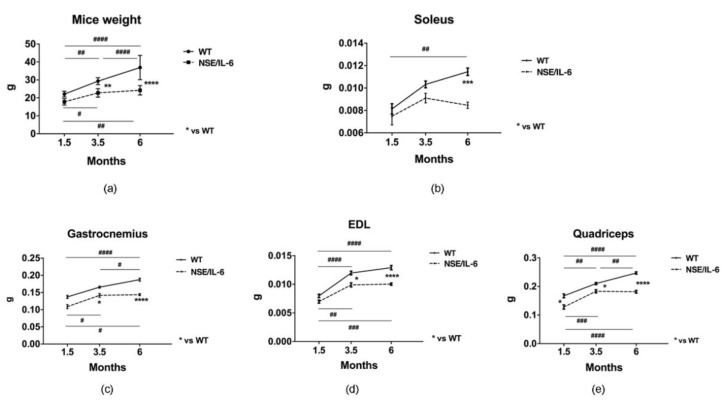
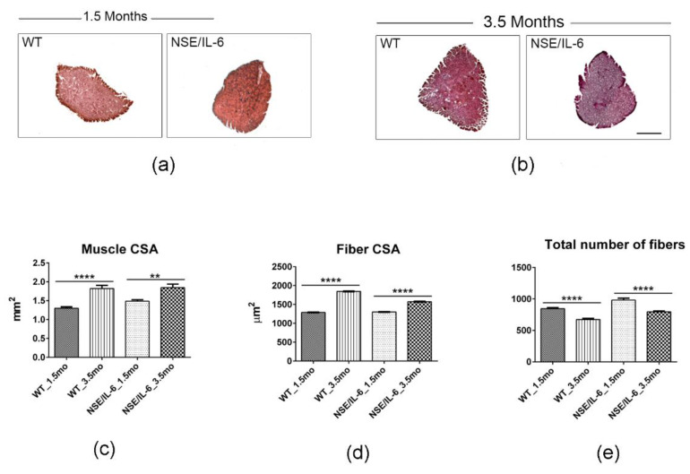
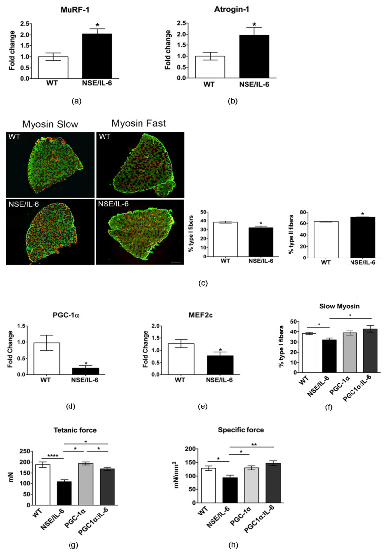
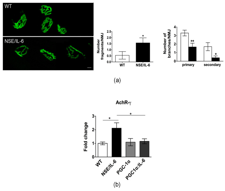
IL-6 is a pleiotropic cytokine that can exert different and opposite effects. The muscle-induced and transient expression of IL-6 can act in an autocrine or paracrine manner, stimulating anabolic pathways associated with muscle growth, myogenesis, and with regulation of energy metabolism. In contrast, under pathologic conditions, including muscular dystrophy, cancer associated cachexia, aging, chronic inflammatory diseases, and other pathologies, the plasma levels of IL-6 significantly increase, promoting muscle wasting. Nevertheless, the specific physio-pathological role exerted by IL-6 in the maintenance of differentiated phenotype remains to be addressed. The purpose of this study was to define the role of increased plasma levels of IL-6 on muscle homeostasis and the mechanisms contributing to muscle loss. Here, we reported that increased plasma levels of IL-6 promote alteration in muscle growth at early stage of postnatal life and induce muscle wasting by triggering a shift of the slow-twitch fibers toward a more sensitive fast fiber phenotype. These findings unveil a role for IL-6 as a potential biomarker of stunted growth and skeletal muscle wasting.

Keywords: PGC-1α; interleukin-6; muscle atrophy; muscle growth; skeletal muscle.


Conflict of interest statement

The authors declare no conflict of interest. The funders had no role in the design of the study; in the collection, analyses, or interpretation of data; in the writing of the manuscript, or in the decision to publish the results.

Figures

Figure 1
Figure 1
IL-6 overexpression affects muscle growth and size in distinct muscle groups. (a) Body weight growth curves. Wild type (WT) and NSE/IL-6 mice were measured to determine the change in body weight from 1.5 to 6 months of age. n = 9; ** p < 0.01, **** p < 0.0001, # p < 0.05, ## p < 0.01, #### p < 0.0001. (be) Growth curve of soleus (b), gastrocnemius (c), extensor digitorum longus (EDL) (d), and quadriceps muscles (e) of indicated genotype.. n = 12; * p < 0.05, *** p < 0.001; **** p < 0.0001, # p < 0.05, ## p < 0.01, ### p < 0.001, #### p < 0.0001. All measurements are presented as means ± SEM. Statistical significance assessed by two-way ANOVA followed by multiple comparisons test.
Figure 2
Figure 2
IL-6 overexpression significantly impairs muscle growth during the postnatal life. (a,b) Hematoxylin and eosin staining of transverse section of soleus muscles from indicated genotypes at 1.5 and 3.5. Scale bar 500μm. (ce) The graphs show quantification of total cross-sectional area (muscle CSA) (c), fibers-cross sectional area (Fiber CSA) (d) and total number of fibers (e) in soleus muscles of wild type (WT) and NSE/IL-6 (IL-6) mice at 1.5 and 3.5 months of age. Data are represented as average ± SEM. n = 10 mice; ** p < 0.01, **** p < 0.0001. Statistical significance assessed by two-way ANOVA.
Figure 3
Figure 3
IL-6 overexpression promotes muscle atrophy and wasting and affects the functional performance of skeletal muscle in adulthood. (a) Hematoxylin and eosin staining of transverse section of soleus muscles from indicated genotypes at 6 months of age. Scale bar 500 μm.(bd) Analysis of morphologic and morphometric parameters in transgenic (NSE/IL-6) and wild type (WT) soleus muscle at 6 months of age. Data are represented as average ± SEM. n = 10; * p < 0.05, **** p < 0.0001. Statistical significance assessed by unpaired Student’s t-test. (eg) Frequency distribution of myofiber-cross sectional area in transgenic (NSE/IL-6) and wild type (WT) soleus at 1.5 (e), 3.5 (f), and 6 (g) months of age. Data are represented as medians. n = 10; **** p < 0.0001. Statistical significance assessed by Mann–Whitney Rank Sum Test. (hk) Physiological properties of soleus muscle from 3.5 (h,i) and 6 (j,k)-month-old wild type (WT) and NSE/IL-6 mice. Tetanic force and Specific force. Data are represented as average ± SEM. n = 15; * p < 0.05, **** p < 0.0001. Statistical significance assessed by unpaired Student’s t-test.
Figure 4
Figure 4
IL-6 overexpression impinges the heterogeneity of muscle fibers. (a,b) Real time PCR of ubiquitin-proteasome markers, Atrogin-1 and MuRF1 in soleus of wild type (WT) and NSE/IL-6 mice at 6 months of age. Values are expressed as fold change variations relative to WT and are presented as means ± SEM and n = 4; * p < 0.05. Statistical significance assessed by Mann–Whitney Rank Sum Test. (c) Immunofluorescence staining of wild type (WT) and NSE/IL-6 soleus muscles immunolabeled with either a mouse anti-myosin slow or a mouse anti-myosin fast antibody at 6 months of age. Scale bar 250μm. At the right, graphs showing the percentage of positive type I (left) or type II (right) muscle fibers. Data are represented as average ± SEM. n = 4 * p< 0.05. Statistical significance assessed by Mann–Whitney Rank Sum Test. (d,e) Real time PCR for the expression of PGC-1α and MEF2c in soleus of wild type (WT) and NSE/IL-6 mice at 6 months of age. Values are presented as means ± SEM and n = 4; * p < 0.05. Statistical significance assessed by Mann–Whitney Rank Sum Test. (f) Quantification of immunofluorescence staining of wild type (WT), NSE/IL-6, PGC-1α and IL-6:MCK/PGC-1α (PGC1α:IL-6) soleus muscles immunolabeled with a mouse anti-myosin slow antibody at 6 months of age. Data are represented as average ± SEM. n = 3, * p< 0.05. Statistical significance assessed by Mann–Whitney Rank Sum Test. (g,h) Physiological properties of soleus muscle from 6-month-old wild type (WT), NSE/IL-6, PGC-1α and IL-6:MCK/PGC-1α (PGC1α:IL-6) mice: Tetanic force and Specific force. Data are represented as average ± SEM. n = 8; * p < 0.05, ** p < 0.01, **** p < 0.0001. Statistical significance assessed by unpaired Student’s t-test.
Figure 5
Figure 5
IL-6 overexpression affects neuromuscular junction (NMJ) destabilization. (a) Confocal microscopy of post-synaptic NMJ stained with a-bungarotoxin; representative images of one spatial series (from 0 to 50 µm) composed of 25 optical sections with a step size of 2 µm, from tibialis anterior of 6-month-old wild type and NSE/IL-6 mice. Scale bar 10 μm. Graphs showing the degree of NMJ fragmentation (Number fragments/NMJ) and the topology of branching pattern (Number of branches/NMJ). Data are represented as average ± SEM n = 3; * p< 0.05, ** p < 0.01. Statistical significance assessed by Mann–Whitney Rank Sum Test. (b) Real time PCR for the expression of AchR-γ in soleus wild type (WT) and NSE/IL-6, PGC-1α, and PGC1α:IL-6 mice at 6 months of age. Values are expressed as fold change variations relative to WT and are presented as means ± SEM and are n = 3; * p < 0.05. Statistical significance assessed by Mann–Whitney Rank Sum Test.

References

    1. Naka T., Narazaki M., Hirata M., Matsumoto T., Minamoto S., Aono A., Nishimoto N., Kajita T., Taga T., Yoshizaki K., et al. Structure and function of a new STAT-induced STAT inhibitor. Nature. 1997;387:924–929. doi: 10.1038/43219. - DOI - PubMed
    1. Tanaka T., Narazaki M., Kishimoto T. IL-6 in inflammation, immunity, and disease. Cold Spring Harb. Perspect. Biol. 2014;6:a016295. doi: 10.1101/cshperspect.a016295. - DOI - PMC - PubMed
    1. Serrano A.L., Baeza-Raja B., Perdiguero E., Jardí M., Muñoz-Cánoves P. Interleukin-6 Is an Essential Regulator of Satellite Cell-Mediated Skeletal Muscle Hypertrophy. Cell Metab. 2008;7:33–44. doi: 10.1016/j.cmet.2007.11.011. - DOI - PubMed
    1. Al-Shanti N., Stewart C.E. Inhibitory effects of IL-6 on IGF-1 activity in skeletal myoblasts could be mediated by the activation of SOCS-3. J. Cell. Biochem. 2012;113:923–933. doi: 10.1002/jcb.23420. - DOI - PubMed
    1. Baeza-Raja B., Muñoz-Cánoves P. p38 MAPK-induced Nuclear Factor-κB Activity Is Required for Skeletal Muscle Differentiation: Role of Interleukin-6. Mol. Biol. Cell. 2004;15:2013–2026. doi: 10.1091/mbc.e03-08-0585. - DOI - PMC - PubMed

Publication types