

Rhabdomyosarcoma of the Cervix in a Post-Menopausal Woman-An Unparalleled Phenomenon
- PMID: 34360144
- PMCID: PMC8345433
- DOI: 10.3390/ijerph18157851
Rhabdomyosarcoma of the Cervix in a Post-Menopausal Woman-An Unparalleled Phenomenon
Abstract
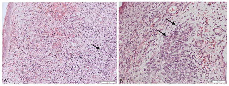
Rhabdomyosarcoma of the cervix is a soft tissue sarcoma that usually occurs in young women. It is very rare in adulthood. We discuss symptoms, the process of diagnosis of rhabdomyosarcoma embryonale of the cervix in a 61-year-old women and differences in treatment dependent on patient's age. A 61-year-old woman with symptoms such as palpable mass in the external cervical opening and post-menopausal hemorrhaging was admitted to the oncology ward where excision of the polyp was performed. Embryonal rhabdomyosarcoma (ERMS) was diagnosed by histopathological examination of obtained tissues. The diagnosis was complemented by chest computed tomography and pelvis magnetic resonance imaging to exclude metastases. A Wertheim-Meigs operation and excision of the ovaries, the fallopian tubes and the surrounding tissue was performed in the course of treatment. In the patient's follow-up of 25 months to date, there have been no signs of recurrence or symptoms connected to ERMS. Based on the therapeutic outcome, the decision to limit the treatment to a surgical resection was adequate for a post-menopausal patient. Because of the rarity of ERMS in the post-menopausal age, we think that the patient should be carefully followed up to further examine this issue and develop diagnostic and treatment guidelines.
Keywords: cervix; gynecology; neoplasm; oncology; post-menopausal; rhabdomyosarcoma; sarcoma; uterus.
Conflict of interest statement
The authors declare no conflict of interest.
Figures


References
-
- Ferrari A., Dirksen U. Sarcomas of Soft Tissue and Bone. Prog. Tumor Res. 2016;43:128–141. - PubMed
Publication types
MeSH terms
LinkOut - more resources
Full Text Sources
