Review
. 2021 Jul 21;22(15):7785.
doi: 10.3390/ijms22157785.

Mechanisms Governing Anaphylaxis: Inflammatory Cells, Mediators, Endothelial Gap Junctions and Beyond

Affiliations
Review

Mechanisms Governing Anaphylaxis: Inflammatory Cells, Mediators, Endothelial Gap Junctions and Beyond

Samantha Minh Thy Nguyen et al. Int J Mol Sci. .

Abstract

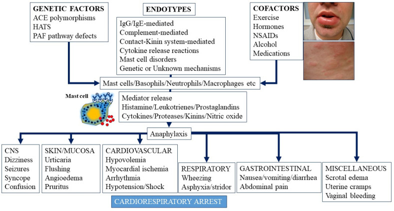
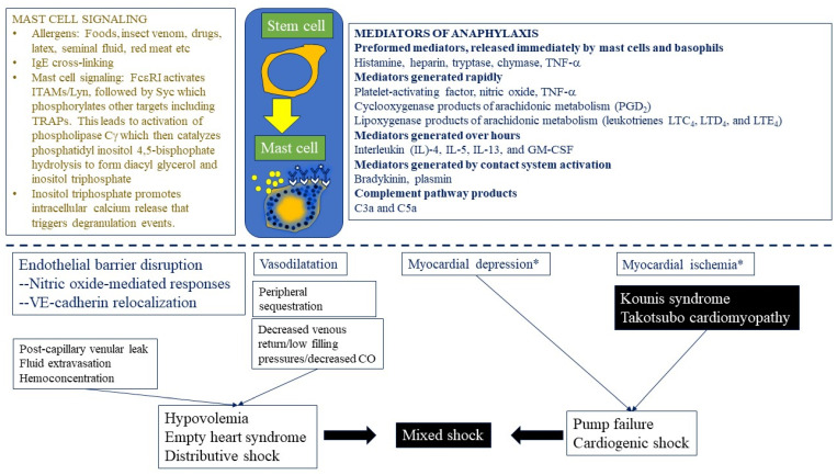
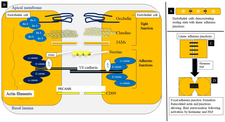
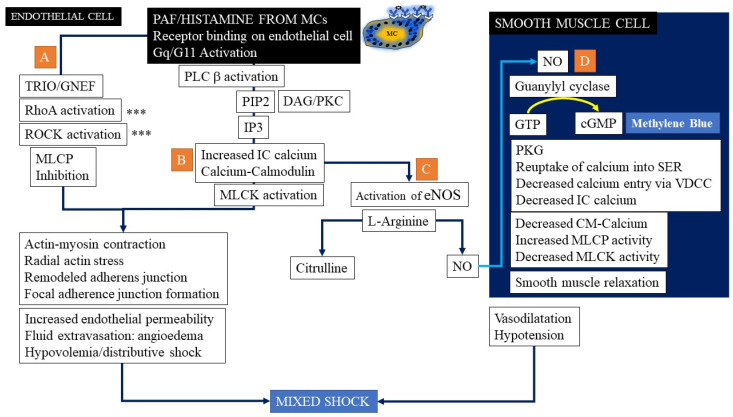
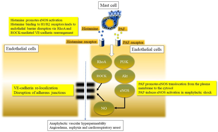
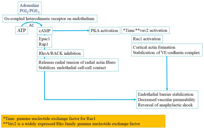
Anaphylaxis is a severe, acute, life-threatening multisystem allergic reaction resulting from the release of a plethora of mediators from mast cells culminating in serious respiratory, cardiovascular and mucocutaneous manifestations that can be fatal. Medications, foods, latex, exercise, hormones (progesterone), and clonal mast cell disorders may be responsible. More recently, novel syndromes such as delayed reactions to red meat and hereditary alpha tryptasemia have been described. Anaphylaxis manifests as sudden onset urticaria, pruritus, flushing, erythema, angioedema (lips, tongue, airways, periphery), myocardial dysfunction (hypovolemia, distributive or mixed shock and arrhythmias), rhinitis, wheezing and stridor. Vomiting, diarrhea, scrotal edema, uterine cramps, vaginal bleeding, urinary incontinence, dizziness, seizures, confusion, and syncope may occur. The traditional (or classical) pathway is mediated via T cells, Th2 cytokines (such as IL-4 and 5), B cell production of IgE and subsequent crosslinking of the high affinity IgE receptor (FcεRI) on mast cells and basophils by IgE-antigen complexes, culminating in mast cell and basophil degranulation. Degranulation results in the release of preformed mediators (histamine, heparin, tryptase, chymase, carboxypeptidase, cathepsin G and tumor necrosis factor alpha (TNF-α), and of de novo synthesized ones such as lipid mediators (cysteinyl leukotrienes), platelet activating factor (PAF), cytokines and growth factors such as vascular endothelial growth factor (VEGF). Of these, histamine, tryptase, cathepsin G, TNF-α, LTC4, PAF and VEGF can increase vascular permeability. Recent data suggest that mast cell-derived histamine and PAF can activate nitric oxide production from endothelium and set into motion a signaling cascade that leads to dilatation of blood vessels and dysfunction of the endothelial barrier. The latter, characterized by the opening of adherens junctions, leads to increased capillary permeability and fluid extravasation. These changes contribute to airway edema, hypovolemia, and distributive shock, with potentially fatal consequences. In this review, besides mechanisms (endotypes) underlying IgE-mediated anaphylaxis, we also provide a brief overview of IgG-, complement-, contact system-, cytokine- and mast cell-mediated reactions that can result in phenotypes resembling IgE-mediated anaphylaxis. Such classifications can lead the way to precision medicine approaches to the management of this complex disease.

Keywords: allergic reaction; allergy; anaphylactic shock; anaphylaxis; angioedema; coagulation; complement; cytokines; epinephrine; food allergy; histamine; hypotension; mast cell; tryptase.


Conflict of interest statement

The authors declare no conflict of interests.

Figures

Figure 1
Figure 1
Anaphylaxis-triggers (including endotypes), genetic predisposition, selected cofactors and role of mast cells and mediators in manifestations and culminating potentially in cardiorespiratory arrest. CNS = central nervous system, NSAIDs = nonsteroidal anti-inflammatory drugs, ACE = angiotensin converting enzyme, HATS = hereditary alpha tryptasemia syndrome, PAF = platelet activating factor, IgE = immunoglobulin E, IgG = immunoglobulin G.
Figure 2
Figure 2
Human MCs, originating from CD34+/CD117+/CD13+ multipotent, hematopoietic progenitors, migrate to peripheral tissues and undergo differentiation and maturation under the influence of growth factors, including stem cell factor (SCF). The figure shows mast cell signaling and mediators expressed in anaphylaxis and their potential relationship to endothelial, blood vessel, myocardial and circulatory effects. A complex sequence of events including endothelial barrier disruption, fluid extravasation, vasodilatation, decreased venous return, myocardial depression, and myocardial ischemia lead to distributive or mixed shock pictures. ITAM = immunoreceptor tyrosine-based activation motifs; Lyn/Syc/TRAPS/PLC/PIP2/DAG/IP3 are signaling molecules (see text for elaboration). CO = cardiac output.
Figure 3
Figure 3
Multiple autocrine and paracrine loops that may play a role in refractory anaphylaxis. 1 = TNF-NFkappaB loop; 2 = Stem cell factor-mast cell growth loop; 3 = mast cell histamine-endothelial nitric oxide synthase (eNOS or NOS3) and barrier disruption loop; 4 = Feedback inhibition of mast cell degranulation by NO.
Figure 4
Figure 4
Structure of the adherens and tight junctions in endothelial cell–cell contact and barrier development. (A) This represents resting state with AJ configuration regulated by mechanical forces and shear stress. (B) The endothelial barrier is regulated by a set of cell–cell adhesions which include tight junctions (TJs), adherens junctions (AJs), gap junctions and other molecules (including nectins, PECAM and CD99) which in turn associate with the actin skeleton. Three types of catenins (α, β and γ [plakoglobin]) bind to and stabilize the VE-cadherin molecule, with α-catenin serving as a communication of cadherins with actin filaments. (C) The VE-cadherin-catenin complex also associates with vinculin (V) which serve as actin-binding protein. 120-Catenin binds directly to the cytoplasmic domain of VE-cadherin, close to the membrane, while the β- and γ-catenins bind to the cytoplasmic tail and helping to anchor α-catenin which in turn is tethered to the actin cytoskeleton by other proteins including vinculin, α-actinin and afadin. (D) Permeability-increasing factors such as histamine and PAF cause remodeling of the actin cytoskeleton and destabilization of the endothelial cell to cell junctions. Induction of radial contractile actin bundles, and associated actin-myosin contraction causes the re-localization of linear VE-cadherin complexes to focal adherence junctions leading to formation of intercellular gaps in endothelial cells and resultant hyperpermeability.
Figure 5
Figure 5
Binding of PAF and histamine to their respective receptors which are G protein-linked leads to activation of Gq/G11. This leads to the activation of the guanine nucleotide exchange factor, Trio, which in sequence activates the small GTPases such as RhoA, which then activate the serine/threonine kinase, ROCK which in turn phosphorylates myosin light chain phosphatase (MLCP), inhibiting its activity (A). Receptor binding activates phospholipase Cβ (B) which then catalyzes PIP2 (phosphatidyl inositol 4,5-bisphophate) hydrolysis to form DAG (diacyl glycerol) and IP3 (inositol triphosphate). Calcium-dependent activation of MLC kinase (MLCK) now occurs, resulting in increased acto-myosin contractility and contributing to changing actin bundle orientation (induction of radial actin stress fibers) with the latter switching from being parallel to the junctions to perpendicular, thereby inducing junctional stress and disrupting integrity and vascular leakiness. Meanwhile, nitric oxide (NO), produced not by inducible nitric oxide synthase (iNOS), but by constitutive endothelial form (eNOS) that is rapidly activated via the PI3K/Akt pathway (C). NO induces the formation of cGMP in smooth muscle cells from sGC (D), which then activates PKG leading to a reuptake of calcium from the cytosol by the sarcoplasmic reticulum (SER) as well diminished calcium influx via voltage-dependent calcium channels (VDCC). This, combined with the opening of potassium channels and the exit of calcium out of the cell leads to drop in intracellular calcium concentrations, inactivation of calmodulin and resultant failure to activate MLCK. MLC phosphatase activity also increases correspondingly, leading to disruption of the actin-myosin cross-bridge and causing vasodilatation of blood vessels. Methylene blue has become a novel treatment for refractory cases of distributive shock due to its inhibitory effects on the eNOS-cGMP pathway.
Figure 6
Figure 6
Activation pathways of histamine and PAF with involvement of their respective endothelial receptors are shown-leading to capillary permeability changes via vascular endothelial-cadherin (VE cadherin) and adherens junction (AJ) disruption. This culminates in fluid leak from postcapillary venules (PCV) contributing to hypovolemic/distributive shock. The elaboration of other mediators can also contribute to bronchospasm, angioedema and respiratory distress leading to asphyxia/hypoxemia and respiratory arrest. Involvement of RhoA and ROCK signaling in VE-cadherin relocalization as well as the PI3kinase-Akt pathway of eNOS activation are shown in cartoon format (adapted from Nakamura and Muraata, 2018) [65].
Figure 7
Figure 7
Epinephrine (or adrenaline), prostacyclin (PGI2) and PGE2 bind to G protein linked receptors, which activate adenyl cyclase (AC), which in turn leads to the formation of cyclic AMP (cAMP). cAMP via Epac1 activates Rap1 which inhibits Rhoa and ROCK and thereby relieves the radial tension of the radial actin fibers. Another pathway via Protein kinase A (PKA)/Tiam-Vav2-Rac1 pathway results in stabilizing VE-cadherin complexes.
Figure 8
Figure 8
Activation of the contact system results in inflammatory responses, coagulation cascades with vascular thrombosis, fibrinolysis and complement activation. 1 = Mast cell derived histamine, PAF and IL-6 can cause disruption of tight and adherens junctions. 2 = mast cell derived heparin or PolyP can activate Factor XII which sequentially activates kallikrein and high molecular weight kininogen (HMWK), resulting in formation of bradykinin (BK) which can bind to BK receptors and increase vascular permeability. 3 = tryptase from mast cells could activate factor XII and kallikrein. 4 = Mast cells can activate fibrinolysis by secretion of tissue plasminogen activator (tPA) or urokinase. 5 = Mast cell tryptase has also been shown to activate fibrinolytic pathways. The net effect of contact pathway activation is increased capillary permeability, angioedema, and hypovolemic shock.

References

    1. LoVerde D., Iweala O.I., Eginli A., Krishnaswamy G. Anaphylaxis. Chest. 2018;153:528–543. doi: 10.1016/j.chest.2017.07.033. - DOI - PMC - PubMed
    1. Krishnaswamy G. Critical Care Management of the Patient With Anaphylaxis: A Concise Definitive Review. Crit. Care Med. 2021;49:838–857. doi: 10.1097/CCM.0000000000004893. - DOI - PubMed
    1. Cardona V., Ansotegui I.J., Ebisawa M., El-Gamal Y., Rivas M.F., Fineman S., Geller M., Gonzalez-Estrada A., Greenberger P.A., Borges M.S., et al. World Allergy Organization Anaphylaxis Guidance 2020. World Allergy Organ. J. 2020;13 doi: 10.1016/j.waojou.2020.100472. - DOI - PMC - PubMed
    1. Sclar D.A., Lieberman P.L. Anaphylaxis: Underdiagnosed, Underreported, and Undertreated. Am. J. Med. 2014;127:S1–S5. doi: 10.1016/j.amjmed.2013.09.007. - DOI - PubMed
    1. Sampson H.A., Muñoz-Furlong A., Campbell R.L., Adkinson N.F., Bock S.A., Branum A., Brown S., Camargo C.A., Cydulka R., Galli S.J., et al. Second symposium on the definition and management of anaphylaxis: Summary report—Second National Institute of Allergy and Infectious Disease/Food Allergy and Anaphylaxis Network symposium. J. Allergy Clin. Immunol. 2006;117:391–397. doi: 10.1016/j.jaci.2005.12.1303. - DOI - PubMed