. 2021 Jul 22;22(15):7852.
doi: 10.3390/ijms22157852.

Mucus Release and Airway Constriction by TMEM16A May Worsen Pathology in Inflammatory Lung Disease

Affiliations

Mucus Release and Airway Constriction by TMEM16A May Worsen Pathology in Inflammatory Lung Disease

Raquel Centeio et al. Int J Mol Sci. .

Abstract

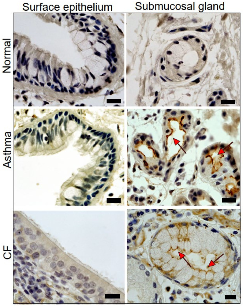
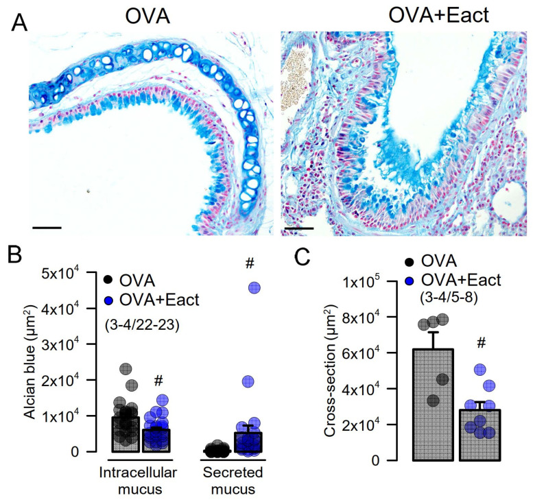
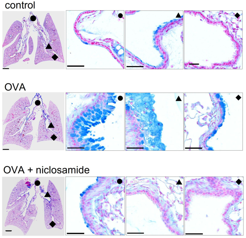
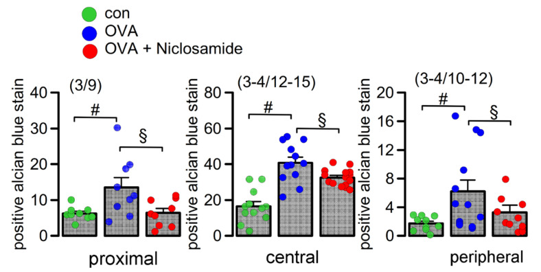
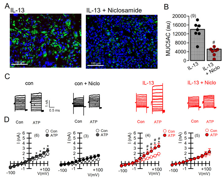
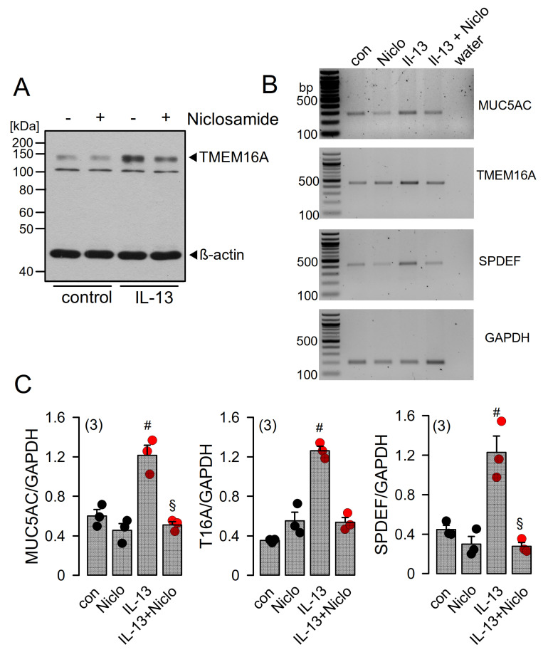
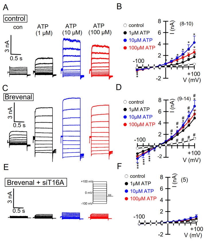
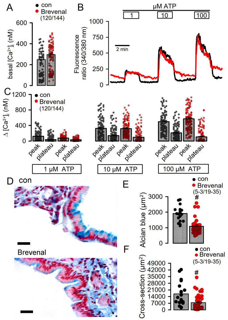
Activation of the Ca2+ activated Cl- channel TMEM16A is proposed as a treatment in inflammatory airway disease. It is assumed that activation of TMEM16A will induce electrolyte secretion, and thus reduce airway mucus plugging and improve mucociliary clearance. A benefit of activation of TMEM16A was shown in vitro and in studies in sheep, but others reported an increase in mucus production and airway contraction by activation of TMEM16A. We analyzed expression of TMEM16A in healthy and inflamed human and mouse airways and examined the consequences of activation or inhibition of TMEM16A in asthmatic mice. TMEM16A was found to be upregulated in the lungs of patients with asthma or cystic fibrosis, as well as in the airways of asthmatic mice. Activation or potentiation of TMEM16A by the compounds Eact or brevenal, respectively, induced acute mucus release from airway goblet cells and induced bronchoconstriction in mice in vivo. In contrast, niclosamide, an inhibitor of TMEM16A, blocked mucus production and mucus secretion in vivo and in vitro. Treatment of airway epithelial cells with niclosamide strongly inhibited expression of the essential transcription factor of Th2-dependent inflammation and goblet cell differentiation, SAM pointed domain-containing ETS-like factor (SPDEF). Activation of TMEM16A in people with inflammatory airway diseases is likely to induce mucus secretion along with airway constriction. In contrast, inhibitors of TMEM16A may suppress pulmonary Th2 inflammation, goblet cell metaplasia, mucus production, and bronchoconstriction, partially by inhibiting expression of SPDEF.

Keywords: ETX001; Eact; TMEM16A; airways; asthma; brevenal; cystic fibrosis.


Conflict of interest statement

The authors declare no conflict of interest.

Figures

Figure 1
Figure 1
Expression of TMEM16A in human healthy, asthmatic, and CF lungs. Lung slices of normal airways indicate little expression of TMEM16A in both surface airway epithelium and airway submucosal glands. Pronounced expression of TMEM16A in apical membranes of surface epithelial cells and airway submucosal glands of patients with asthma and CF (brown precipitation, DAP staining). Submucosal glands were often found to be hypertrophic. Representative stainings of four patients each. Bars indicate 20 µm.
Figure 2
Figure 2
Activation of TMEM16A by Eact induces mucus release and airway contraction. (A) Mucus staining by alcian blue in OVA-treated asthmatic mice. Acute application of the activator of TMEM16A, Eact (4.8 µg/mL intratracheal), induced acute release of mucus from goblet cells and increased intraluminal mucus. Bars indicate 50 µm. (B) Summary of intracellular and intraluminal mucus. (C) Summary of cross sectional area indicating airway contraction by Eact. Mean ± SEM (number of animals/number of measurements). # significant difference when compared to control (p < 0.05, unpaired t-test).
Figure 3
Figure 3
Mucus production and mucus secretion is inhibited by niclosamide. Whole mouse lung analysis by stitching microscopy. Low level mucus production and mucus secretion in proximal (●), central (▲), and peripheral (♦) airways of control mice. Bars = 1000 (very left panels) and 50 µm (higher resolution panels), respectively. Pronounced upregulation of mucus production and intraluminal mucus accumulation in lungs of OVA-treated (asthmatic) mice. Mucus production and mucus secretion is strongly suppressed in asthmatic mice treated with niclosamide (13 µg/mL, 4 days).
Figure 4
Figure 4
Quantification of airway mucus. Quantification of mucus in proximal, central, and peripheral airways. Mean ± SEM (Number of mice/number of airways analyzed). # significant difference when compared to control (p < 0.05, ANOVA). § significant difference when compared to OVA (p < 0.05, ANOVA).
Figure 5
Figure 5
Expression of MUC5AC and activation of TMEM16A in Calu-3 airway epithelial cells is inhibited by niclosamide. (A) Expression of MUC5AC induced by IL-13 (20 ng/mL; 72 h) in Calu-3 airway epithelial cells was inhibited by simultaneous incubation with niclosamide (1 µM). Bar = 100 µm. (B) Quantification of MUC5AC expression indicating inhibition by niclosamide (Niclo). (C) Current overlays from whole-cell patch clamp experiments before and after induction of MUC5AC expression by IL-13. Activation of whole-cell currents by purinergic stimulation (ATP, 100 µM) was enhanced by IL-13, which was completely inhibited by acute application of niclosamide (1 µM). (D) Corresponding current/voltage relationships. The inhibitor of Ca2+-activated KCNN4 K+ channels, TRAM-34 (100 nM), was present in all patch clamp experiments to avoid potential activation of Ca2+-activated K+ channels. Mean ± SEM (number of cells). * significant activation by ATP (p < 0.05, paired t-test). # significant difference when compared to the absence of IL-13 (p < 0.05, unpaired t-test).
Figure 6
Figure 6
Niclosamide inhibits expression of MUC5AC and SPDEF in Calu-3 cells. (A) Western blot indicating upregulation of TMEM16A in Calu-3 cells by IL-13 (20 ng/mL, 72 h) and inhibition of expression by niclosamide (1 µM). Blots were performed in triplicates. (B) RT-PCR analysis of the expression of MUC5AC, TMEM16A, and SAM pointed domain-containing ETS transcription factor (SPDEF) in Calu-3 airway epithelial cells. SPDEF is an integrator of goblet cell differentiation and pulmonary Th2 inflammation. (C) Low cycle numbers (20×) were chosen for quantification of expression by relating specific signals to expression of the housekeeper protein GAPDH. IL-13 (20 ng/mL) leads to upregulation of expression of MUC5AC, TMEM16A, and SPDEF. Niclosamide (Niclo, 1 µM, 72 h) strongly inhibits expression of MUC5AC, TMEM16A, and SPDEF. Mean ± SEM (number of assays). #,§ significant increase by IL-13 and inhibition by niclosamide, respectively (p < 0.05, ANOVA).
Figure 7
Figure 7
Activation of TMEM16A whole-cell currents is potentiated by brevenal. (A) Dose-dependent activation of endogenous TMEM16A whole-cell currents in CFBE airway epithelial cells, by the purinergic agonist ATP. Clamp voltages ± 100 mV in steps of 20 mV. (B) Corresponding current/voltage relationships. (C,D) Activation of whole-cell currents obtained from cells pre-incubated with brevenal (500 nM, 15 min) and corresponding current/voltage relationships. (E) Activation of whole-cell currents in brevenal-incubated cells, in which expression of TMEM16A has been knocked down by treatment with siRNA-TMEM16A (c.f. Supplementary Figure E2). (F) Corresponding current/voltage relationships. Mean ± SEM (number of cells). * significant activation by ATP (p < 0.05, ANOVA). The inhibitor of Ca2+-activated KCNN4 K+ channels, TRAM-34 (100 nM), was present in all patch clamp experiments to avoid potential activation of Ca2+-activated K+ channels. # significant difference when compared to the absence of brevenal (p < 0.05, ANOVA). No currents were activated in siRNA-TMEM16A-treated cells.
Figure 8
Figure 8
Brevenal does not increase intracellular Ca2+ but releases mucus from goblet cells and induces airway contraction. (A) Comparable basal intracellular Ca2+ levels in airway epithelial cells in the absence or presence of brevenal (500 nM, 15 min preincubation), as measured by the Ca2+ sensor Fura-2. (B) Concentration-dependent rise in intracellular Ca2+ by ATP in the absence or presence of brevenal. (C) Concentration-dependent increase in peak and plateau Ca2+ by ATP in the absence or presence of brevenal. (D) Effects of acute application of brevenal (3.2 µg/100 µL, 10 min) in mouse airways by tracheal instillation. Alcian blue staining of mucus indicated reduced intracellular mucus in airways treated with brevenal. Brevenal-exposed airways appeared contracted. Bars = 15 µm. (E) Summary of alcian blue staining in airway epithelial cells reflecting the amount of intracellular mucus, which is significantly reduced upon exposure to brevenal. (F) Summary of airway cross sectional area obtained in airways of control mice and mice treated with brevenal, suggesting airway contraction by brevenal. Mean ± SEM; (number of animals/number of measurements). # significant difference when compared to control mice (p < 0.05, ANOVA).

References

    1. Danahay H.L., Lilley S., Fox R., Charlton H., Sabater J., Button B., McCarthy C., Collingwood S.P., Gosling M. TMEM16A potentiation: A novel therapeutic approach for the treatment of cystic fibrosis. Am. J. Respir. Crit. Care Med. 2020 doi: 10.1164/rccm.201908-1641OC. - DOI - PMC - PubMed
    1. Knowles M.R., Clarke L.L., Boucher R.C. Activation by extracellular nucleotides of chloride secretion in the airway epithelia of patients with cystic fibrosis. N. Engl. J. Med. 1991;325:533–538. doi: 10.1056/NEJM199108223250802. - DOI - PubMed
    1. Mall M., Gonska T., Thomas J., Schreiber R., Seydewitz H.H., Kuehr J., Brandis M., Kunzelmann K. Modulation of Ca2+ activated Cl− secretion by basolateral K+ channels in human normal and cystic fibrosis airway epithelia. Pediatr. Res. 2003;53:608–618. doi: 10.1203/01.PDR.0000057204.51420.DC. - DOI - PubMed
    1. Son M., Ito Y., Sato S., Ishikawa T., Kondo M., Nakayama S., Shimokata K., Kume H. Apical and basolateral ATP-induced anion secretion in polarized human airway epithelia. Am. J. Respir. Cell Mol. Biol. 2004;30:411–419. doi: 10.1165/rcmb.2003-0183OC. - DOI - PubMed
    1. Kunzelmann K., Mall M. Pharmacotherapy of the ion transport defect in cystic fibrosis: Role of purinergic receptor agonists and other potential therapeutics. Am. J. Respir. Med. 2003;2:299–309. doi: 10.1007/BF03256658. - DOI - PubMed