. 2021 Jul 23;22(15):7882.
doi: 10.3390/ijms22157882.

Tumor Promoting Effect of BMP Signaling in Endometrial Cancer

Affiliations

Tumor Promoting Effect of BMP Signaling in Endometrial Cancer

Tomohiko Fukuda et al. Int J Mol Sci. .

Abstract

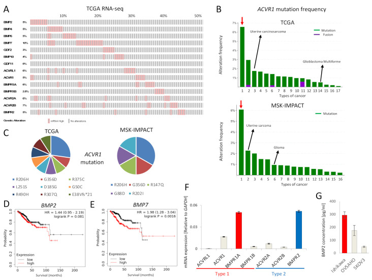
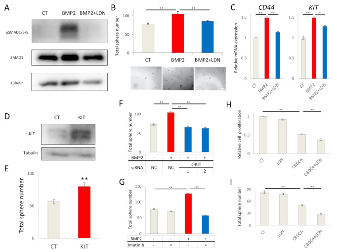
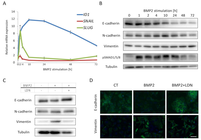
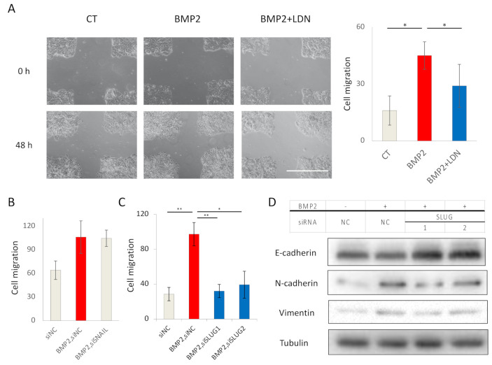
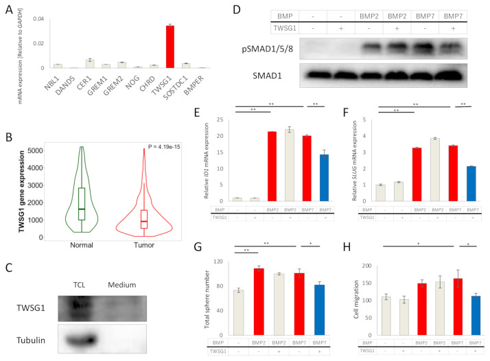
The effects of bone morphogenetic proteins (BMPs), members of the transforming growth factor-β (TGF-β) family, in endometrial cancer (EC) have yet to be determined. In this study, we analyzed the TCGA and MSK-IMPACT datasets and investigated the effects of BMP2 and of TWSG1, a BMP antagonist, on Ishikawa EC cells. Frequent ACVR1 mutations and high mRNA expressions of BMP ligands and receptors were observed in EC patients of the TCGA and MSK-IMPACT datasets. Ishikawa cells secreted higher amounts of BMP2 compared with ovarian cancer cell lines. Exogenous BMP2 stimulation enhanced EC cell sphere formation via c-KIT induction. BMP2 also induced EMT of EC cells, and promoted migration by induction of SLUG. The BMP receptor kinase inhibitor LDN193189 augmented the growth inhibitory effects of carboplatin. Analyses of mRNAs of several BMP antagonists revealed that TWSG1 mRNA was abundantly expressed in Ishikawa cells. TWSG1 suppressed BMP7-induced, but not BMP2-induced, EC cell sphere formation and migration. Our results suggest that BMP signaling promotes EC tumorigenesis, and that TWSG1 antagonizes BMP7 in EC. BMP signaling inhibitors, in combination with chemotherapy, might be useful in the treatment of EC patients.

Keywords: ACVR1; BMP; EMT; cancer stem cells; endometrial cancer.


Conflict of interest statement

The authors have no conflict of interest to declare.

Figures

Figure 1
Figure 1
BMP signaling is activated in EC. (A) mRNAs for BMP ligands or receptors are over-expressed in EC. RNA-seq of the TCGA endometrial cancer dataset containing 177 EC tumors was analyzed via cBioPortal. RNA expression cutoff Z score was adjusted to 2.0. The results of 90 tumors are shown. (B) ACVR1 is more frequently mutated in EC compared to other cancers. ACVR1 mutation frequency of the TCGA pancancer atlas studies and of the MSK-IMPACT clinical sequencing cohort was assessed by cBioPortal. Red arrows point to EC. Lanes 1 to 17 (in the top panel); endometrial carcinoma, skin cutaneous melanoma, uterine carcinosarcoma, colorectal adenocarcinoma, bladder urothelial carcinoma, lung adenocarcinoma, mesothelioma, stomach adenocarcinoma, adrenocortical carcinoma, head and neck squamous cell carcinoma, ovarian serous cystadenocarcinoma, lung squamous cell carcinoma, liver hepatocellular carcinoma, glioblastoma multiforme, kidney renal clear cell carcinoma, brain lower grade glioma and breast invasive carcinoma, Lanes 1 to 16 (in the bottom panel); endometrial cancer, uterine sarcoma, melanoma, mesothelioma, cancer of unknown primary, glioma, esophagogastric cancer, colorectal cancer, hepatobiliary cancer, head and neck cancer, mature B-cell neoplasms, germ cell tumor, non-small cell lung cancer, renal cell carcinoma, bladder cancer and breast cancer. (C) Details of ACVR1 mutations found in EC are shown. Eleven cases of the TCGA dataset (out of 244 cases) and six cases of the MSK-IMPACT dataset (out of 113 cases) had the ACVR1 mutations indicated. (D,E) Overall survival was analyzed using RNA-Seq data of KM plotter, which contained 542 EC patients. Patients were divided into two groups, i.e., above or below median mRNA expression. The effects of expression of BMP2 (D) and BMP7 (E) on the survival of EC patients, are shown. (F) mRNA expression levels of BMP receptors in Ishikawa EC cells, as determined by qRT-PCR and normalized relative to GAPDH. (G) BMP2 secretion by Ishikawa cells, and by OVSAHO and SKOV3 ovarian cancer cells for comparison. Confluent cell cultures were incubated in serum-free medium for 24 h; thereafter, the conditioned medium was analyzed for BMP2 by an ELISA. BMP2 concentration was normalized to 1 mg total protein in lysates. The results in panel F and G are shown as the mean ± SE.
Figure 2
Figure 2
BMP2 promotes EC cell stemness by c-KIT induction. (A) BMP2 stimulation induces SMAD1/5/8 phosphorylation in Ishikawa cells. Cells were treated in the absence (CT) or presence of 20 ng/mL BMP2 and 200 nM LDN193189 (LDN) for 24 h. α-tubulin was used as an internal control. (B) BMP2 induces stemness of Ishikawa cells, as determined by a sphere formation assay. Cells were cultured with stem cell medium containing 20 ng/mL BMP2 and 200 nM LDN in 96-well ultra-low attachment plates for eight days; thereafter, sphere numbers per well were counted using a microscope. Images of spheres are shown at the bottom of the graphs. Scale bar = 200 µm. (C) BMP2 induces expression of CD44 and c-KIT mRNA in Ishikawa cells. Cells were treated with PBS (CT), BMP2 (20 ng/mL) or LDN, alone or in combination, for 72 h. mRNA expression was determined by RT-PCR and is shown as fold change relative to control (CT). (D) c-KIT expression was quantified by immunoblots in Ishikawa cells 72 h after transfection with empty vector (CT) or c-KIT (KIT) plasmids. α-tubulin was used as an internal control. (E) Overexpression of c-KIT by transfection induces stemness of Ishikawa cells, as determined by a sphere formation assay. (F,G) BMP2-induced stemness of Ishikawa cells is dependent on c-Kit. Cells were transfected with siNC, siKIT-1, or siKIT-2 for 48 h; thereafter, cells were cultured for an additional eight days in the presence and absence of 20 ng/mL BMP2 (F), or incubated in the presence and absence of 20 ng/mL BMP2 and 10 µM imatinib (G). Cancer stemness was determined by the formation of spheres. (H,I) Ishikawa cells were incubated in the absence (CT) or presence of 200 nM LDN and 500 µM carboplatin (CBDCA). Cell proliferation was determined after 72 h by an MTS assay, and is expressed relative to CT (H), and stemness by a sphere formation assay after eight days (I). The results in panels B, C, E–I are shown as the mean ± SE. * p-value < 0.05, ** p-value < 0.01.
Figure 3
Figure 3
BMP2 induces EMT of EC cells. (A,B) Ishikawa cells were cultured in serum-free medium overnight and treated with 20 ng/mL BMP2 for the indicated time periods. Expression of SNAIL, SLUG, and ID1 mRNA was analyzed by qRT-PCR and normalized relative to 0 h (A), and expression E-cadherin, N-cadherin, vimentin and phospho-SMAD1/5/8 was analyzed by immunoblots, using α-tubulin as a loading control (B). (C,D) Ishikawa cells were incubated in the absence (CT) or presence of 20 ng/mL BMP2 and 200 nM LDN, in 1% FBS-containing medium for 48 h, and then subjected to immunoblotting (C) and immunofluorescent staining (D) for EMT markers. Scale bar = 10 µm. The results in panel A are shown as the mean ± SE.
Figure 4
Figure 4
BMP2 enhances EC cell migration via SLUG induction. (A) Migration of Ishikawa cells was evaluated by a scratch assay. Confluent cell cultures were scratched by a 10 µL pipette tip and incubated in the absence (CT) or presence of 20 ng/mL BMP2 and 200 nM LDN193189 (LDN) in 3% FBS-containing medium for 48 h. Cell motility was determined by measuring the gaps between the cell sheets at 0 and 48 h. Scale bar = 100 µm. (BD) BMP2-induced EC cell migration is dependent on SLUG, but not on SNAIL. Ishikawa cells transfected with siNC, siSNAIL (B), or siSLUG-1 or siSLUG-2 (C) for 48 h, were incubated in the absence (CT) and presence of 20 ng/mL BMP2 in 3% FBS-containing medium for an additional 48 h. Cell migration was analyzed by scratch assays (B,C), and expression of EMT markers was determined by immunoblotting (D). The results in panels A, B, and C are shown as the mean ± SE. * p-value < 0.05, ** p-value < 0.01.
Figure 5
Figure 5
TWSG1 antagonizes BMP7 in EC cells. (A) TWSG1 mRNA is abundantly expressed in Ishikawa cells. mRNA expression of ten BMP antagonists was determined by qRT-PCR and normalized relative to GAPDH. (B) TWSG1 mRNA expressions of EC and non-cancerous endometrium were analyzed by submitting a query to TNM plot with selection of RNA Seq data. (C) Ishikawa cells secrete TWSG1. TWSG1 protein was detected by immunoblotting in both total cell lysates (TCL) and cultured medium (Medium) of Ishikawa cells. (D) TWSG1 inhibits BMP7-induced SMAD1/5/8 phosphorylation. Ishikawa cells were cultured overnight in serum-free medium, and then treated with or without 20 ng/mL BMP2, 50 ng/mL BMP7, and 1000 ng/mL TWSG1 for 3 h. Cell lysates were subjected to immunoblotting for P-SMAD1/5/8 and SMAD1. (E,F) TWSG1 suppresses BMP7-induced ID1 and SLUG expression. RNA was extracted from Ishikawa cells cultured under the same conditions as (D). ID1 (E) and SLUG (F) mRNA expression was evaluated with qRT-PCR. mRNA expression was normalized relative to no stimulation. (G) TWSG1 decreases BMP7-enhanced sphere formation. Ishikawa cells were incubated with or without 20 ng/mL BMP2, 50 ng/mL BMP7 and 1000 ng/mL TWSG1 for eight days, where spheres were later counted using a microscope. (H) TWSG1 suppresses BMP7-induced EC cell migration. Ishikawa cell cultures were subjected to a scratch, after which cells were incubated in the absence or presence of 20 ng/mL BMP2, 50 ng/mL BMP7 and 1000 ng/mL TWSG1 in 3% FBS-containing medium; after 48 h, the widths of the scratches were determined. The results in panels A, E–H are shown as the mean ± SE. * p-value < 0.05, ** p-value < 0.01.

References

    1. Bray F., Ferlay J., Soerjomataram I., Siegel R.L., Torre L.A., Jemal A. Global cancer statistics 2018: GLOBOCAN estimates of incidence and mortality worldwide for 36 cancers in 185 countries. CA Cancer J. Clin. 2018;68:394–424. doi: 10.3322/caac.21492. - DOI - PubMed
    1. Johnson A.L., Medina H.N., Schlumbrecht M.P., Reis I., Kobetz E.N., Pinheiro P.S. The role of histology on endometrial cancer survival disparities in diverse Florida. PLoS ONE. 2020;15:e0236402. doi: 10.1371/journal.pone.0236402. - DOI - PMC - PubMed
    1. Gonzalez Bosquet J., Terstriep S.A., Cliby W.A., Brown-Jones M., Kaur J.S., Podratz K.C., Keeney G.L. The impact of multi-modal therapy on survival for uterine carcinosarcomas. Gynecol. Oncol. 2010;116:419–423. doi: 10.1016/j.ygyno.2009.10.053. - DOI - PubMed
    1. de Jong R.A., Nijman H.W., Wijbrandi T.F., Reyners A.K., Boezen H.M., Hollema H. Molecular markers and clinical behavior of uterine carcinosarcomas: Focus on the epithelial tumor component. Mod. Pathol. 2011;24:1368–1379. doi: 10.1038/modpathol.2011.88. - DOI - PubMed
    1. Miyazono K., Katsuno Y., Koinuma D., Ehata S., Morikawa M. Intracellular and extracellular TGF-β signaling in cancer: Some recent topics. Front. Med. 2018;12:387–411. doi: 10.1007/s11684-018-0646-8. - DOI - PubMed

MeSH terms