. 2021 Jul 26;22(15):7938.
doi: 10.3390/ijms22157938.

Atrazine Inhalation Causes Neuroinflammation, Apoptosis and Accelerating Brain Aging

Affiliations

Atrazine Inhalation Causes Neuroinflammation, Apoptosis and Accelerating Brain Aging

Tiziana Genovese et al. Int J Mol Sci. .

Abstract

Background: exposure to environmental contaminants has been linked to an increased risk of neurological diseases and poor outcomes. Chemical name of Atrazine (ATR) is 6-chloro-N-ethyl-N'-(1-methylethyl)-1,3,5-triazine-2,4-diamine, and it is the most commonly used broad-spectrum herbicide in agricultural crops. Several studies have demonstrated that ATR has the potential to be harmful to the brain's neuronal circuits. Until today nobody has explored the effect of ATR inhalation on young and aged mice.

Methods: young and aged mice were subject to 25 mg of ATR in a vehicle made with saline and 10% of Dimethyl sulfoxide (DMSO) every day for 28 days. At the end of experiment different behavioral test were made and brain was collected.

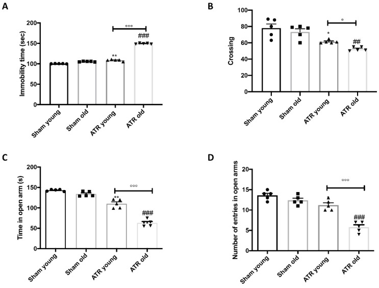
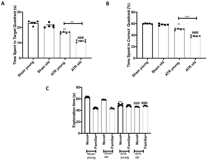
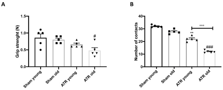
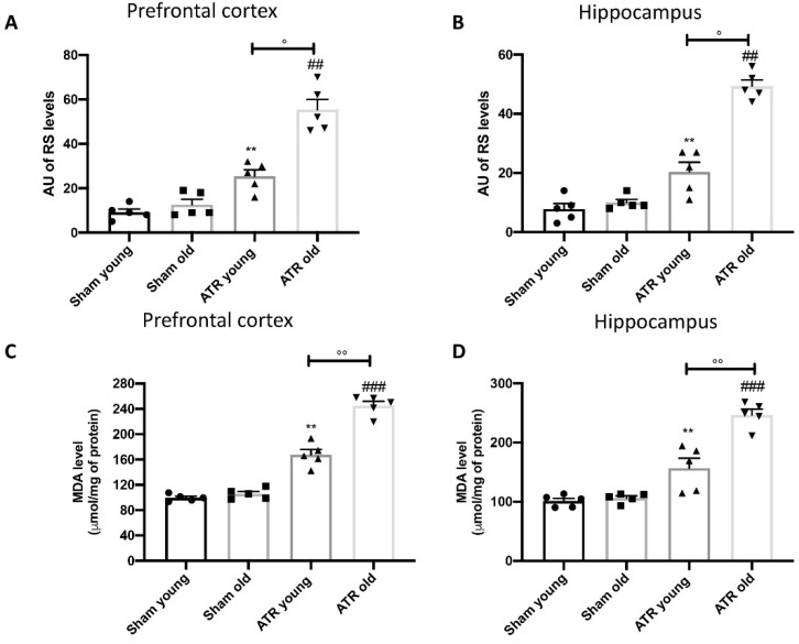
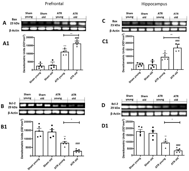
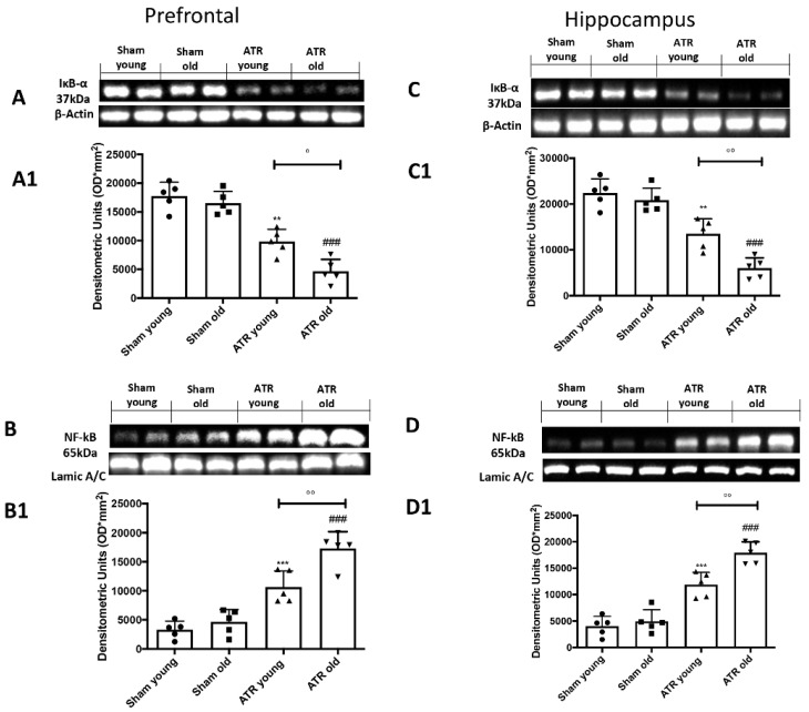
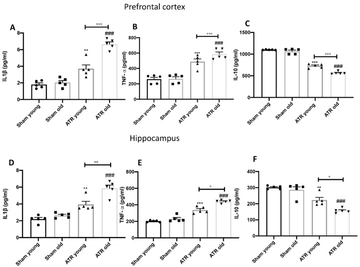
Results: exposure to ATR induced the same response in terms of behavioral alterations and motor and memory impairment in mice but in aged group was more marked. Additionally, in both young and aged mice ATR inhalations induced oxidative stress with impairment in physiological antioxidant response, lipid peroxidation, nuclear factor kappa-light-chain-enhancer of activated B cells (nf-κb) pathways activation with consequences of pro-inflammatory cytokines release and apoptosis. However, the older group was shown to be more sensitive to ATR inhalation.

Conclusions: our results showed that aged mice were more susceptible compared to young mice to air pollutants exposure, put in place a minor physiologically response was seen when exposed to it.

Keywords: aging; atrazine; brain alterations; endocrine disruptor; inflammation; oxidative stress.


Conflict of interest statement

The authors declare no conflict of interest.

Figures

Figure 1
Figure 1
Effect of ATR inhalation on anxiety and depression. Forced Swim Test (A); Open Field test (B); Time in open arms (C) and number of entries in open arms (D) was recorded during Elevated Plus Maze test. During the behavioral tests we found that aged mice were more susceptibility compared to young to show anxiety and depression after ATR inhalation. Data is expressed as the mean ± SEM of n = 5 animals for each group. * p < 0.05 vs. Sham young; ** p < 0.01 vs. Sham young; ## p < 0.01 vs. Sham old; ### p < 0.001 vs. Sham old; ° p < 0.05 ATR old vs. ATR young; °°° p < 0.001 ATR old vs. ATR young.
Figure 2
Figure 2
Effect of ATR inhalation on spatial learning and memory function. Morris Water Maze Test (A,B); Novel object recognition (C). MWM test showed that aged mice were more susceptible compared to young to develop spatial and memory deficits after ATR inhalation. No significant difference were found between young and aged mice during NOR. Data is expressed as the mean ± SEM of n = 5 animals for each group. ** p < 0.01 vs. Sham young; *** p < 0.001 vs. Sham young; ### p < 0.001 vs. Sham old; °° p < 0.01 ATR old vs. ATR young; °°° p < 0.001 ATR old vs. ATR young.
Figure 3
Figure 3
Effect of ATR inhalation on motor functions. Catalepsy test (A); Rotarod test (B); Time to turn (C) and Total time (D) for pole test. Aged mice were more susceptibility compared to young to develop motor deficits after ATR inhalation. Data is expressed as the mean ± SEM of n = 5 animals for each group. ** p < 0.01 vs. Sham young; ### p < 0.001 vs. Sham old; °°° p < 0.001 ATR old vs. ATR young.
Figure 4
Figure 4
Effect of ATR inhalation on grip strength and sociability. Grip strength (A); Social interaction test (B). Aged mice were more susceptibility to develop a reduction in grip strength and social interaction compared to young mice after ATR inhalation. Data is expressed as the mean ± SEM of n = 5 animals for each group. ** p < 0.01 vs. Sham young; # p < 0.05 vs. Sham old; ### p < 0.001 vs. Sham old; °°° p < 0.001 ATR old vs. ATR young.
Figure 5
Figure 5
Effect of ATR inhalation on oxidative stress and lipid peroxidation. RS levels in prefrontal cortex (A); RS levels in hippocampus (B); MDA levels in prefrontal cortex (C); MDA levels in hippocampus (D). After ATR exposure, both young and aged mice showed a significant increase in RS production as well as in lipid peroxidation in prefrontal cortex and hippocampus. Statistical analysis reveal that aged mice were more susceptibility compared to young mice to develop RS and lipid peroxidation. Data is expressed as the mean ± SEM of n = 5 animals for each group. ** p < 0.01 vs. Sham young; ## p < 0.01 vs. Sham old; ### p < 0.001 vs. Sham old; ° p < 0.05 ATR old vs. ATR young; °° p < 0.01 ATR old vs. ATR young.
Figure 6
Figure 6
Effect of ATR inhalation on SOD, CAT and GPx activity. By ELISA kit we investigated the effect of ATR inhalation on SOD, CAT and GPx activity in prefrontal cortex (A–C) as well as in the hippocampus (D–F). After ATR exposure, both young and aged mice showed a significant increase in physiological antioxidant response in prefrontal cortex and hippocampus. Statistical analysis revealed that older mice showed less physiological response than younger mice. Data is expressed as the mean ± SEM of n = 5 animals for each group. ** p < 0.01 vs. Sham young; *** p < 0.001 vs. Sham young; ## p < 0.01 vs. Sham old; °°° p < 0.001 ATR old vs. ATR young.
Figure 7
Figure 7
Impact of ATR inhalation on brain apoptosis. Western blots and respectively quantification of Bax in prefrontal cortex (A,A1) and hippocampus (C,C1) and Bcl-2 in prefrontal cortex (B,B1) and hippocampus (D,D1) We found that those aged mice are more subjected to brain apoptosis compared to young mice after 28 days of ATR inhalation. Data is expressed as the mean ± SEM of n = 5 animals for each group. ** p < 0.01 vs. Sham young; *** p < 0.01 vs. Sham young; ### p < 0.001 vs. Sham old; ° p < 0.05 ATR old vs. ATR young; °° p < 0.01 ATR old vs. ATR young.
Figure 8
Figure 8
Impact of ATR inhalation on brain inflammation. Western blots and respectively quantification of ik-bα in prefrontal cortex (A,A1) and hippocampus (C,C1) and nf-kb in prefrontal cortex (B,B1) and hippocampus (D,D1). We found that aged mice are more subjected to brain inflammation compared to young mice after 28 days of ATR inhalation. Data is expressed as the mean ± SEM of n = 5 animals for each group. ** p < 0.01 vs. Sham young; *** p < 0.001 vs. Sham young; ### p < 0.001 vs. Sham old; ° p < 0.05 ATR old vs. ATR young; °° p < 0.01 ATR old vs. ATR young.
Figure 9
Figure 9
Impact of ATR inhalation on cytokines production. ELISA kit of IL-1β (A) and TNF-α (B) and IL-10 (C) in prefrontal cortex. ELISA kit of IL-1β (D) and TNF-α (E) and IL-10 (F) in hippocampus. After ATR exposure, both young and aged mice showed a significantly increase in pro-inflammatory cytokines production and a significantly decrease in IL-10 release in prefrontal cortex as well as in hippocampus. Statistical analysis reveal that older mice showed a major inflammatory response than younger mice. Data is expressed as the mean ± SEM of n = 5 animals for each group. ** p < 0.01 vs. Sham young; *** p < 0.001 vs. Sham young; ### p < 0.001 vs. Sham old; ° p < 0.05 ATR old vs. ATR young. °° p < 0.01 ATR old vs. ATR young; °°° p < 0.001 ATR old vs. ATR young.

References

    1. Bondy S.C. Anthropogenic pollutants may increase the incidence of neurodegenerative disease in an aging population. Toxicology. 2016;341–343:41–46. doi: 10.1016/j.tox.2016.01.007. - DOI - PubMed
    1. Sharman E.H., Bondy S.C., Sharman K.G., Lahiri D., Cotman C.W., Perreau V.M. Effects of melatonin and age on gene expression in mouse CNS using microarray analysis. Neurochem. Int. 2007;50:336–344. doi: 10.1016/j.neuint.2006.09.001. - DOI - PMC - PubMed
    1. Bondy S.C., Sharman E.H. Melatonin and the aging brain. Neurochem. Int. 2007;50:571–580. doi: 10.1016/j.neuint.2006.12.014. - DOI - PubMed
    1. Bondy S.C., Lahiri D.K., Perreau V.M., Sharman K.Z., Campbell A., Zhou J., Sharman E.H. Retardation of brain aging by chronic treatment with melatonin. Ann. N. Y. Acad. Sci. 2004;1035:197–215. doi: 10.1196/annals.1332.013. - DOI - PubMed
    1. Sharman E.H., Vaziri N.D., Ni Z., Sharman K.G., Bondy S.C. Reversal of biochemical and behavioral parameters of brain aging by melatonin and acetyl L-carnitine. Brain Res. 2002;957:223–230. doi: 10.1016/S0006-8993(02)03551-5. - DOI - PubMed