Review
. 2021 Jul 26;22(15):7984.
doi: 10.3390/ijms22157984.

Sex Steroids and the Shaping of the Peripubertal Brain: The Sexual-Dimorphic Set-Up of Adult Neurogenesis

Affiliations
Review

Sex Steroids and the Shaping of the Peripubertal Brain: The Sexual-Dimorphic Set-Up of Adult Neurogenesis

Sara Trova et al. Int J Mol Sci. .

Abstract

Steroid hormones represent an amazing class of molecules that play pleiotropic roles in vertebrates. In mammals, during postnatal development, sex steroids significantly influence the organization of sexually dimorphic neural circuits underlying behaviors critical for survival, such as the reproductive one. During the last decades, multiple studies have shown that many cortical and subcortical brain regions undergo sex steroid-dependent structural organization around puberty, a critical stage of life characterized by high sensitivity to external stimuli and a profound structural and functional remodeling of the organism. Here, we first give an overview of current data on how sex steroids shape the peripubertal brain by regulating neuroplasticity mechanisms. Then, we focus on adult neurogenesis, a striking form of persistent structural plasticity involved in the control of social behaviors and regulated by a fine-tuned integration of external and internal cues. We discuss recent data supporting that the sex steroid-dependent peripubertal organization of neural circuits involves a sexually dimorphic set-up of adult neurogenesis that in turn could be relevant for sex-specific reproductive behaviors.

Keywords: adult neurogenesis; gonadal hormones; pheromones; puberty; sexual dimorphism.


Conflict of interest statement

The authors declare no conflict of interest.

Figures

Figure 1
Figure 1
Sex hormones and adult neurogenesis in the mouse brain. In (A) is represented the brain sensitivity to sex hormones and pheromones and the time course of endogenous secretions and adult neurogenesis from prepubertal to postpubertal life. According to the “two-stage model” proposed in Schulz and Sisk (2016), the gray to white nuance in the picture illustrates the progressive postnatal reduction of structural brain sensitivity to sex steroids, which peaks during the early prepubertal life (0–3.5 weeks) and peripuberty (3.5–7 weeks), in coincidence with GnRH, testosterone and estradiol release (solid-colored lines). The onset of puberty is triggered by GnRH increase secretion, which results in a rise of circulating sex steroids. In the peripubertal period, adult neurogenesis (solid black line) halves, coinciding with the increase in sex steroids, and in females starts to be modulated by male pheromones (dotted black line). In (B) is depicted a representation of adult neurogenesis in the SVZ/OB region during postnatal life and its pheromonal-dependent modulation in females. In the prepubertal period, the level of AN is elevated (green and red cells) and it does not change upon exposure to opposite-sex pheromones. Starting from the late peripubertal period male pheromones stimulate AN and secretion of gonadal hormones, which reciprocally interact to optimize reproduction. Note that peripubertal life is regarded as an important phase of structural and functional hormone-dependent refinement of sexually dimorphic circuits, which includes a set-up of adult neurogenesis in both male and female mice. Abbreviations: AN, adult neurogenesis; E, estradiol; GnRH, gonadotropin-releasing hormones; T, testosterone; SVZ/OB, subventricular zone/olfactory bulb.
Figure 2
Figure 2
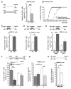
Time course and modulation of adult neurogenesis during peripubertal life and adulthood. (A) Injections of the cell birth marker BrdU before puberty (P21) results in double the number of 28-day-old newborn granule cell (GC) neurons in the AOB when compared with BrdU treatments performed after the onset of puberty (at P28) (unpaired Student’s t-test, P21 vs. P28, p = 0.0004), indicating a sharp decrease of adult neurogenesis during peripubertal life. (B) Graph showing the onset of puberty in female mice determined by examination of vaginal opening and occurrence of the first estrus. (C,D) Daily exposure of female mice to pheromones (male soiled bedding, MB) increases GC survival only after the onset of the first estrus cycle (P35–P42). Bedding stimulations were timed during the 2nd week after GC genesis and their survival evaluated 28 dpi (unpaired Student’s t-test, P28–P35 ctrl vs. MB, p = 0.669, P35–P42 ctrl vs. MB, ⇤ p = 0.016) (see Oboti et al. 2017 for major details). (E) Bedding exposure in ovariectomized (OVX) prepubertal females (P21) does not modulate GC survival as significantly as in SHAM-operated animals (two-way ANOVA, factors: ovariectomy, F(2,10) = 6.766, p = 0.017; stimulus, F(2,10)= 12.14, p = 0.002; interaction, F(2,10)= 0.520, p = 0.479; Tukey’s post hoc, SHAM ctrl vs. SHAM MB, ⇤ p = 0.045; OVX ctrl vs. OVX MB, p = 0.206). (F) In the GnRH::cre/DicerloxP/loxP mice, GnRH is significantly downregulated starting from P21. Adult neurogenesis in these animals was quantified by BrdU injections at 3–4 months of age. The density of progenitors cells (Ki67+ cells) in the dorsolateral subventricular zone (dlSVZ) is significantly reduced in males, whereas no differences were found in females. (G) By contrast, in the main olfactory bulb (MOB), only females showed a reduction in newborn cell survival (density of BrdU+ cells). Interestingly, wild-type females have a higher density of BrdU cells compared with wild-type males, indicating sexually dimorphic neurogenesis in this region. (H). Survival of 28-day-old newborn granule cells in the MOB of control females ovariectomized at P21 (when GnRH in GnRH::cre/DicerloxP/loxP mice is downregulated) and injected with BrdU at P28 shows significant reduction when compared with SHAM operated females (Student’s t-test, * p = 0.032), suggesting that the alteration of adult neurogenesis identified in GnRH::cre/DicerloxP/loxP females are mostly attributable to impaired sex steroids secretion (see the text and Trova et al. 2020 for major details). * p < 0.05; ** p < 0.01; *** p < 0.001. Abbreviations: ctrl, control; MB, male bedding; MOB, main olfactory bulb; AOB, accessory olfactory bulb; GrL, granule cell layer; OVX, ovariectomy; dpi, days post-injection. All graphs were taken or modified from [14,88].

References

    1. Fox S.E., Levitt P., Nelson C.A. How the timing and quality of early experiences influence the development of brain architecture. Child. Dev. 2011;81:28–40. doi: 10.1111/j.1467-8624.2009.01380.x. - DOI - PMC - PubMed
    1. Stoka A.M. Phylogeny and evolution of chemical communication: An endocrine approach. J. Mol. Endocrinol. 1999;22:207–225. doi: 10.1677/jme.0.0220207. - DOI - PubMed
    1. Phoenix C.H., Goy R.W., Gerall A.A., Young W.C. Organizing action of prenatally administered testosterone propionate on the tissues mediating mating behavior in the female guinea pig. Endocrinology. 1959;65:369–382. doi: 10.1210/endo-65-3-369. - DOI - PubMed
    1. Bakker J. Sexual differentiation of the neuroendocrine mechanisms regulating mate recognition in mammals. J. Neuroendocrinol. 2003;15:615–621. doi: 10.1046/j.1365-2826.2003.01036.x. - DOI - PubMed
    1. Baum M.J., Bakker J. Roles of sex and gonadal steroids in mammalian pheromonal communication. Front. Neuroendocrinol. 2013;34:4. doi: 10.1016/j.yfrne.2013.07.004. - DOI - PubMed

Substances