Review
. 2021 Jul 27;22(15):7999.
doi: 10.3390/ijms22157999.

Human Mitochondrial RNA Processing and Modifications: Overview

Affiliations
Review

Human Mitochondrial RNA Processing and Modifications: Overview

Marta Jedynak-Slyvka et al. Int J Mol Sci. .

Abstract

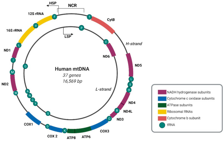
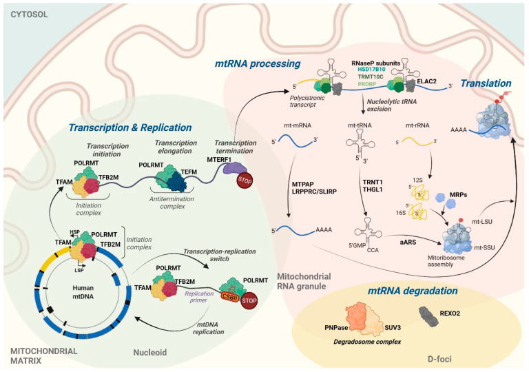
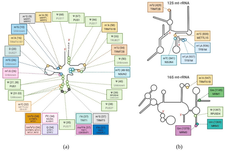
Mitochondria, often referred to as the powerhouses of cells, are vital organelles that are present in almost all eukaryotic organisms, including humans. They are the key energy suppliers as the site of adenosine triphosphate production, and are involved in apoptosis, calcium homeostasis, and regulation of the innate immune response. Abnormalities occurring in mitochondria, such as mitochondrial DNA (mtDNA) mutations and disturbances at any stage of mitochondrial RNA (mtRNA) processing and translation, usually lead to severe mitochondrial diseases. A fundamental line of investigation is to understand the processes that occur in these organelles and their physiological consequences. Despite substantial progress that has been made in the field of mtRNA processing and its regulation, many unknowns and controversies remain. The present review discusses the current state of knowledge of RNA processing in human mitochondria and sheds some light on the unresolved issues.

Keywords: RNA decay; RNA modifications; RNA processing; mitochondria; mitochondrial genome; mitochondrial transcription.


Conflict of interest statement

The authors declare no conflict of interest.

Figures

Figure 1
Figure 1
Map of the human mitochondrial genome. A double-stranded circular mitochondrial DNA (mtDNA) molecule includes 37 genes encoding 13 subunits of the OXPHOS system, 2 rRNAs, and 22 tRNAs. Transcription of both mtDNA strands initiates within the non-coding regulatory region (NCR), and the black arrows indicate transcription direction. HSP, LSP–transcription promoter of H- and L-strand, respectively. Open reading frames of ATP8/ATP6 and ND4L/ND4 are marked. Created with BioRender.com.
Figure 2
Figure 2
General overview of human mitochondrial gene expression. mtDNA replication and transcription occur within nucleoid structures (represented in light green) that spatially co-localize with mtRNA granules (MRGs, represented in light pink), which are suggested to be the place of post-transcriptional mtRNA processing and mitoribosome assembly. Degradation of mtRNA, mediated by the degradosome complex that involves SUV3 helicase and PNPase, occurs in D-foci that partially co-localize with MRGs. REXO2 exonuclease mediates the subsequent decay of RNA oligonucleotides. Aminoacyl-tRNA synthetases are abbreviated as aARS; mitochondrial ribosomal proteins are referred to as MRPs. Created with BioRender.com.
Figure 3
Figure 3
Summary of post-transcriptional base modification of human mt-tRNAs and mt-rRNAs. Schematic representation of the secondary structure of: (a) generic mt-tRNA; and (b) 12S and 16S mt-rRNA with indicated post-transcriptional base modifications (circles) identified in humans. Chemical modification, base position number (in brackets), and enzyme responsible (if known) for each position are depicted in boxes. The modifying enzymes predicted to be responsible for the indicated modification, but not experimentally confirmed, are indicated in gray and followed by a question mark. Modifications shown: 1-methyladenosine (m1A), 1-methylguanosine (m1G), N2-methylguanosine (m2G), dihydrouridine (D), N2,N2-dimethylguanosine (m22G), pseudouridine (Ψ), 3-methylcytidine (m3C), 5-taurinomethyluridine (τm5U), 5-taurinomethyl-2-thiouridine (τm5s2U), 5-formylcytidine (f5C), Queuosine (Q), N6-isopentenyladenosine (i6A), 2-methylthio-N6-isopentenyladenosine (ms2i6A), N6-threonylcarbamoyladenosine (t6A), 5-methylcytidine (m5C), 5-methyluridine (m5U), N4-methylcytidine (m4C), N6,N6-dimethyladenosine (m62A), 2′-O-methylguanosine (Gm), and 2′-O-methyluridine (Um). Created with BioRender.com.

References

    1. Robberson D.L., Clayton D.A. Replication of Mitochondrial DNA in Mouse L Cells and Their Thymidine Kinase- Derivatives: Displacement Replication on a Covalently-Closed Circular Template. Proc. Natl. Acad. Sci. USA. 1972;69:3810–3814. doi: 10.1073/pnas.69.12.3810. - DOI - PMC - PubMed
    1. Anderson S., Bankier A.T., Barrell B.G., de Bruijn M.H.L., Coulson A.R., Drouin J., Eperon I.C., Nierlich D.P., Roe B.A., Sanger F., et al. Sequence and organization of the human mitochondrial genome. Nature. 1981;290:457–465. doi: 10.1038/290457a0. - DOI - PubMed
    1. Istiaq Alam T., Kanki T., Muta T., Ukaji K., Abe Y., Nakayama H., Takio K., Hamasaki N., Kang D. Human mitochondrial DNA is packaged with TFAM. Nucleic Acids Res. 2003;31:1640–1645. doi: 10.1093/nar/gkg251. - DOI - PMC - PubMed
    1. Garrido N., Griparic L., Jokitalo E., Wartiovaara J., van der Bliek A.M., Spelbrink J.N. Composition and dynamics of human mitochondrial nucleoids. Mol. Biol. Cell. 2003;14:1583–1596. doi: 10.1091/mbc.e02-07-0399. - DOI - PMC - PubMed
    1. Bogenhagen D.F., Wang Y., Shen E.L., Kobayashi R. Protein components of mitochondrial DNA nucleoids in higher eukaryotes. Mol. Cell. Proteom. 2003;2:1205–1216. doi: 10.1074/mcp.M300035-MCP200. - DOI - PubMed

MeSH terms

LinkOut - more resources