. 2021 Jul 28;22(15):8066.
doi: 10.3390/ijms22158066.

Photoreceptor Compartment-Specific TULP1 Interactomes

Affiliations

Photoreceptor Compartment-Specific TULP1 Interactomes

Lindsey A Ebke et al. Int J Mol Sci. .

Abstract

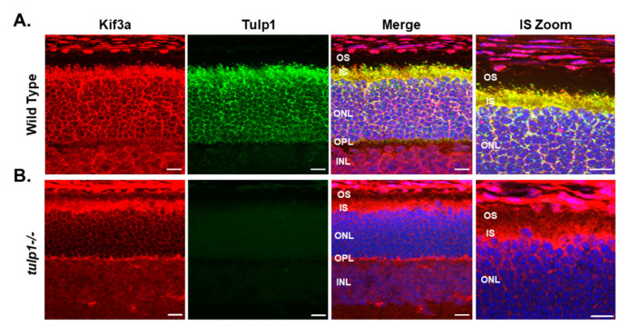
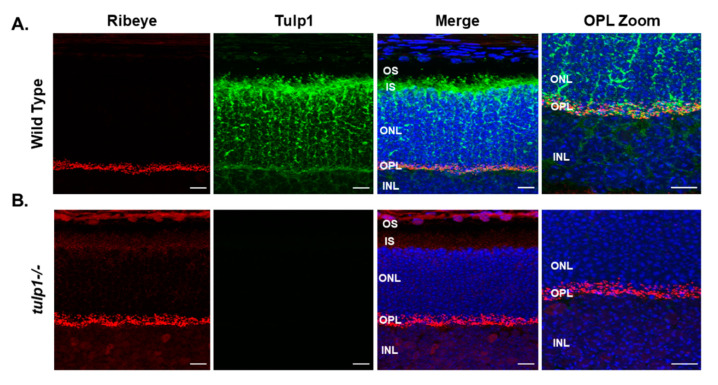
Photoreceptors are highly compartmentalized cells with large amounts of proteins synthesized in the inner segment (IS) and transported to the outer segment (OS) and synaptic terminal. Tulp1 is a photoreceptor-specific protein localized to the IS and synapse. In the absence of Tulp1, several OS-specific proteins are mislocalized and synaptic vesicle recycling is impaired. To better understand the involvement of Tulp1 in protein trafficking, our approach in the current study was to physically isolate Tulp1-containing photoreceptor compartments by serial tangential sectioning of retinas and to identify compartment-specific Tulp1 binding partners by immunoprecipitation followed by liquid chromatography tandem mass spectrometry. Our results indicate that Tulp1 has two distinct interactomes. We report the identification of: (1) an IS-specific interaction between Tulp1 and the motor protein Kinesin family member 3a (Kif3a), (2) a synaptic-specific interaction between Tulp1 and the scaffold protein Ribeye, and (3) an interaction between Tulp1 and the cytoskeletal protein microtubule-associated protein 1B (MAP1B) in both compartments. Immunolocalization studies in the wild-type retina indicate that Tulp1 and its binding partners co-localize to their respective compartments. Our observations are compatible with Tulp1 functioning in protein trafficking in multiple photoreceptor compartments, likely as an adapter molecule linking vesicles to molecular motors and the cytoskeletal scaffold.

Keywords: Tulp1; cilia; photoreceptor degeneration; protein trafficking; proteomics; synapse.


Conflict of interest statement

The authors declare no conflict of interest.

Figures

Figure 1
Figure 1
Isolation of Tulp1-containing photoreceptor compartments. (A) OCT image of a retinal flat mount preparation to assess the quality of the dissected retina. Any disturbed or folded peripheral edges, highlighted by a star, were removed prior to tangential sectioning. The red rectangle encompasses the nitrocellulose support. (B) A dot blot analysis of all serial sections probed with antibodies against rhodopsin. A blot in which only the outermost 4–5 sections corresponding to the photoreceptor OSs were strongly positive for rhodopsin indicated optimally sectioned retinas. (C) A schematic illustration of retina showing each cell type. The protein content of each tangential section was analyzed by Western blot probed for known compartment-specific proteins. The five marker proteins included Phosducin, Tulp1, Synaptophysin, uMtCK and rhodopsin. Each lane of the gel represents the protein content of a single 10 μm section starting from the GCL and progressing through the photoreceptor OSs. The GCL-, IS-, OPL-containing samples used for further analysis are circled in red.
Figure 2
Figure 2
IP of retinal lysate. The top panels show Western blot analysis of the MAP1B IP experimental samples probed with Tulp1 antibodies. In the IP product lane, a band corresponding to Tulp1 is detected. A corresponding band is seen in the rat retinal lysate and wt mouse retinal homogenate but not in the tulp1−/− retinal lysate, liver lysate or non-specific IgG IP lanes. The bottom panels show Western blot analysis of the MAP1B IP experimental samples probed with MAP1B antibodies. In the IP product, rat retinal lysate, wt mouse retinal lysate and tulp1−/− retinal lysate lanes, a band corresponding to MAP1B is detected. No bands are seen in the liver or non-specific IgG IP sample lanes.
Figure 3
Figure 3
Immunolocalization of MAP1B and Tulp1 in P17 mouse retinas. (A) Wt retinal sections stained with MAP1B (red) and Tulp1 (green). (B) Tulp1−/− retinal sections stained with MAP1B (red) and Tulp1 (green). Sections were counterstained with DAPI (blue). Scale bar: 50 µm. INL, inner nuclear layer; OPL, outer plexiform layer; ONL, outer nuclear layer; IS, inner segment layer; OS, outer segment layer.
Figure 4
Figure 4
IP of retinal lysate. The top panels show Western blot analysis of the Kif3a IP experimental samples probed with Tulp1 antibodies. In the IP product lane, a band corresponding to Tulp1 is detected. A corresponding band is seen in the rat retinal lysate and wt mouse retinal homogenate but not in the tulp1−/− retinal lysate, heart lysate or non-specific IgG IP lanes. The bottom panels show Western blot analysis of the Kif3a IP experimental samples probed with Kif3a antibodies. In the IP product, rat retinal lysate, wt mouse retinal lysate and tulp1−/− retinal lysate lanes, a band corresponding to Kif3a is detected. No bands are seen in the heart or non-specific IgG IP sample lanes.
Figure 5
Figure 5
Immunolocalization of Kif3a and Tulp1 in P17 mouse retinas. (A) Wt retinal sections stained with Kif3a (red) and Tulp1 (green). (B) Tulp1−/− retinal sections stained with Kif3a (red) and Tulp1 (green). Sections were counterstained with DAPI (blue). Scale bar: 50 µm. INL, inner nuclear layer; OPL, outer plexiform layer; ONL, outer nuclear layer; IS, inner segment layer; OS, outer segment layer.
Figure 6
Figure 6
IP of retinal lysate. The top panels show Western blot analysis of the Ribeye IP experimental samples probed with Tulp1 antibodies. In the IP product lane, a band corresponding to Tulp1 is detected. A corresponding band is seen in the rat retinal lysate and wt mouse retinal homogenate but not in the tulp1−/− retinal lysate, spleen lysate or non-specific IgG IP lanes. The bottom panel shows Western blot analysis of the Ribeye IP experimental samples probed with Ribeye antibodies. In the IP product, rat retinal lysate, wt mouse retinal lysate and tulp1−/− retinal lysate lanes, a band corresponding to Ribeye is detected. No bands are seen in the spleen or non-specific IgG IP sample lanes.
Figure 7
Figure 7
Immunolocalization of Ribeye and Tulp1 in P17 mouse retinas. (A) Wt retinal sections stained with Ribeye (red) and Tulp1 (green). (B) Tulp1−/− retinal sections stained with Ribeye (red) and Tulp1 (green). Sections were counterstained with DAPI (blue). Scale bar: 50 µm. INL, inner nuclear layer; OPL, outer plexiform layer; ONL, outer nuclear layer; IS, inner segment layer; OS, outer segment layer.

References

    1. Pearring J.N., Salinas R.Y., Baker S.A., Arshavsky V.Y. Protein sorting, targeting and trafficking in photoreceptor cells. Prog. Retin Eye Res. 2013;36:24–51. doi: 10.1016/j.preteyeres.2013.03.002. - DOI - PMC - PubMed
    1. Palczewski K. Chemistry and biology of vision. J. Biol. Chem. 2012;287:1612–1619. doi: 10.1074/jbc.R111.301150. - DOI - PMC - PubMed
    1. Arshavsky V.Y., Burns M.E. Photoreceptor signaling: Supporting vision across a wide range of light intensities. J. Biol. Chem. 2012;287:1620–1626. doi: 10.1074/jbc.R111.305243. - DOI - PMC - PubMed
    1. Young R.W. The renewal of photoreceptor cell outer segments. J. Cell Biol. 1967;33:61–72. doi: 10.1083/jcb.33.1.61. - DOI - PMC - PubMed
    1. Young R.W., Bok D. Participation of the retinal pigment epithelium in the rod outer segment renewal process. J. Cell Biol. 1969;42:392–403. doi: 10.1083/jcb.42.2.392. - DOI - PMC - PubMed

LinkOut - more resources